
Greeting
Good day, dear Professor Doctor …,
I would now like to present the patient Karin Weber to you.
She is 43 years old, one meter sixty-eight tall and weighs about 80 kg. The body mass index (BMI) is 28.3 kg/m² and thus falls into the overweight range.
Current history
The 43-year-old patient, Ms. Weber, presented with colicky, colicky, acute pain in the right flankthat had been increasing for about three hours.
The pain radiates into the vaginaas well as into the right inguinal region .
According to her statements, the pain intensity was estimated at about 7 out of 10.
She had not experienced such pain previously.
The following was reported: Hematuria, Dysuriaas well as alguria in the form of burning. In addition there is nausea, a single episode of emesiswith food remnants, pollakisuria (with subjectively low urine volume; five micturitions since the morning), a general feeling of weakness as well as hyperhidrosis.
The following questions were denied: fever, urgency, incontinence, weakening or interruption of the urinary stream, newly occurring change in bowel movements, pathological vaginal bleeding, vaginal discharge, dyspnea, tachycardia, dizziness, skin changes, edema.
Autonomic history
The autonomic history is unremarkable, apart from stress-related insomniain the form of both difficulty falling asleep and maintaining sleep in connection with family stresses in the context of a divorce.
Past medical history
According to the history there has been a known type 2 diabetes mellitus for two years, which the patient regularly self-monitors.
Fasting blood glucose values are around 110 mg/dl, postprandial values rise to approx. 155 mg/dl.
Diabetes-associated complications are not known so far.
Regular ophthalmologic check-ups have not been performed to date.
Furthermore, there has been arterial hypertension for about two years, which is currently well controlled under ongoing therapy. Blood pressure values are approximately 125/85 mmHg.
The patient reports a recurring tendovaginitisof the right wrist for three years, which is job-related.
The complaints have so far been treated symptomatically with a brace and intermittent use of metamizole.
About six months ago there was a herpes zosterinfection on the right thoracic side, which was successfully treated with aciclovir.
According to the patient, no childhood vaccinations were received, which at the age of eight led to a pronounced measles infection (morbilli) with accompanying pneumonia. Due to the course of the illness, a two-day inpatient admission was required.
Previous surgeries
Fifteen years ago the patient sustained a comminuted fracture of the right olecranon. Surgical treatment was performed with osteosynthetic stabilization using plates and screws. Postoperatively, immobilization with a splint was required for several weeks, followed by physiotherapeutic aftercare. The metal removal took place about one year later. The course was uncomplicated and there are currently no complaints.
Five years ago the patient underwent a hysterectomy due to atypical uterine bleeding and pain.
The cause was uterine fibroids.
A hysterectomy (subtotal or total) was performed, with the adnexa left in place.
Gynecology
Eight and seventeen years ago respectively she had spontaneous vaginal deliveries. She currently has no gynecological complaints.
Medications
The patient is currently taking the following medications:
Zentrum® Multivitamin, 1 tablet daily
Atacand® 4 mg, 1 × daily
Metformin 500 mg, 2 × daily
Metamizole 500 mg, as needed
and administers the EpiPen® as needed
———–
Aciclovir (400 mg, 1 tablet 5× daily)
Allergies
The patient reported the following allergies:
Known wasp venom allergy with urticaria and dyspnea in the sense of an anaphylactic reaction.
Noxae
The patient smoked about five cigarettes daily for approximately ten years (equivalent to about 2.5 pack years). She has not smoked for three years.
She drinks one to two bottles of beer every evening to sleep better. She reports no drug use.
Social history
The patient works as a self-employed pastry chef and owns her own business. She currently lives alone and is in the process of separation; a divorce from her husband is planned.
The patient is the mother of two children: her 17-year-old daughter suffers from anorexia nervosa, her 8-year-old son from an attention deficit/hyperactivity disorder (ADHD).
Family history
The patient’s father was a heavy smoker and, as a result of advanced peripheral arterial occlusive disease, underwent a forefoot amputation with removal of the three anterior toes on the right foot.
The patient’s 70-year-old mother suffers from age-related scoliosis, which was treated conservatively with brace therapy and physiotherapy. She currently lives in a nursing home. In addition, there is a bilaterally surgically treated cataract.
The patient’s 51-year-old brother suffered a carotid dissection, at the age of 49, which was treated surgically.
She has no pets.
Three months ago she travelled to Italy
Suspected and differential diagnosis
In my opinion, the anamestic information – in particular the character of the pain, the location and the presence of dysuria and hematuria – most likely indicate a urolithiasis .
Differential diagnoses include a pyelonephritis (e.g. with additional fever), a lumbar back pain (with radiating pain and anamnestically reported physical overexertion).
In this case an extrauterine pregnancy (e.g. tubal = ectopic pregnancy) is not possible due to the hysterectomy performed.
With pain in the hypogastrium, differential considerations also include a diverticulitis (left-sided) or an appendicitis (right-sided).
Diagnostic work-up
After administration of an analgesic, it should be checked whether there is an obstruction to urinary flow. If such an obstruction exists, the patient should be admitted for inpatient care and further diagnosed and treated accordingly. If there is no obstruction, further management depends on the size and location of the stones.
physical examination
After the history, a detailed physical examination should be performed. First the vital parameters are recorded, including heart rate, blood pressure, respiratory rate, oxygen saturation and body temperature.
Subsequently the physical examination focuses on the described complaints. It begins with inspection, looking for possible abnormalities such as skin changes or swelling (e.g. bladder distension).
This is followed by palpation of the flanks and abdomen to assess tenderness or other pathological findings. To palpate the urinary bladder, place the fingers of one hand extended cranial to the symphysis over the lower abdomen and apply brief pressure to the bladder region.
To assess the kidneys, a percussion pain test can be performed: the clinician taps both flanks with the fist or the edge of the hand – initially gently and orienting, if negative, somewhat more forcefully. Normally the patient feels only a jolt, but no pain.
Percussion is best suited for estimating the size of the urinary bladder.
Finally auscultation is performed. In renal artery stenosis, aneurysms of the renal arteries or arteriovenous shunts, a flow murmur may occasionally be auscultable on the right or left at the costal margin.
Since the patient has both arterial hypertension and diabetes mellitus, initial diagnostics should also include first indications of sequelae such as retinopathy, neuropathy or peripheral arterial occlusive disease.
|
Additional knowledge |
|
The kidneys and the upper urinary tract are retroperitoneal, whereas the urinary bladder and the internal female genital organs are extraperitoneal. The left kidney is anatomically located slightly lower than the right. With maximal bladder filling (approx. 500 ml) it rises into the abdominal cavity. |
Laboratory tests
These would include a complete blood count, blood glucose, electrolytes (sodium, potassium), renal retention parameters (creatinine, urea), C-reactive protein (CRP) and the erythrocyte sedimentation rate (ESR).
In addition, a urinalysis and a urine culture should be performed. Urinalysis may reveal leukocyturia, hematuria or proteinuria. The urine culture serves to detect a urinary tract infection.
For reliable results certain conditions must be met when collecting urine: if possible the urine should be obtained as first-morning urine and analyzed within 45 minutes. Urine from collection bags or indwelling catheters is not suitable. The first portion is discarded and the midstream urine is collected (midstream specimen). In certain cases targeted urine collection using a two-glass test, catheterization or suprapubic aspiration may be required.
Urinalysis is performed using dipsticks.
For urine culture the urine is collected under sterile conditions and microbiologically examined using a culture method.
Imaging modalities
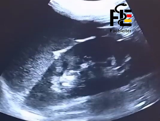
Abdominal ultrasound (first choice)
Ultrasound is an imaging modality that uses sound waves to examine internal organs. You should lie flat and remain as still as possible. First a special gel is applied to the skin. I then move the transducer over the skin to examine the desired areas. The examination generally requires no special preparation, is painless and takes about 20 minutes.
In nephrolithiasis, renal stones can be detected as bright echoes. Enlarged renal calyces or a dilated renal pelvis (hydronephrosis) can also be visualized as an indication of urinary outflow obstruction.
Abdominal CT – standard procedure for detection of urolithiasis
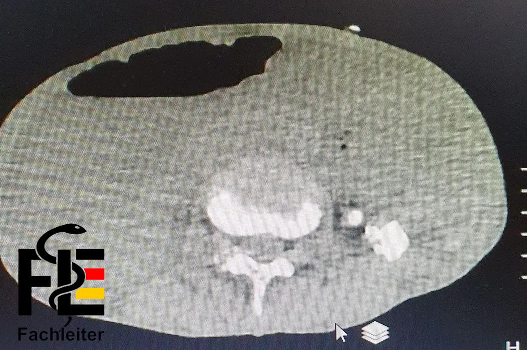
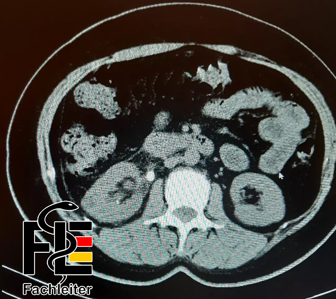
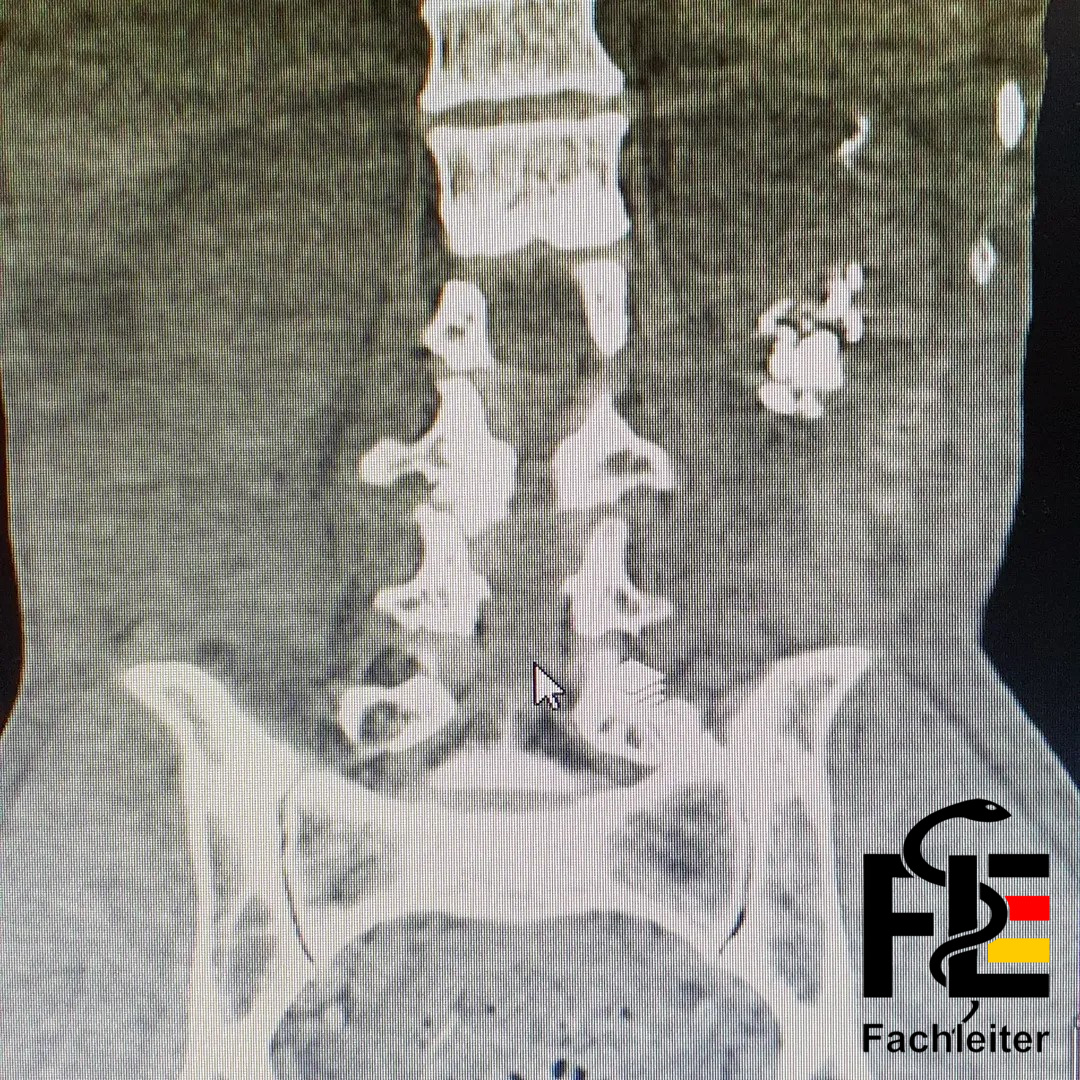
Computed tomography (CT) is an imaging modality that visualizes internal organs using X-rays and generates cross-sectional images of the body. The patient should lie flat while being moved into a tubular scanner. During the examination, which lasts only a few minutes, the patient must remain still. CT is painless, but in some cases special preparation may be necessary. Sometimes contrast medium is used to better delineate structures. When administering contrast, existing allergies, possible dehydration, urinary tract obstruction, pre-existing renal insufficiency and the intake of nephrotoxic medications such as metformin should be considered.
In nephrolithiasis, urinary stones typically appear as hyperdense (bright) structures within the renal pelvis, ureters or bladder. Secondary signs such as dilation of the pelvicalyceal system (hydronephrosis) or ureteral dilatation may also be observed.
Because sonography involves no radiation exposure, it is preferred over CT in pregnant patients.
X-ray
Conventional radiographs such as the kidney plain radiograph and intravenous urography with contrast are today considered diagnostically less informative and, due to their limited sensitivity, are increasingly regarded as obsolete.
Only radiopaque stones – for example calcium-containing stones – are visible
Treatment
NSAIDs (e.g. ketorolac, ibuprofen) are excellent for treating renal colic and are considered first-line therapy.
• Ketorolac i.m. 15–30 mg
• Morphine 2–5 mg i.v. slowly titrated (alternatively 5–10 mg i.m. if no i.v. access is available)
• Ondansetron 4 mg slowly i.v. → 4 mg i.m.
• Intravenous fluid administration
If a urinary tract infection is suspected in combination with an obstructing stone, urgent renal decompression is required – either by placement of a ureteral stent (double-J stent) by a urologist or by percutaneous nephrostomy – in combination with antibiotic therapy.
In patients with a small stone (particularly in the distal ureter) and a high probability of spontaneous passage, medical expulsive therapy with α-blockers (e.g. tamsulosin) can be helpful.
Therapy overview by location and size:
Proximal ureteral stones < 1 cm diameter:
• shock wave lithotripsy (SWL) or ureterorenoscopy (URS)
Proximal ureteral stones > 1 cm diameter:
• SWL or URS
Distal ureteral stones:
• URS
acute pyelonephritis
Outpatient
Ciprofloxacin 500 mg twice daily for 7 days
(or levofloxacin 750 mg once daily for 5 days)
Inpatient
Ceftriaxone 1–2 g i.v. once daily
After improvement of the general condition switch to oral therapy with ciprofloxacin, generally after 48–72 hours.
Complications
Urinary outflow obstruction / urinary retention (hydronephrosis)
Pyelonephritis / Urosepsis
Chronic renal insufficiency
Consultations
Urology
|
Area |
Designation |
Main symptoms |
|
Lower urinary tract |
Lower UTI (Cystitis, urethritis) |
Dysuria, pollakisuria, alguria, suprapubic pain, cloudy urine, no fever |
|
Upper urinary tract |
Upper UTI (Pyelonephritis) |
Fever, flank pain, chills, nausea, vomiting |
Extra
Names:
Katherine Weiss, Frau Eiermann, Hubert Winkelmann, Friedrich Stein, Ulli Stockmann, Toni Teurek
Professions:
She owns her own cheese shop
Owner of a pub
Details:
She comes to us because her general practitioner is on vacation.
He doesn’t know whether he has gained weight; he eats a lot because he has a food truck.
He lives with his poodle.
History of cesarean section 40, 38 and 36 years ago, followed by sterilization surgery (the patient cannot remember the exact type of procedure).
Previous illnesses:
Tenosynovitis due to work in a butcher’s shop
Hyperuricemia
Contusions (occupation-related as an auto mechanic)
He burned his arm with hot water yesterday and only applied a bandage.
Migraine
COPD
Benign prostatic hyperplasia (BPH): transurethral resection of the prostate
Appendectomy
Medications
Melatonin
Diclofenac ointment as needed
Hormone patch
Allopurinol 300 mg 1-0-0
Sumatriptan
Symbicort
Allergies
Grass pollen allergy with rhinoconjunctivitis
Contrast agent allergy (erythema and edema)
Drug history
Tried drugs at parties at age 17–18, no further attempts at consumption thereafter
Family history:
Father: CAD, stent implantation, arterial hypertension
Mother: PAD, depression
Brother: bypass surgery
Questions
1. What do the following medical terms mean?
Polyuria → increased urine output (> 2–3 liters/24 h) (e.g., in diabetes mellitus, diabetes insipidus)
Oliguria → decreased urine output (< 500 ml/24 h) (e.g., in dehydration, renal insufficiency)
Anuria → severely reduced or absent urine output (< 100 ml/24 h) (e.g., in acute renal failure, obstruction)
Nocturia → increased nighttime urination (e.g., in heart failure)
2. Which spasmolytics do you know?
Spasmolytics are medications that relax smooth muscle and relieve cramp-like pain (especially in the gastrointestinal and urogenital tracts). Known active substances include:
Butylscopolamine (e.g., Buscopan®)
Mebeverine
Trospium chloride (especially for overactive bladder)
3. Can one get measles in adulthood?
Yes, measles can also occur in adulthood—especially if adequate vaccination protection is lacking.
Measles are not solely a childhood disease.
Adults without vaccination protection or without a previous infection can become infected at any time.
Complications such as: pneumonia, otitis media, hepatitis
4. What are the specialist titles of the following physicians?
Gynecologist → gynecologist
Pediatrician → pediatrician
Dermatologist → dermatologist
Neurologist → neurologist
Ophthalmologist → ophthalmologist
5. Briefly explain what gout is.
Arthritis urica, also known as gouty arthritis, is an inflammatory joint disease caused by deposition of uric acid crystals (sodium urate) in the joints and surrounding tissues. It occurs when blood uric acid levels (hyperuricemia) rise and crystals precipitate
Therapy: Avoid red meat, seafood, alcohol, sugar-rich beverages, weight control
Uric acid–lowering medications: allopurinol, febuxostat
6. How is an EpiPen applied correctly?
The EpiPen is used for severe allergic reactions (anaphylaxis). It is placed with the orange tip perpendicular to the outer thigh—can be used through clothing—and pressed firmly until a click is heard. It must then be held in that position for about 10 seconds to ensure the adrenaline is fully injected. Emergency services should be contacted immediately afterwards.
7. The fluid balance is the comparison of fluid intake and output over a certain period of time.
8. Which clinical findings on palpation can indicate appendicitis? Briefly explain.
McBurney’s point → tenderness between the right anterior superior iliac spine and the navel (typical location of the appendix)
Rovsing’s sign → pain in the right lower abdomen when palpating the left lower abdomen (indirect sign of peritoneal irritation)
Psoas sign → pain in the right lower abdomen when extending the right leg against resistance (suggests retrocecal position of the appendix)
Obturator sign → pain on internal rotation of the right flexed leg (indicates appendix located near the internal obturator muscle)
Notes
BMI
Calculation of the body mass index
The body mass index (BMI) is a simple, dimensionless index for classifying body weight in relation to height. It is calculated by dividing body weight in kilograms by the square of height in meters. The BMI serves as a rough guide to the relationship of mass to height, but does not take body composition or fat distribution into account.
colicky
Colicky pain quality
Colicky describes pain that occurs in waves: phases of high intensity alternate with phases of relative relief. Typical are sudden, cramp-like pain episodes that vary markedly in intensity and often show high but not constantly sustained intensity.
Flank
Flank
The flank refers to the lateral region of the abdomen between the lower margin of the thorax (rib area) and the pelvic brim. This region contains, among other structures, the kidneys and retroperitoneal structures as well as soft tissues of the abdominal wall. Clinically, flank pain is an important localized symptom that may indicate renal, retroperitoneal, or musculoskeletal causes.
Vagina
Vagina (vagina)
The vagina is a muscular and elastic tube that extends from the external genitalia (vestibulum) to the cervix. It serves as a connection between the external body surface and the uterus and fulfills functions during sexual intercourse, menstruation and as a birth canal, among others.
Inguinal region
Inguinal region
The inguinal region (groin) is located on both sides at the transition between the lower abdomen and the thigh. Important structures include the inguinal ligament, the inguinal canal running above it with the spermatic cord in men or the round ligament of the uterus in women, as well as nerves and inguinal lymph nodes. Below the inguinal ligament run, among others, the A. and V. femoralis and the N. femoralis. Clinically the region is particularly relevant for inguinal and femoral hernias, enlarged lymph nodes, palpation of the femoral pulse and vascular access.
Hematuria
Hematuria
Refers to the presence of blood in the urine. A distinction is made between macroscopic hematuria, in which the blood is visible to the naked eye, and microscopic hematuria, which is detectable only in the laboratory.
Dysuria
Dysuria
Describes difficult or painful bladder emptying. Affected individuals often report burning, a feeling of pressure or an increased urinary urge during micturition.
Alguria
Alguria
Refers to pain associated with urination. The complaints can be perceived as burning, stabbing or pressing and often occur together with dysuria or hematuria.
Nausea
Nausea
Subjective sensation of nausea and urge to vomit without mandatory evacuation of stomach contents. It is often accompanied by upper abdominal discomfort and loss of appetite and can precede vomiting.
Emesis
Emesis
Acute process of expulsion of stomach contents via the mouth and esophagus, medically termed vomiting. Clinically this can occur once or repeatedly; in the present report a single episode with food remnants was stated.
Pollakiuria
Pollakiuria
Increased frequency of bladder emptying, usually associated with only small urine volumes each time. Typically affected individuals feel the need to urinate more frequently; in the present case a subjective low urine volume and five micturitions since the morning were also reported.
Hyperhidrosis
Hyperhidrosis
Excessive sweating that exceeds the amount expected for temperature regulation. It can be localized or generalized and was described in the report as increased general sweating.
Urgency
Urgency (strong urinary urge)
Short description: Sudden, hardly suppressible need to urinate.
Incontinence
Incontinence
Definition: Involuntary loss of urine in varying degrees.
Causes and differential diagnoses: Causes include urgency-related incontinence in overactive bladder or urinary tract infections, sphincter-related disorders, neurogenic causes as well as functional or anatomical problems. The specific classification (stress, urge, overflow or functional incontinence) depends on history and findings.
Weakened or interrupted urinary stream
Weakened or interrupted urinary stream
Description: Reduced force or intermittent urinary stream during micturition.
Key causes: Such symptoms often indicate urinary outflow obstruction; benign prostatic hyperplasia is a common cause in older men, and urethral strictures or bladder emptying disorders may also underlie it.
New onset change in bowel habits
New onset change in bowel habits
Definition: Any recent change in frequency, consistency, color or accompanying symptoms of bowel movements.
Possible causes and significance: Such changes can indicate various diseases, including colorectal neoplasms, chronic inflammatory bowel diseases, functional disorders like irritable bowel syndrome or infectious enteritides; the prioritization of differential diagnoses depends on accompanying symptoms and risk factors.
Approach: For relevant changes, initiate targeted history, physical examination and, if necessary, further diagnostics.
Pathological vaginal bleeding
Pathological vaginal bleeding
Abnormal bleeding from the vagina that occurs outside the normal menstruation or in unusual intensity or duration. Such bleeding can have various gynecological causes. Common relevant differential diagnoses include benign uterine muscle tumors (uterine fibroids), the presence of endometrial tissue outside the uterus (endometriosis) and ectopic pregnancy. When it occurs, the clinic must investigate these and other causes.
Genital discharge
Genital discharge
Refers to abnormal vaginal discharge that differs from normal vaginal secretion in color, odor or quantity. Common causes are inflammatory diseases of the vagina (vaginitis), inflammation of the cervix (cervicitis) and inflammatory processes of the adnexal structures (adnexitis). Characteristics of the discharge can provide clues to the underlying disease and should be considered in the clinical assessment.
Dyspnea
Dyspnea
Describes the subjective experience of shortness of breath or difficult breathing. Dyspnea can indicate problems of the lungs, heart, circulation or metabolic disturbances and is an important parameter for assessing the general condition. When present, attention should be paid to severity, accompanying symptoms and possible systemic complications.
Tachycardia
Tachycardia
Refers to an increased heart rate above the expected resting value. Tachycardia can be a sign of circulatory disturbance, a compensatory response or a systemic disease and provides information about the hemodynamic situation. Clinical evaluation should assess heart rate in the context of symptoms and possible systemic complications.
Edema
Edema — definition and clinical features
An edema arises from a pathological accumulation of fluid in the interstitium or extracellular space of the tissue, not inside the blood vessels. Causes can be increased hydrostatic pressure, decreased colloid osmotic pressure, lymphatic drainage disorders or increased vascular permeability. Clinically an edema appears as visible and palpable swelling, possibly with pitting and a typical distribution (e.g., peripheral, generalized, sacral). In the present context an edema was denied.
Insomnia
Insomnia — features and context
Insomnia describes persistent problems falling asleep, staying asleep or early awakening with impairment of daytime functioning. Causes are varied and include psychological stress. In the present history there are stress-related sleep disturbances affecting both sleep onset and maintenance and described in connection with family stress due to a divorce. The rest of the autonomic history: unremarkable.
Complications
Complications of diabetes — overview
In diabetes mellitus, micro- and macrovascular complications are typically distinguished. Microangiopathic complications include diabetic retinopathy, diabetic nephropathy and diabetic polyneuropathy. Macrovascular consequences include, for example, coronary heart disease, cerebrovascular disease and peripheral arterial occlusive disease.
mmHg
mmHg — meaning of the unit
The abbreviation mmHg stands for “millimeter of mercury column”.
Tenosynovitis
Tenosynovitis
Definition and cause: Tenosynovitis refers to inflammation of the tendon sheath that surrounds the tendon and ensures its gliding ability. Common triggers are repetitive strain, monotonous movements or local overuse.
Therapy and course: Treatment is primarily based on rest of the affected structures, targeted physiotherapy to normalize tendon function and local measures for pain relief. If needed, orthopedic splints or braces are used for offloading. In selected cases further interventional measures such as local corticosteroid injections may be considered. For purely symptomatic analgesia it should be noted that metamizole is primarily analgesic and antipyretic but does not have pronounced anti-inflammatory effects like NSAIDs.
Herpes zoster
Herpes zoster (shingles)
Cause and development:
Reactivation of the varicella-zoster virus, which remained dormant in the ganglia of the spinal or cranial nerves after a prior chickenpox infection. Upon reactivation the virus spreads along a nerve to the overlying skin.
Clinical picture:
Typical are unilateral, dermatomal pain and sensory disturbances followed by grouped vesicles on an erythematous base. Thoracic dermatomes are among the most common locations. Burning or stabbing pain may occur before or concurrently with the skin rash.
Acute treatment:
Therapy includes early antiviral treatment, analgesic measures and local skin care. Involvement of the eye or severe courses require immediate specialist care.
Complications and course:
Major complications are persistent neuropathic pain (post-zoster neuralgia) as well as secondary skin infections or, depending on location, neurological manifestations. Risk and severity can increase with advancing age or immunosuppression.
Prevention:
A vaccine is available for risk groups that can reduce the occurrence of zoster and severe courses.
Note on context:
The statement that a thoracic zoster was treated with aciclovir about six months ago is consistent with usual acute antiviral therapy; further management depends on the current symptom picture.
Measles infection
Measles infection (morbilli)
Pathogen and transmission: Measles are transmitted by a highly contagious RNA virus, mainly via droplet infection when coughing or sneezing.
Clinical picture: The disease often begins with fever, cough, runny nose and conjunctival irritation; characteristic is a subsequent generalized exanthem, usually appearing first on the face. In severe courses, respiratory involvement such as pneumonia can occur.
Complications and prevention: Possible complications include otitis media, pneumonia and, in rare cases, meningitis or encephalitis. The most effective prevention is vaccination against measles (part of the MMR vaccine). In light of the history of missing childhood vaccinations and a severe measles infection with pneumonia, a pronounced early course is plausible.
Comminuted fracture
Comminuted fracture
Definition: A comminuted fracture occurs when a bone shatters into several fragments at the fracture site.
Olecranon
Anatomy:
The olecranon is the posterior bony prominence of the ulna at the elbow joint and serves as the attachment point for the triceps tendon.
Injuries and treatment:
A comminuted fracture of the olecranon often requires open reduction and internal fixation, for example with plate-and-screw osteosynthesis, to reconstruct the joint surfaces and extension function. This is usually followed by a limited period of immobilization and subsequent physiotherapeutic mobilization and functional training.
Long-term course:
After uncomplicated healing, pain relief and recovery of elbow function can be expected. In individual cases the implant is removed after completed bone healing.
Atacand
Class and mechanism of action:
Atacand contains candesartan, an angiotensin II receptor antagonist that blocks the effect of angiotensin II at AT1 receptors, thereby reducing vascular tone and blood pressure.
Indications:
Candesartan is primarily used to treat arterial hypertension and in certain situations of heart failure to reduce hemodynamic load.
Important risks and contraindications:
Possible side effects include dizziness, headache and hypotension; increases in potassium levels and impaired renal function may occur. Use is contraindicated in pregnancy. Caution is advised in severe liver disease, biliary obstruction, known intolerance to the active substance and bilateral renal artery stenosis.
Wasp venom allergy
Definition
IgE-mediated hypersensitivity to wasp venom (a wasp is a flying insect notable for its black-and-yellow coloring. It can sting).
Clinical manifestations
The spectrum ranges from locally limited reactions to generalized urticaria and systemic symptoms such as shortness of breath, bronchospasm, circulatory compromise and potentially life-threatening anaphylaxis.
Care aspects
Systemic reactions require immediate emergency medical treatment; affected individuals with a systemic history should have an emergency plan and carry an adrenaline autoinjector. Specific immunotherapy against wasp sting allergy can be considered for long-term risk reduction.
Pack years
Definition
Pack years = (number of cigarettes smoked per day ÷ 20) × number of years of regular smoking.
Explanation
One pack year corresponds to the daily consumption of one pack (20 cigarettes) over one year. Example: 5 cigarettes per day over 10 years equals (5/20) × 10 = 2.5 pack years.
Anorexia
Anorexia nervosa is an eating disorder characterized by severely restricted food intake, an intense fear of weight gain and a distorted perception of one’s body. Some affected individuals additionally attempt to control their weight through excessive exercise, self-induced vomiting or misuse of laxatives.
Treatment includes psychotherapy, nutritional therapy and medical care. In adults, cognitive behavioral therapy is an appropriate psychotherapeutic approach; in children and adolescents, family-based approaches play an important role.
ADHD
Attention-deficit/hyperactivity disorder (ADHD)
Core feature: Persistent inattention, increased activity and/or impulsive behavior that impair daily functioning and typically begin in childhood.
Diagnostics: Assessment is based on the overall clinical evaluation, including history, collateral history and, if applicable, standardized instruments; other causes must be excluded.
Treatment approach: Multimodal therapy with behavioral interventions, psychoeducation and school-based measures; occupational therapy can support daily skills. If needed, pharmacological treatment with central stimulants (e.g., methylphenidate) or other approved substances is considered, with benefits and possible side effects monitored by a physician.
Scoliosis
Scoliosis
Definition: Lateral curvature of the spine often associated with rotation of the vertebral bodies.
Causes and course: Forms range from idiopathic variants (common in adolescents) to degenerative or neuromuscular causes in older age; course and symptoms vary depending on severity and cause.
Clinical consequences and treatment: Minor deformities can be observed and treated with physiotherapy. Orthoses are considered for growth-related progression; for pronounced or symptomatic deformities, surgical correction may be indicated. Pain management and functional rehabilitation are important components of care.
Cataract
Cataract (gray cataract)
Description: A clouding of the eye lens that can lead to blurred vision, increased sensitivity to glare and muted color perception; diplopia may occur in individual cases.
Causes and diagnosis: Most commonly age-related changes of the lens; other contributing factors are eye injuries, intense UV exposure, certain medications such as corticosteroids and metabolic diseases like diabetes. Assessment is performed by ophthalmologic examination, typically including slit lamp findings.
Therapy: Definitive treatment is surgical removal of the clouded lens with implantation of an artificial intraocular lens; the decision for surgery is based on functional limitation and subjective impairment.
Carotid dissection
Definition/pathology
A carotid dissection occurs when the vessel wall of the carotid artery tears or a split forms within the wall so that blood accumulates between the layers and an intramural hematoma or false lumen develops. This can impair normal blood flow and trigger thromboembolic events.
Causes
It can occur after trauma or arise spontaneously without prior injury; predisposing factors may include connective tissue disorders or vascular changes.
Clinical presentation
Typical complaints are sudden, usually unilateral headache or neck pain; focal neurological deficits and a Horner syndrome may also be observed.
Therapeutic note
Antithrombotic treatment is often used to reduce stroke risk; in selected cases endovascular or surgical procedures are considered.
Urolithiasis
Classification and types
Urolithiasis refers to the formation of urinary stones in the kidney or urinary tract. Four typical stone types with different pathophysiological mechanisms are often distinguished:
Calcium stones
The most common group includes predominantly calcium oxalate and calcium phosphate stones. Risk factors are increased calcium excretion (e.g., in hyperparathyroidism) and increased oxalate excretion; dietary oxalate sources can promote excretion.
Uric acid stones
These stones preferentially form in acidic urine; promoting factors are increased purine production or intake and metabolic disorders that lead to hyperuricemia.
Struvite stones
Struvite stones typically develop in association with urinary tract infections by urease-producing pathogens (e.g., Proteus spp., occasionally other bacterial germs).
Cystine stones
This rare form is due to a genetic metabolic disorder (cystinuria) in which increased cystine is excreted in the urine and can form crystals and stones.
Pyelonephritis
Definition
Pyelonephritis is a bacterial inflammation of the renal pelvis and renal parenchyma.
Clinical differentiation
Clinically, pyelonephritis can cause flank pain and thus mimic nephrolithiasis. Characteristically, however, there are more frequent systemic signs such as fever, chills and general malaise, which are less pronounced with uncomplicated urinary stones.
Diagnostic note
Urinalysis provides clues; definitive microbiological confirmation is obtained by urine culture.
Low back pain
Term and typical presentation
Lumbalgia refers to non-specific pain in the lumbar spine area, often dependent on strain and associated with a stressful activity or overuse.
Accompanying symptoms
Some affected individuals report radiating pain as well as sensory abnormalities such as paresthesias or hypoesthesias, particularly when nerve root involvement is present.
Diagnostics
Assessment is primarily based on the medical history and clinical examination; imaging procedures are used selectively when a specific cause is suspected or when red flags are present.
Ectopic pregnancy
In an ectopic pregnancy, vaginal bleeding often occurs. In addition, patients report a missed menstruation (amenorrhea). Diagnostic evaluation is performed by measuring β-hCG in the serum and by transvaginal sonography.
The treatment of an ectopic pregnancy depends on the symptoms, hemodynamic status, the course of β-hCG, and the sonographic findings. Depending on the findings, options include close monitoring, methotrexate, or usually laparoscopic surgery. If rupture is suspected, significant internal bleeding or hemodynamic instability is present, an immediate emergency operation is required.
Diverticulitis
Clinical features:
Typical complaints are pain in the left lower abdomen, often accompanied by fever and changes in bowel habits such as constipation or diarrhea.
Diagnostics:
The suspected diagnosis is based on history and physical examination. Imaging procedures such as abdominal ultrasound or abdominal computed tomography are used to confirm the diagnosis and assess possible complications.
Appendicitis
Clinical features:
Characteristic is pain that begins periombilically with later migration to the right lower abdomen (McBurney region). Accompanying complaints may include nausea, vomiting and loss of appetite; on palpation there are often tenderness and rebound pain.
Diagnostics:
The suspected diagnosis arises from history and clinical examination. Ultrasound or CT are used to confirm the diagnosis and to clarify possible complications.
Heart rate
Normal range:
In adults at rest, a heart rate of approximately 60 to 100 beats per minute is considered typical.
Blood pressure
Blood pressure measurement
For blood pressure determination, an inflatable cuff is used, which is inflated using a hand pump or an integrated pumping system. The manometer displays the pressure; with manual auscultation, Korotkoff sounds are heard with a stethoscope to determine systolic and diastolic values. There are also automatic devices that record the values electronically.
Respiratory rate
Respiratory rate
Respiratory rate denotes the number of breaths per minute at rest; in adults the normal range is typically about 12 to 20 breaths per minute. For measurement, observe thoracic or abdominal movements ideally for a full minute.
Oxygen saturation
Oxygen saturation
Oxygen saturation is measured noninvasively with a pulse oximeter on the finger, earlobe, or toe and gives the proportion of hemoglobin bound with oxygen as a percentage. Measurements can be affected by poor peripheral blood flow, movement, nail polish or certain hemoglobin variants; therefore interpretation in the clinical context is important.
Body temperature
Body temperature
Measured body temperature depends on the measurement site. Rectal measurements are considered an approximate reference value for core body temperature.
Axillary: add +0.5 °C,
Oral: add +0.3–0.5 °C
Rectal: considered the reference value
Shock wave lithotripsy
Procedure:
Extracorporeal shock wave lithotripsy (ESWL) is a non-invasive, externally applied procedure to fragment kidney and upper ureteral stones using focused shock waves.
Indication and effect:
The method is used for appropriately located and sized stones; the high-energy waves break the calculi into smaller fragments that can then be passed through the urinary tract.
Course:
The patient lies on a special table during the treatment; the actual treatment session usually lasts approximately half an hour to an hour but can vary. Pain management or sedation is provided depending on the condition and findings.
Preparation and aftercare:
Prior to the procedure, fasting for the time period recommended by the treating physician and weighing the discontinuation of anticoagulant medications are common. Adequate fluid intake after the procedure supports passage of the fragments. Further individually tailored measures are decided by the treating team.
Ureterorenoscopy
Procedure:
Ureterorenoscopy (URS) is an endoscopic, minimally invasive procedure for diagnosis and treatment of conditions of the upper urinary tract, often for the targeted removal of urinary stones.
Approach:
A thin optical instrument is advanced through the urethra and bladder into the ureter so that stones can be directly visualized and, if necessary, fragmented or retrieved.
Anesthesia and duration:
The examination is generally performed under regional or general anesthesia; the duration is typically about half an hour to one and a half hours, depending on the findings and the scope of the procedure.
Preparation:
Before the procedure, fasting according to the physician’s instructions and, if applicable, temporary discontinuation of anticoagulant medications should be considered. The treating team provides specific recommendations individually.
PatientSalutation: Mr./Ms. Senior Physician ……….., I would like to present the patient Karin Weber to you. |
Date30/06/2025 |
| Age 43 years | Weight 80 kg | Height 168 cm |
Allergies (keywords)
Wasp venom: urticaria, dyspnea (anaphylaxis)
Substances / drugs (keywords)
Nicotine abuse for 10 years (5 cigarettes/day, approx. 2.5 pack-years), smoke-free for 3 years
Alcohol: 1–2 bottles of beer daily as a sleep aid
Drugs: none
Social history (keywords)
Self-employed confectioner, owner of her own business
Lives alone, divorce planned
Two children: daughter (17) with anorexia nervosa, son (8) with ADHD
Family history (keywords)
Father: heavy smoker, PAD → forefoot amputation on the right (3 toes)
Mother (70): age-related scoliosis, treated conservatively, lives in a nursing home; bilateral cataract surgery
Brother (51): carotid dissection at 49, treated surgically
Medications (keywords)
Zentrum multivitamin tablets, 1×/day
Metformin 500 mg, 1×/day
Metamizole 500 mg as needed
Atacand® 4 mg
EpiPen
History (full sentences)
The 43-year-old patient, Ms. Weber, presented with acute, colicky pain in the right flank that had been increasing for about three hours.
The pain radiates to the vagina as well as to the right inguinal region.
According to her statements, the pain intensity was estimated at about 7 out of 10.
She had not experienced similar pain previously.
The following was reported: hematuria, dysuria as well as alguria in the form of burning. In addition there is nausea, a single emesis with food remnants, pollakiuria (with subjectively small urine volume; five micturitions since the morning), a general feeling of weakness and hyperhidrosis.
The following were denied: fever, urgency, incontinence, tachycardia, dizziness, edema
The vegetative history is unremarkable, except for stress-related insomnia (difficulty falling and staying asleep) in the context of family strain due to an impending divorce.
Pre-existing conditions
• Type 2 diabetes mellitus for 2 years, blood glucose self-monitored (fasting approx. 110 mg/dl, postprandial approx. 155 mg/dl), no known secondary complications
• Arterial hypertension for 2 years, well controlled under therapy (approx. 125/85 mmHg)
• Recurrent tendovaginitis on the right for 3 years (occupationally related, symptomatic treatment with brace and metamizole)
• Herpes zoster on the right thorax 6 months ago
• Measles infection with accompanying pneumonia at age 8 (2-day inpatient treatment)
Previous surgeries
• Comminuted fracture of the right olecranon (15 years ago, osteosynthetically treated, uncomplicated course)
• Hysterectomy for uterine fibroids 5 years ago (uterus removed only, adnexa preserved)
Gynecology
• Two spontaneous vaginal births (8 and 17 years ago)
• No current gynecological complaints
Suspected diagnosis – Differential diagnosis (full sentences)
Most likely is urolithiasis, based on the character of the pain, the location and the accompanying symptoms. Differential diagnoses include pyelonephritis and lumbalgia.
Further management – Therapy (full sentences)
For further clarification, a physical examination, laboratory diagnostics—especially urine analysis and urine culture—as well as sonography should be performed. At the same time the patient will receive an analgesic, for example ketorolac.
Signature
First name Last name
Yours sincerely
1. Main complaint
1. Suddenly occurring, colicky flank/back pain radiating to the labia in women, to the scrotum in men, and to the inguinal region.
2. Associated symptoms
2. Hematuria, dysuria, alguria, nausea, emesis, pollakisuria, urinary urgency, decreased urinary stream, interrupted urinary stream, urinary retention
3. Important negatives
3. Fever, newly onset change in bowel habits, pathological vaginal bleeding, genital discharge, menstrual disorder, dyspnea, tachycardia, dizziness, skin changes, edema
4. Risk factors
4. High-sodium and high-protein diet (animal protein), low or high urine pH (diabetes mellitus, fasting), hypercalciuria (e.g., in hyperparathyroidism), hypocitraturia, hyperoxaluria (noticeably high oxalate intake, e.g., rhubarb), hyperuricosuria (gout), metabolic syndrome (e.g., diabetes mellitus, overweight).
5. Physical Examination
5. Vital signs
Inability to find a comfortable position (often bent forward), bladder distension, costovertebral angle tenderness.
Auscultation: flow murmurs in renal artery stenosis, renal artery aneurysms or arteriovenous shunts.
6. Differential diagnoses
6. Urolithiasis
Pyelonephritis (with additional fever)
a lumbago (with radiating pain and after physical overexertion)
ectopic pregnancy, pelvic inflammatory disease
pain in the hypogastrium: diverticulitis (left-sided) or appendicitis (right-sided)
7. Laboratory
Complete blood count, electrolytes (sodium, potassium), renal retention parameters (creatinine, urea), C-reactive protein (CRP)
Urinalysis (pyuria, hematuria, etc.)
Urine culture
Urine collection: first morning urine, midstream urine
8. Further management
Abdominal ultrasound (first-line): renal stones as bright echoes, dilated renal calyces (calix renalis) or a dilated renal pelvis (pelvis renalis) (hydronephrosis)
CT of the abdomen (standard procedure for detection of urolithiasis): hyperdense (bright) structures
9. Pathobiology
9. Calcium stones:
most common form
Causes: hypercalciuria (e.g., hyperparathyroidism), oxalate-rich diet (spinach, rhubarb), low urine pH (metabolic acidosis)
Uric acid stones (gout), struvite stones (urinary tract infections; Proteus), cystine stones
10. Therapy
10. Analgesia: NSAIDs (e.g., ketorolac, ibuprofen)
if necessary opioids
Obstructing stone: ureteral stent (double-J stent) or percutaneous nephrostomy
Small stones (distal ureter): tamsulosin (medical expulsive therapy)
Interventional procedures: SWL/URS extracorporeal shock wave lithotripsy or ureterorenoscopy
11. Now imagine: How would you typically report about a urolithiasis?
11. The patient presented with a three-hour history of suddenly occurring colicky pain in the right flank, radiating to the right inguinal region and into the vagina. She also reported terminal hematuria, dysuria/alguria, pollakisuria with low urine volume, nausea, an episode of emesis, weakness and hyperhidrosis.
General term:
1. Fever
2. Nausea
3. Vomiting
4. Difficult / painful urination
5. Frequent urination of small volumes
nocturia
Technical term:
1. Pyrexia
2. Nausea
3. Emesis
4. Dysuria
5. Alguria
General term:
6. Frequent urination of small volumes
7. Inability to empty the bladder
8. Involuntary urine loss
9. Sudden, strong urinary urgency with urine loss
10. Urine loss with exertion (e.g., coughing, sneezing, laughing)
Technical term:
6. Pollakisuria
7. Urinary retention
8. Urinary incontinence
9. Urge incontinence
10. Stress incontinence
General term:
11. nocturnal urination
12. involuntary bedwetting during sleep (common in children)
13. painful, cramp-like urination in small volumes
Medical term:
11. Nocturia
12. Nocturnal enuresis
13. Strangury
Pyelonephritis
fachleiterinfo@gmail.com
instagram.com/fachleiterinfo
Therapy
• Outpatient: e.g. Ciprofloxacin 500 mg 2× daily for 7 days
• Inpatient
fachleiterinfo@gmail.com
instagram.com/fachleiterinfo
Pyelonephritis
Ascending bacterial infection of the kidney
More common in women
Most common pathogen: E. coli
Symptoms: flank pain, fever, dysuria, nausea
Urine culture + antibiogram
Kidney stone + fever/chills → exclude complicated pyelonephritis / urinary obstruction
To run an AI simulation, download the PDF with the prompt, then upload it to an AI application such as ChatGPT.
Page 1 · 1–20 / 102
DOCTOR: Hello,
My name is Doctor Winkelmann.
I am working today as the junior doctor on the ward.
I would like to take a history from you.
If you did not understand something correctly or have questions, please let me know.
First I need some personal information, then we can continue.
What is your name?
PATIENT: My name is Karin Weber.
DOCTOR: Could you please spell your first and last name slowly?
PATIENT: Of course,
my first name: K, A, R, I, N
and my last name: W, E, B, E, R
DOCTOR: First of all I would like to find out whether you consider the pain or complaints to be tolerable.
PATIENT: The pain is really severe.
DOCTOR: We will take care of this immediately and I will ask you a few more questions, then we can prescribe something for the pain.
How old are you, Ms Weber?
PATIENT: I am forty-three years old.
DOCTOR: When exactly were you born?
PATIENT: My birthday is the twenty-fifth of June 1978.
DOCTOR: How tall are you?
PATIENT: I am one sixty-eight tall.
DOCTOR: How much do you weigh?
PATIENT: I weigh about eighty kilos.
DOCTOR: What is the name of your primary care physician?
PATIENT: My GP is Doctor Karla Scholz.
DOCTOR: Exactly where does it hurt?
PATIENT: I have back pain, more precisely in the lower right part of my back.
DOCTOR: Since when exactly have you had the pain?
PATIENT: For about three hours now.
DOCTOR: Is the pain constant or does it come in phases?
PATIENT: The pain is actually constant, but it comes in very strong waves. It almost feels like labor… gets a bit better briefly and then very intense again.
DOCTOR: Did the pain start abruptly or develop gradually?
PATIENT: The pain began suddenly.
DOCTOR: Has the pain improved, worsened or remained unchanged over time?
PATIENT: They have worsened.
DOCTOR: How would you describe the pain?
Is it more dull, pulling, burning, or cramp-like?
PATIENT: The pain is dull and comes in waves, almost like contractions.
DOCTOR: Does the pain radiate anywhere?
PATIENT: Yes, the pain goes into the labia and into the right groin.
DOCTOR: How would you rate the intensity of your pain on a scale from 1 to 10, where 1 is mild and 10 is unbearable?
PATIENT: I would say about 7 out of 10. I can hardly move.
DOCTOR: Is there anything that relieves or worsens your pain – for example certain movements or positions, coughing, bowel movements, or urination?
PATIENT: Yes, the pain gets worse when I move.
At rest it sometimes gets a bit better, but it never goes away completely. I have to bend forward the whole time, otherwise it hurts too much.
When I urinate I also have a burning sensation.
DOCTOR: What did you do when the pain first started?
PATIENT: I was at home on the sofa.
DOCTOR: Have you had such pain before?
Have the pain’s duration, frequency, or intensity changed?
PATIENT: No, this is new to me.
I have never had such pain before.
DOCTOR: Have you tried anything for it?
PATIENT: No, I haven’t tried anything yet.
DOCTOR: Have you noticed blood or blood clots in the urine?
Does this occur at the beginning, in the middle, or at the end of urination?
PATIENT: Yes, about two hours ago when I peed it was dark at the beginning, then it became lighter again and at the end completely normal.
Page 2 · 21–40 / 102
DOCTOR: Has your urinary pattern changed?
Do you need to go to the toilet more or less often?
And does more or less than usual come out?
PATIENT: Yes, I have to go to the bathroom more often than usual, but usually only a little comes out.
DOCTOR: What do you mean by ‘more often’?
PATIENT: Since this morning I’ve already been to the toilet five times.
DOCTOR: Do you have sudden, strong urgency to urinate?
PATIENT: No, I don’t have sudden or strong urgency.
DOCTOR: Do you sometimes leak urine unintentionally – for example when coughing or suddenly?
PATIENT: No, that hasn’t happened so far.
I don’t leak urine – not even when coughing or so.
DOCTOR: Do you have difficulty starting to urinate?
PATIENT: Urination starts completely normally.
DOCTOR: Is the urine stream weak or interrupted?
PATIENT: The urine stream is neither weak nor interrupted.
DOCTOR: Have you noticed anything – for example an unusual smell?
PATIENT: No, nothing at all.
DOCTOR: Have you noticed changes in your stool? For example diarrhea, constipation, bloody or mucous stool?
PATIENT: No, everything is normal there.
DOCTOR: Are you nauseous?
PATIENT: Yes, I feel nauseous.
My neighbor recommended I drink a lot of water, but I can’t manage that.
DOCTOR: Have you vomited?
How often?
PATIENT: Yes, I vomited once.
DOCTOR: What did you vomit – for example food remnants?
PATIENT: It was mostly food remnants and some stomach acid.
DOCTOR: When exactly did you vomit?
PATIENT: About an hour ago.
DOCTOR: Have you noticed anything unusual – for example bleeding or discharge from the genital area?
PATIENT: No, that was not the case.
I have no bleeding outside my period and have not noticed any unusual discharge.
DOCTOR: When was the last time you had your period?
PATIENT: About five years ago I had unusual bleeding and pelvic pain. It turned out the cause was benign muscle nodules called fibroids. For that reason my uterus was removed.
DOCTOR: Do you have any known allergies, for example to painkillers?
PATIENT: No, none at all.
DOCTOR: Do you have heart problems, kidney problems or other illnesses?
PATIENT: No, as far as I know I don’t have any problems there.
DOCTOR: All right, you will now receive an injection of a painkiller called Ketorolac. That should relieve the pain soon.
PATIENT: Thank you, that would be good.
DOCTOR: How do you feel?
PATIENT: I feel a bit weak, but otherwise I’m fine.
DOCTOR: Are you thirsty?
PATIENT: A little, but because of the nausea I can barely drink or eat anything.
DOCTOR: Do you have dizziness, a racing heart, palpitations or shortness of breath?
PATIENT: No, I don’t have those symptoms.
DOCTOR: Did you experience any blackouts?
PATIENT: No, that didn’t happen.
Page 3 · 41–60 / 102
DOCTOR: Are you sweating more than usual?
PATIENT: Yes, especially at rest or without physical exertion I sweat more.
DOCTOR: Have you noticed any swelling?
PATIENT: Nothing special.
DOCTOR: How did you get to the hospital?
PATIENT: My brother brought me.
DOCTOR: Do you have fever, chills or sweating attacks?
PATIENT: No, none of that.
DOCTOR: How is your appetite?
PATIENT: I like to eat everything.
DOCTOR: Have you recently lost or gained weight unintentionally?
PATIENT: No, not really. But honestly I’m thinking about losing a few kilos. How can I achieve that?
DOCTOR: The most important thing is lifestyle – that is, diet and exercise.
For diet you should cook as fresh as possible, eat plenty of vegetables, fruit and whole grain products and avoid highly processed foods, sugar and fatty foods.
And exercise is just as important – preferably be active at least three times a week for about 30 minutes, for example walking, cycling or swimming.
Do you have difficulty falling asleep or staying asleep?
PATIENT: Yes, I have problems both falling asleep and staying asleep.
DOCTOR: For what reason are you suffering from this?
Since when?
PATIENT: I have a lot of stress.
I have been living alone and separated from my family for six months.
We plan to officially divorce in the coming months.
I worry a lot.
DOCTOR: That really sounds stressful. No wonder you’re sleeping poorly.
What have you done about it?
PATIENT: In the evenings I drink one to two glasses of beer so I can sleep better.
DOCTOR: Although alcohol initially appears calming, it is not an appropriate option because it is both harmful to health and has a high potential for dependence.
We can gladly discuss this in more detail in a conversation – I’m available to you at any time.
PATIENT: Thank you very much, doctor.
DOCTOR: How often do you wake up at night?
How long does it take you to fall asleep?
PATIENT: Usually I wake up two to three times a night.
On weekdays it sometimes happens that I only wake once or twice.
Falling asleep often takes two to three hours – that’s why I feel tired and not rested in the mornings.
DOCTOR: Do you suffer from chronic diseases such as high blood pressure, diabetes or high blood lipids?
PATIENT: For 2 years I have suffered from high blood pressure and diabetes.
DOCTOR: How was the diagnosis made?
PATIENT: The diagnosis was made during a preventive examination with my family doctor. After three measurements in the practice she said I have high blood pressure. The values were about 140 over 90 at that time.
DOCTOR: Do you also measure your blood pressure and blood sugar at home?
What are the values?
PATIENT: Yes, I regularly measure my blood pressure and blood sugar at home.
My blood pressure is usually around 125 over 85 millimeters of mercury.
For blood sugar it’s about 110 fasting, and after meals around 155.
DOCTOR: Do you have complications such as kidney damage, visual disturbances or cardiovascular diseases – for example a stroke or the so-called Schaufensterkrankheit?
PATIENT: No, not so far.
DOCTOR: Have you ever been hospitalized because of high or low blood sugar or blood pressure values?
PATIENT: No, I haven’t been to the hospital for that.
DOCTOR: Are you taking regular medications for these conditions?
PATIENT: Yes, I take Atacand for my blood pressure and Metformin for my blood sugar.
DOCTOR: In what dosage and how often do you take them?
PATIENT: The package says Atacand 4 and Metformin 500. I take Atacand once daily in the morning and Metformin twice a day.
DOCTOR: When did you last see an ophthalmologist?
PATIENT: Never.
Why is that actually important?
DOCTOR: That’s an important question – for example, high blood pressure or diabetes can damage the eyes, often without you noticing at first.
An ophthalmologist can detect early changes to the retina before permanent damage occurs.
That’s why we recommend regular check-ups, even if you have no symptoms.
Is there anything else I should know?
PATIENT: I’ve been having recurrent problems with my right wrist for 3 years – the tenosynovitis keeps coming back.
DOCTOR: How often does the tenosynovitis occur?
Is there a specific trigger?
PATIENT: The symptoms occur approximately every three months.
I work as a pastry chef and I suspect that lifting heavy baking trays repeatedly triggers it.
Page 4 · 61–80 / 102
DOCTOR: What treatments have been performed so far?
PATIENT: So far I’ve taken painkillers, specifically Novalgin, and worn a splint.
DOCTOR: What dosage of Novalgin do you take?
And how often per day?
PATIENT: I take 500, usually once to three times a day – depending on how severe the pain is.
DOCTOR: Have you had any unpleasant or serious side effects so far?
PATIENT: Nothing remarkable.
DOCTOR: Do you have any other diseases or pre-existing conditions?
PATIENT: About six months ago I had shingles – that’s when it all started.
Even before that I had stress due to family matters.
At first I felt a burning, fungus-like sensation on the skin, and shortly afterwards the rashes appeared.
DOCTOR: Where exactly is it located?
PATIENT: On the right side of the chest wall.
DOCTOR: How was the shingles treated?
PATIENT: I was given a medication for it, but I can’t remember the name.
DOCTOR: Was it probably Aciclovir or Valaciclovir?
PATIENT: Yes, I think it was Aciclovir.
DOCTOR: Have you ever had an accident?
PATIENT: Yes, about 15 years ago I had an accident – I broke my elbow so badly that it was a comminuted fracture.
DOCTOR: How did the accident happen back then?
PATIENT: While playing handball – I fell awkwardly and severely injured my elbow.
DOCTOR: What treatments did you receive at that time?
PATIENT: I had surgery back then – the elbow had to be stabilized with plates and screws.
Afterwards I had a splint for several weeks and had to go to physiotherapy.
DOCTOR: When were the screws removed?
PATIENT: The screws were removed about a year after the operation, when the bone had healed well.
DOCTOR: Did the surgery proceed without complications?
Do you have problems with it now?
PATIENT: Yes, the surgery went well.
And no, I don’t have any problems with it now.
DOCTOR: Are you taking any other medications?
PATIENT: Yes, I take a daily multivitamin tablet – it’s called Zentrum.
DOCTOR: Do you have a prescription for your medications?
PATIENT: Yes, I do.
DOCTOR: Did you have any pregnancies? Were the births natural or by cesarean section?
PATIENT: Yes, two. All births were natural.
DOCTOR: Did you have any problems during your pregnancies?
PATIENT: No, I had no problems.
DOCTOR: When did you last consult a gynecologist?
PATIENT: I last saw a gynecologist two years ago, and no abnormal findings were detected.
DOCTOR: Are you fully vaccinated, meaning have you received all vaccinations since childhood?
PATIENT: Unfortunately not – I’m not entirely sure.
My parents were against vaccinations back then.
For example, I did not receive the measles vaccine and then had a severe measles infection with pneumonia.
But I am fully vaccinated against COVID.
DOCTOR: How was that treated at the time?
PATIENT: That was a long time ago – I can’t remember the details.
I only know that I was in the hospital for two days.
DOCTOR: When exactly was that?
PATIENT: That was when I was 4 or 5 years old.
Page 5 · 81–100 / 102
DOCTOR: Do you have any known allergies, for example to foods, medications, animals or other substances?
PATIENT: I once had a severe allergic reaction after a wasp sting. That was the first time, in the Englischer Garten – that’s where it all started.
DOCTOR: What do you mean by ‘severe’?
PATIENT: I then get an itchy rash all over my body and shortness of breath.
That’s why I always carry an Epipen with me – in case it happens again.
DOCTOR: Do you smoke?
How many cigarettes do you smoke daily and since when?
PATIENT: I quit 3 years ago.
Before that I smoked for about 10 years – approximately 5 cigarettes a day.
DOCTOR: It’s great that you decided to quit smoking.
Could you tell me what motivated you to do so?
PATIENT: I quit smoking back then because my father had an amputation of the forefoot because of it.
He used to smoke almost 2 packs daily.
DOCTOR: Which toes were amputated?
PATIENT: The three front toes on the right foot.
DOCTOR: Do you have hoarseness or cough particularly at night?
PATIENT: No
DOCTOR: Has it ever happened that you had symptoms of alcohol poisoning due to excessive alcohol consumption?
PATIENT: No, that has never happened.
DOCTOR: May I ask if you have ever used drugs?
PATIENT: Never
DOCTOR: Do you exercise regularly?
What kind and how often?
PATIENT: I exercise regularly, mainly I go jogging.
DOCTOR: Do you work as self-employed or are you employed?
PATIENT: The shop belongs to me.
DOCTOR: How many children do you have?
PATIENT: I have 2 children.
DOCTOR: Are they girls or boys?
How old are they?
PATIENT: I have a daughter – 17 years old – and a son who is 8.
DOCTOR: Are your children healthy?
PATIENT: According to his teacher, my son had problems at school – he couldn’t concentrate properly and was constantly moving.
That’s why the pediatrician prescribed him a medication.
My daughter hardly eats anymore – she constantly says she’s too fat, even though she is already very thin.
DOCTOR: What is the name of the medication your son received?
PATIENT: It’s called methylphenidate.
DOCTOR: Is your son already showing improvement?
PATIENT: Yes, he has visibly improved.
DOCTOR: Has your daughter ever been to a doctor because of this?
PATIENT: Yes, but she still sticks to her opinion.
DOCTOR: I understand. Adolescents are generally more sensitive, so you should be particularly careful with the subject.
We can gladly talk about it in more detail at the end.
PATIENT: Thank you, Doctor.
DOCTOR: Are there any important illnesses in your family, for example in your parents, siblings, or other close relatives?
PATIENT: My mother is 70 years old.
She has a curved back – according to the doctor this is age-related.
The treatment consisted of a brace and physiotherapy.
She now lives in a nursing home.
She also suffered from bilateral cataracts and had surgery on both eyes.
she saw everything as if through a veil.
My brother is 51. Two years ago – so at 49 – he had surgery.
DOCTOR: Why did he have surgery?
PATIENT: He had a tear in an artery in his neck on the right side. The pain was stabbing and pulling and extended from the neck into the head.
DOCTOR: Have you traveled recently?
PATIENT: I traveled to Italy three months ago.
DOCTOR: Do you have pets?
PATIENT: I don’t have any pets.
Page 6 · 101–102 / 102
DOCTOR: We are finished with the medical history.
First I would like to know: How well did the painkiller work?
PATIENT: I feel much better now. The pain is not completely gone, but much more tolerable.
DOCTOR: Do you have any other questions?
PATIENT: Could you please explain to me what is wrong with me?
And do I have to stay in the hospital?
DOCTOR: To make an accurate diagnosis, we still need to perform further examinations – including a physical examination and laboratory tests.
However, your previous symptoms suggest kidney stones.
Once the results are available, we can say more precisely whether an inpatient stay is necessary.
Until then it is best if you initially stay in the hospital so that we can monitor you closely.
Afterwards we will discuss the further course of action and possible treatment steps together.