
- Greeting and personal data
- Current complaints
- Medical history and previous examinations
- Medications
- Allergies and intolerances
- Substance use and social history
- Family history
👨⚕️ Hello, my name is Dr. Schneider. I am responsible for you today. How may I address you?
👩 My name is Monika Weber.
👨⚕️ Ms. Weber, I would like to ask you some questions about your complaints. Is that okay?
👩 Yes.
👨⚕️ How old are you?
👩 I am 60 years old.
👨⚕️ How tall are you and approximately how much do you weigh?
👩 I am 1.64 meters tall and weigh about 74 kilos.
Current complaints
👨⚕️ What brings you to us today?
👩 I have pain in the upper stomach and I feel nauseous.
👨⚕️ Since when have you had the pain?
👩 For about five or six hours.
👨⚕️ Did the pain start suddenly and very strong, or did it increase gradually?
👩 It did not start abruptly. It was mild at first and then became stronger.
👨⚕️ Can you point with a finger where it hurts most?
👩 Here in the middle, just below the breastbone.
👨⚕️ How would you describe the pain?
👩 More pressing. Sometimes it also feels like burning.
👨⚕️ Does the pain radiate to the back, chest, arms or jaw?
👩 No, it basically stays here in the upper abdomen.
👨⚕️ How severe is the pain on a scale from zero to ten?
👩 Earlier it was about seven. Now it is a bit better.
👨⚕️ Have you had such pain before?
👩 Yes, occasionally. So far they were milder and resolved more quickly.
👨⚕️ Have you noticed whether the pain is related to eating or physical exertion?
👩 No, I haven’t noticed a clear connection. Today it started a few hours after eating.
👨⚕️ Have you vomited?
👩 No, I’m just nauseous.
👨⚕️ Have you had acid reflux, diarrhea, constipation or blood in the stool?
👩 No.
Cardiac and respiratory complaints
👨⚕️ Do you also have chest pain or a feeling of pressure in the chest?
👩 Not directly in the chest. It presses mainly here under the breastbone.
👨⚕️ Have you been sweating, had palpitations, dizziness or felt faint?
👩 No.
👨⚕️ Are you getting breathless more than usual?
👩 Due to my COPD I often have some breathlessness with exertion. Today I didn’t notice a significant change.
👨⚕️ Are you coughing more than usual or has your sputum changed?
👩 No.
👨⚕️ Do you know your usual oxygen values? Do you use oxygen at home?
👩 I don’t know my usual values exactly. I don’t use oxygen at home.
Accompanying complaints and possible infection
👨⚕️ Have you had fever, a cold, flu symptoms or recently a gastrointestinal infection?
👩 No.
👨⚕️ Is anyone in your environment currently ill?
👩 No one that I know of.
👨⚕️ Which diseases are known in your medical history?
👩 I have diabetes, high blood pressure and COPD.
👨⚕️ Is a heart disease known to you? Have you ever had a heart attack or an investigation of the coronary arteries?
👩 No, nothing is known to me.
👨⚕️ Has your stomach been examined before?
👩 Yes, I had a gastroscopy. They told me at the time that everything was fine.
👨⚕️ Do you remember when that was?
👩 About two years ago.
👨⚕️ Have you ever had abdominal surgery?
👩 No.
👨⚕️ Which medications do you take regularly?
👩 Metformin for diabetes, ramipril for blood pressure and two inhaler sprays for the lungs. I don’t know the names of the sprays at the moment.
👨⚕️ Have you taken anything for the pain today?
👩 No. I was given pantoprazole here, and with the oxygen I feel somewhat better.
👨⚕️ Do you take analgesics regularly like ibuprofen or diclofenac?
👩 No.
Allergies and intolerances
👨⚕️ Do you have allergies or are you intolerant to certain medications?
👩 I am not aware of any allergies.
👨⚕️ Do you smoke?
👩 I used to smoke but stopped five years ago. Before that I smoked about one pack a day for roughly 20 years.
👨⚕️ Do you drink alcohol?
👩 Rarely, maybe an occasional glass of wine.
👨⚕️ Who do you live with?
👩 With my husband.
👨⚕️ Were there heart attacks or other heart diseases in your family?
👩 My father had a heart attack in his early 60s.
- Info
- Allergies and intolerances
- Substance use and drugs
- Social history
- Family history
- Medications
- History
- Suspected diagnosis and differential diagnoses
Patient: Monika Weber
Age: 60 years · Weight: approx. 74 kg · Height: 164 cm
No allergies are known for the patient.
The patient is a former smoker with about 20 pack‑years and has been abstinent for five years. She rarely drinks alcohol. No information on drug use is available.
The patient lives with her husband. No further information on the social situation is available.
The father suffered a myocardial infarction in his early 60s.
The patient regularly takes metformin and ramipril. For COPD she uses two inhaler sprays, the names of which she could not specify. Dosages and regimen remain to be queried.
Ms. Weber presented with epigastric pain and nausea for about five to six hours. The pain did not start suddenly at maximum intensity but increased over time. She describes it as pressing, at times burning. The complaints are localized in the epigastrium, immediately below the breastbone, and did not radiate. She rated the maximum intensity as 7 out of 10.
Similar pains have occurred occasionally before but were milder and resolved more quickly. The patient did not note a clear association with food intake or physical exertion. She denied vomiting, stool changes, cold sweats, dizziness and syncope.
With known COPD she has exertional dyspnea; she has not noticed a marked increase. She denied increased cough or changed sputum. She does not know her usual oxygen saturation exactly. There is no home oxygen therapy.
She denied fever, recent cold or influenza symptoms and gastroenteritis. She is not aware of ill contacts. A gastroscopy performed about two years ago was, according to her report, unremarkable. Known comorbidities are diabetes mellitus, arterial hypertension and COPD. She is not aware of heart disease or prior surgeries.
After administration of pantoprazole and oxygen her condition improved.
There is a suspicion of an acute coronary syndrome in this patient. The upper abdominal pain and nausea can be manifestations of myocardial ischemia. Diabetes mellitus, arterial hypertension and the family history increase cardiovascular risk. Without an ECG finding and troponin result the suspicion cannot currently be confirmed or excluded.
Differentially, gastritis or gastroesophageal reflux disease are possible, as the pain is partly burning and had occurred episodically before. Given the upper abdominal pain with nausea, cholecystitis and acute pancreatitis should also be considered; typical additional signs have not been elicited so far.
With known COPD the patient’s usual oxygen saturation is about 79 %
Vital signs
| Parameter | Finding |
|---|---|
| Blood pressure | 140/80 mmHg |
| Heart rate | 104/min, regular |
| Respiratory rate | 24/min |
| Body temperature | 36.8 °C |
| Oxygen saturation | Initial 73 %, on repeat measurement 78–79 %. According to the patient her usual value is about 79 %. |
| Blood glucose | 200 mg/dl |
- Repeat oxygen saturation measurement immediately; check measurement quality.
- Start controlled oxygen administration, for example via a 24% Venturi mask with 2–3 l/min.
- Initially target SpO₂ 88–92%; monitor saturation continuously.
Physical examination
General condition: The patient is awake, oriented and responsive. She appears mildly exhausted but can speak in full sentences. Skin is warm and dry; no marked cold sweating.
Lungs: Breath sounds are reduced bilaterally. Expiration is prolonged; occasional expiratory wheeze is audible. No focal crackles are auscultated.
Heart and circulation: Heart sounds are rhythmic. No pathological heart murmur is heard. No peripheral edema.
Abdomen: Abdomen is soft. There is mild tenderness in the epigastrium without guarding. The right upper quadrant is not tender to palpation; Murphy’s sign is negative. No rebound tenderness.
Orders
Monitoring and access
- Transfer the patient immediately to the monitoring area.
- Connect continuous ECG and SpO₂ monitoring.
- Measure blood pressure and respiratory rate every 15 minutes initially. Assess consciousness, work of breathing and upper abdominal pain simultaneously; document pain intensity from 0 to 10.
- Place a peripheral IV access 18 G (green).
Oxygen and blood gas analysis
- Repeat SpO₂ measurement immediately and ensure a reliable pulse signal. Document whether the measurement was on room air or oxygen.
- Apply a 24% Venturi mask with 2–3 l/min. Initially titrate oxygen to an SpO₂ of 88–92%.
- Draw an arterial blood gas now including pH, pCO₂, pO₂, bicarbonate and lactate. Recheck blood gases after oxygen adjustment or with clinical deterioration.
- If SpO₂ remains below 88% despite oxygen, inform physician immediately.
ECG and blood draws
- Obtain a 12‑lead ECG now, time‑stamp it and present to the physician immediately. Request a prior ECG for comparison. Repeat ECG if symptoms increase or new findings occur.
- Take blood now: CBC, CRP, sodium, potassium, creatinine with eGFR, urea, liver enzymes, bilirubin, lipase and high‑sensitivity troponin.
- Repeat high‑sensitivity troponin according to the institution’s 0/1‑ or 0/2‑hour protocol; document the time of first blood draw precisely.
- Measure capillary blood glucose now and then every 4 hours initially.
Imaging and further measures
- Order chest X‑ray; continue oxygen therapy and monitoring during transport.
- If upper abdominal pain persists, request abdominal ultrasound.
- Keep the patient nil by mouth until the first physician reassessment. Reevaluate after ECG, blood gas and initial lab results.
Medications and reporting
- Document already administered pantoprazole with dose, route and time. Check what exactly was given before further doses.
- If pain persists or nausea continues, report to the physician before giving medication so agent and dose can be chosen based on ECG, blood gases and clinical status.
- Inform the physician immediately in case of changes in consciousness, increasing dyspnea, new chest pain, circulatory instability or falling SpO₂.
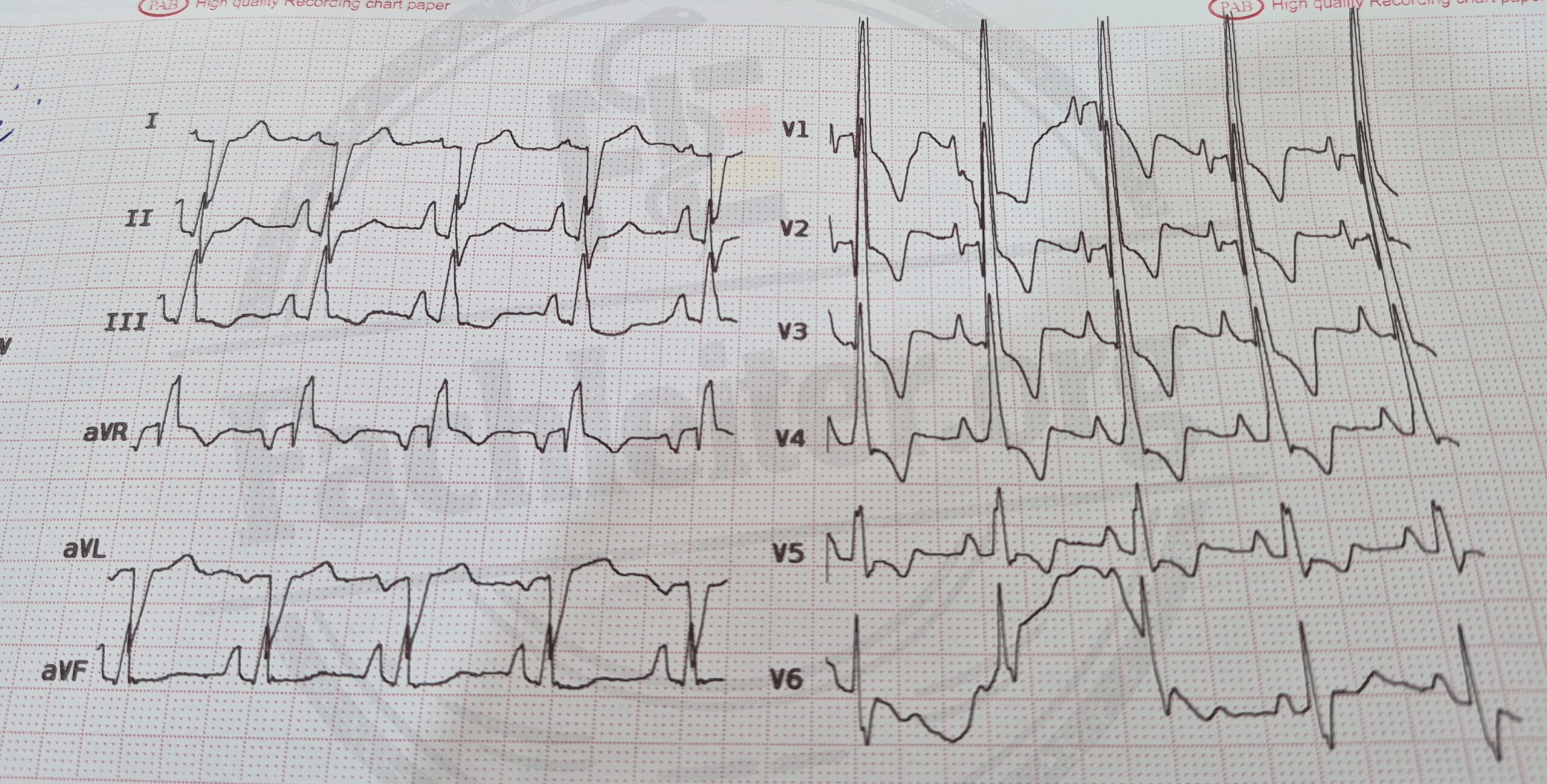
ECG summary
There is a regular sinus rhythm at approximately 88/min. The PQ interval is 120 ms, the QT interval 388 ms. Tall, peaked P waves suggest a P pulmonale.
In V1 the R wave measures five large boxes (25 mm), in V6 the S wave one large box (5 mm). Together with ST depressions and T‑wave inversions in the precordial leads this speaks for right ventricular hypertrophy with a strain pattern.
For an accurate QT assessment the rate‑corrected QT (QTc) should also be provided; at 88/min Bazett correction yields approximately 470 ms from 388 ms.
Regarding the cardiac axis: Lead II is approximately isoelectric. The electrical axis is therefore roughly perpendicular to lead II, whose axis is at +60°. Two directions are possible:
- +60° − 90° = −30°
- +60° + 90° = +150°
Since the QRS complex is negative in lead I and positive in lead III, the cardiac axis is approximately +150°. This corresponds to a marked rightward deviation of the electrical axis (extreme right axis).
Initial laboratory values
| Test | Result |
|---|---|
| Leukocytes | 8.7 /nl |
| Hemoglobin | 16.4 g/dl |
| Hematocrit | 49 % |
| Platelets | 254 /nl |
| CRP | 4 mg/l |
| Sodium | 139 mmol/l |
| Potassium | 4.2 mmol/l |
| Creatinine | 0.9 mg/dl |
| Blood glucose | 203 mg/dl |
| AST | 24 U/l |
| ALT | 21 U/l |
| Total bilirubin | 0.6 mg/dl |
| Lipase | 38 U/l |
| High‑sensitivity troponin T, first measurement | 8 ng/l |
Arterial blood gas analysis on 24% oxygen
| Parameter | Result |
|---|---|
| pH | 7.37 |
| pCO₂ | 57 mmHg |
| pO₂ | 55 mmHg |
| Bicarbonate | 32 mmol/l |
| Lactate | 1.3 mmol/l |
| SpO₂ at time of sampling | 88 % |
The arterial blood gas on 24% oxygen shows hypoxemia and hypercapnia. The elevated bicarbonate with near‑normal pH is compatible with chronic compensated CO₂ retention in the context of known COPD.
Course and further management
The chest X‑ray shows no acute finding. Nausea and upper abdominal pain have decreased markedly. The patient is awake and hemodynamically stable. Under controlled oxygen administration the SpO₂ is 88%. A repeat blood gas is being performed to assess oxygenation, CO₂ and pH over time.
Additionally she received two puffs of salbutamol spray (100 µg each) via a spacer due to wheeze.
High‑sensitivity troponin T is 9 ng/l after two hours compared with 8 ng/l initially. There is no relevant rise. Together with the clinical course and ECG there is currently no indication of an acute myocardial infarction.
Cardiology and pulmonology are being consulted. Before discharge oxygen was tapered under monitoring. Oxygen saturation and blood gases were rechecked under the documented oxygen flow. Since the patient is respiratory stable and no cause requiring inpatient treatment was found, further pulmonological and cardiological evaluation will be performed on an outpatient basis. This includes review of COPD therapy and an echocardiography to assess the suspected right heart strain.