
Greeting
Good day, Mr./Mrs. Senior Physician ………..,
I would now like to present the patient Susanne Holst to you.
She is 63 years old, 1.70 m tall and weighs about 88 kg. The body mass index (BMI) is 30.4 and therefore in the range of obesity.
Presenting history
The patient presented today due to a 2-month history of exertional dyspnea and a thoracic tightness and pressure sensation .
She stated that the course had been slowly progressive and had particularly worsened over the past four days.
The patient’s symptoms increase markedly when climbing stairs up to the first floor . After that she needs to pause for two to three minutes before she can continue. Walking longer distances is also limited. The usual distance between two bus stops she can now cover only half of (about 200 meters) without a break.
In addition, according to her report, retrosternal, stabbing as well as pressing pains occur, radiating into the left arm and the neck. The pain intensity is about 2 to 3 out of 10.
Furthermore, she complained of orthopnea (sleeping with two pillows) and of a bilateral leg edema that has existed for 4 days.
(She added that her shoes especially hurt in the evening.)
She noted the following accompanying symptoms:
Fatigue, Lack of drive, Tachypnea, Irritative cough, Dizziness on position change, Hyperhidrosis, Nocturia with 2 to 3 nocturnal toilet visits as well as nocturnal snoring (sleep apnea?!).
Questions about Nausea, Vomiting, tachycardia, loss of consciousness, panic attacks, pallor and leg pain were denied.
She stated that there is no elevator.
According to the patient, she has not yet seen a doctor because she simply lacked the energy to do so.
Autonomic history
The autonomic history revealed the following abnormalities: Loss of appetite for about two months, yet an unintentional weight gain of approximately 3 kg within the last week.
The patient reported difficulty falling and staying asleep (insomnia), due to work-related stress as well as nocturnal nocturia and orthopnea.
She also suffers from long-standing constipation.
Past medical history
Her past medical history includes the following:
The patient has had type 2 diabetesfor about ten years. According to her report, the diabetes mellitus is currently poorly controlled. Blood glucose values fluctuate. Six months ago there was a possible severe decompensation with a blood glucose level of 700 mg/dL, for which she had to be treated in hospital for two days. No sequelae have occurred to date.
|
No diabetes |
Prediabetes |
Diabetes |
|
|
Fasting glucose |
< 100 mg/dl |
100–125 mg/dl |
≥ 126 mg/dl |
|
Random glucose |
– |
– |
≥ 200 mg/dl |
|
HbA1c |
< 5,7 % |
5,7–< 6,5 % |
≥ 6,5 % |
|
OGTT (2 h value) |
< 140 mg/dl |
140–199 mg/dl |
≥ 200 mg/dl |
According to the patient, she has had arterial hypertension for 10 years, which is currently inadequately controlled. The diagnosis was made by 24-hour blood pressure monitoring. No sequelae have occurred to date.
|
systolic (mmHg) |
diastolic (mmHg) |
Treatment |
|
|
Normal blood pressure |
< 120 |
< 70 |
– |
|
Elevated blood pressure |
120–139 |
70–89 |
+ |
A hypercholesterolemia has been known for 10 years.
She added that she has had atrial fibrillationfor three years. The current INR value is 2.6.
She was hospitalized 20 years ago for pancreatitis. The cause is not documented.
About 35 years ago she sustained a rib fracture while playing volleyball , which was treated conservatively with a bandage.
Previous operations
Three years ago she had a total endoprosthesis Total endoprosthesis (TEP) of the right hip implanted.
Note: When a statement can be objectively confirmed by a medical examination – for example by visible scars – the indicative is appropriate. (No physical examination takes place in the specialist clinical exam.)
Gynecology
In the gynecological history the patient reported two spontaneous, uncomplicated vaginal deliveries about 40 and 35 years ago.
Menopause has existed for about 12 years, with occasional mild hot flashes.
Three months ago, according to the patient, a suspicious finding was detected in the right breast on mammography; further diagnostics are planned for next week.
Medications
Currently, according to the current prescription, the patient is taking the following medication:
• Bisoprolol 5 mg once daily
• Lisinopril 10 mg once daily
• Metformin 500 mg twice daily
• Atorvastatin 40 mg in the evening
• Marcumar ¼ tablet daily, ½ tablet on Sundays (change to Eliquis planned)
• Laxoberal as needed
• Iron supplement (OTC, drugstore), taken every 2–3 days.
Note: When a statement can be objectively confirmed by a medical observation – for example based on a prescription – the indicative is appropriate.
Allergies:
The patient reported the following allergies:
After a bee sting she experienced an anaphylactic shock, for which reason she carries an EpiPen.
She also has an allergy to amoxicillin, which manifested as urticaria and dyspnea. This reaction occurred 20 years ago during treatment for pneumonitis.
Noxae
In the noxae history the patient stated that she has been a non-smoker for 4 years; previously she smoked one pack of cigarettes daily for over 30 years (30 pack-years). Current alcohol consumption is about one glass of sparkling wine per day. In the past she occasionally used THC.
Social history
The patient is a saleswoman in a clothing store and lives alone in an apartment.
In the social history the patient stated she is a saleswoman in a clothing store and lives alone in an apartment. She has two children and three grandchildren. One of the grandchildren has a small congenital Atrial septal defect (ASD).
Physical activity: –
Family history
In the family history the patient reported that her father (83 years) had a pancreatic carcinoma that was surgically treated one year ago; he died six months after the procedure.
The mother (84 years) is demented and lives in a nursing home; at the age of 52 she underwent surgery for a abdominal carcinoma due to bleeding and abdominal pain.
Suspected and differential diagnosis
From my point of view the anamestic information – namely exertional dyspnea, chest pain, fatigue, orthopnea, leg edema and nocturia – most likely indicate heart failure. The patient has multiple risk factors: obesity, lack of exercise, hypertension, diabetes mellitus and dyslipidemia.
Based on the history the patient can be classified as NYHA class III.
In the context of a chronic obstructive pulmonary disease a chronically productive cough is typically expected as the leading symptom.
An edema can also occur in the context of diabetic nephropathy with long-standing diabetes mellitus and arterial hypertension; however, this does not explain the other symptoms.
A pulmonary embolism usually occurs acutely; a deep vein thrombosis (DVT) is often present as well.
Diagnostic workup
physical examination
Because the symptoms and risk factors strongly indicate a decompensated heart failure, in this case inpatient admission should be arranged already after the history, followed by a thorough physical examination. First the vital signs are recorded – including heart rate, blood pressure, respiratory rate, oxygen saturation and body temperature. A targeted physical examination then follows.
We begin with inspection and look for possible edema, pallor, jugular venous distension or other visible abnormalities. This is followed by palpation, for example to assess tenderness on pressure and peripheral pulses (e.g., A. radialis or A. tibialis posterior). The presence of pitting edema can also be checked. In the next step percussion is performed percussion – among other things to assess possible pulmonary changes or hepatosplenomegaly. Finally, we auscultate heart and lungs to detect pathological breath sounds or abnormal heart sounds.
In heart failure we may, for example, hear crackles at the lung bases – indicating pulmonary congestion – as well as a third heart sound (gallop rhythm), which suggests a diastolic dysfunction.
The cardiac apex is generally located in the 5th intercostal space at the midclavicular line on the left. Palpation of the apical impulse provides information about heart size and contractility. A displaced or prominent palpable apical impulse can indicate left heart failure.
In addition, signs of jugular venous distension and a positive hepatojugular reflux should be checked, as they can indicate right heart failure.
Laboratory tests
With suspected heart failure a complete blood count, C-reactive protein, electrolytes, the C-reactive proteinelectrolytes, the lipid profile, renal function parameters, liver values, TSH (thyroid-stimulating hormone), HbA1c, fasting blood glucose, coagulation parameters, D-dimers?! as well as the B-type natriuretic peptide (NT-proBNP) should be measured.
To exclude an acute coronary syndrome, troponin I or T as well as CK-MB should be measured.
Anatomical diagnosis
Electrocardiogram
An ECG is performed by placing electrodes on the skin of the chest, arms, and legs to record the heart’s electrical activity.
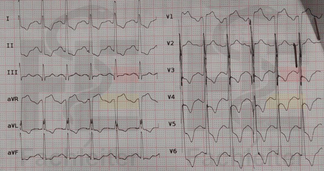
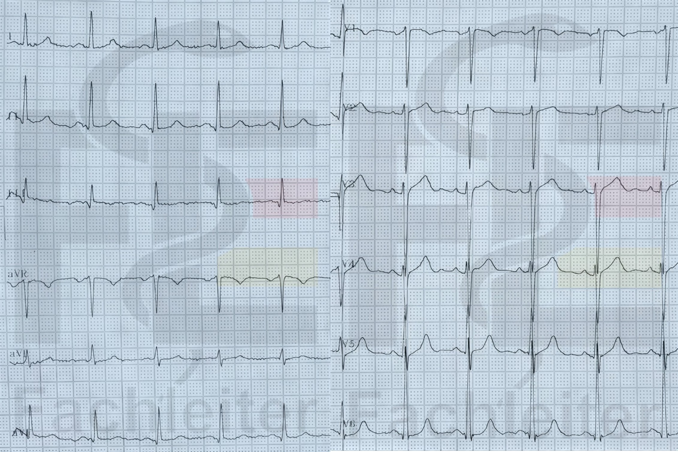
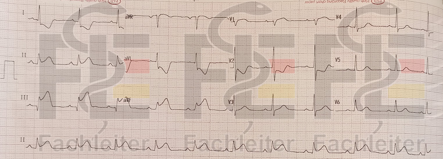
On ECG, heart failure often shows nonspecific changes such as tachycardia (e.g., atrial fibrillation), signs of left ventricular hypertrophy, QRS widening, ST-segment depressions or T-wave inversions, as well as indications of previous myocardial infarctions (e.g., pathological Q waves).
Evaluations
Sinus tachycardia with a heart rate of 120/min. A widened and deepened negative terminal portion of the P wave in V1 may indicate left atrial enlargement. Tall R waves in V5/V6 and deep S waves in V1/V2 as well as ST depressions and negative T waves in I and V3–V6 are consistent with left ventricular hypertrophy. Anatomical confirmation is obtained by echocardiography.
Sinus rhythm at a normal rate with a heart rate of 75/min. A biphasic P wave in V1 can be normal; a widened and deepened negative terminal portion may indicate left atrial enlargement. Tall R waves in V5/V6 and deep S waves in V1/V2 may suggest left ventricular hypertrophy. Anatomical confirmation is obtained by echocardiography.
Inferoposterior myocardial infarction with suspected right ventricular involvement. For further assessment, additionally record V7–V9 and V3R/V4R.
Chest X-ray
The patient removes jewelry and clothing in the chest area. He stands or sits facing the X-ray plate. During the exposure he is asked to take a deep breath and briefly hold it. Among other things, the cardiac silhouette (e.g. in cardiac hypertrophy), the vascular markings (signs of pulmonary congestion or congestion) and possible signs of a pulmonary edema are assessed.
Echocardiography:
Echocardiography is an imaging technique used to assess cardiac structure and function, wall motion and heart valve motion using ultrasound. For transthoracic echocardiography (TTE) no special preparation is required. The patient usually lies in the left lateral position while the probe is placed on the chest wall to display various cardiac planes. Among other things, the Ejection fraction (EF) can be measured, an important parameter for assessing the pump function of the left ventricle.
An echocardiogram, also called a cardiac ultrasound, will show that the patient either has a reduced ejection fraction (less than 40%), a moderately reduced ejection fraction (between 40 and 50%), or a preserved ejection fraction.
(For transesophageal echocardiography (TEE) the patient must fast for about six hours. Before the examination information is provided and, if necessary, sedation is given. During TEE an ultrasound probe is introduced via the esophagus to obtain detailed images of the heart, particularly the valves and atria.)
Treatment
Lifestyle modification
• Weight reduction
• regular physical activity
• reduction of salt intake
General acute measures
• Monitoring: ECG monitoring, blood pressure, heart rate, oxygen saturation
• Oxygen therapy: Only if SpO₂ < 90% or clinical signs of hypoxia
Therapy
|
Classification |
Treatment strategy (allegedly not required) |
|
|
Stage A |
Weight control, regular physical exercise, smoking cessation, abstinence from alcohol and drugs, treatment of arterial hypertension, diabetes and dyslipidemia |
|
|
Stage B |
NYHA 1 |
Stage A + ACEI or ARB ± beta blocker ± implantable cardioverter-defibrillator (ICD) |
|
Stage C |
NYHA 2 NYHA 3 |
Stage A + diuretic + ACE or ARNI + beta blocker + mineralocorticoid receptor antagonists ± cardiac glycosides ± isosorbide dinitrate/ hydralazine ± ivabradine ± implantable cardioverter-defibrillator (ICD) or biventricular pacemaker systems |
|
Stage D |
NYHA 4 |
|
|
New York Heart Association classification of heart failure |
|
|
NYHA I |
Patients have no physical limitations and show no symptoms during everyday physical activities. |
|
NYHA II |
Ordinary physical activity causes mild symptoms and leads to a slight limitation of exercise tolerance. |
|
NYHA III |
Patients are comfortable only at rest; less than normal physical activity causes symptoms. |
|
NYHA IV |
Heart failure symptoms occur even at rest, and even slight physical activity causes symptoms. |
|
Stage A |
At-risk patients without structural heart disease or symptoms |
|
Stage B |
Structural heart disease without symptoms. |
|
Stage C |
Structural heart disease with symptoms. |
|
Stage D |
Therapy-refractory symptoms, often requiring specialized measures. |
Coronary angiography:
Coronary angiography enables assessment of the coronary arteries and targeted treatment of stenoses or occlusions.
Angiography is a minimally invasive procedure to image and, if necessary, treat arteries using X-rays and contrast medium. After disinfection of the puncture site the contrast medium is injected via a large artery such as the femoral artery or the radial artery. X-ray images are then taken to visualize the blood flow and possible thromboses or stenoses. In some cases this procedure is replaced by duplex sonography because it is non-invasive and does not require contrast medium. The procedure usually lasts 30 to 60 minutes and does not require elaborate preparation. If necessary, a therapeutic measure can be performed at the same time, for example by using a balloon catheter or a stent to treat vascular stenoses. Angiography carries risks such as bleeding, infection, allergic reactions to contrast medium, and rarely vascular injury or renal dysfunction.
Extra
Names:
Mr. Haas, Mr. Schieler, Mrs. Fischer, Herbert Nordheide
Occupatione:
Gardener, cooper
Details:
I visited my great-granddaughter today because it was her birthday.
He experienced a pressure-like feeling in the chest while working.
Past medical history:
Endoscopic surgery for benign prostatic hyperplasia 5 years ago
Tension headaches
Knee TEP (total knee arthroplasty)
Hypothyroidism
Femoral neck fracture due to a fall
Breast biopsy for a tumor, result: benign
Status post nucleotomy 3 years ago due to a lumbar disc prolapse. As a consequence there is low back pain, which is treated if necessary with ibuprofen 400 mg.
Allergies:
House dust mites: rhinoconjunctivitis
Birch allergy with rhinoconjunctivitis
Medications:
xarelto
Family history:
Father: lung carcinoma
Mother: dementia
Mother died of cervical carcinoma
Social:
widowed 2 years ago (breast carcinoma)
Husband died of sepsis.
Questions
1. Do I have to stay overnight here? I have to take care of my son – he cannot stay alone. What should I do now?
I understand that your son is very important to you. Hospital admission is medically necessary, but we are happy to support you in finding a good solution together with social services or your family.
2. How much alcohol is too much daily?
For men: no more than 24 g pure alcohol/day
For women: no more than 12 g pure alcohol/day
Approximately 10–12 g pure alcohol are contained in 0.25 l beer, 0.1 l sparkling wine, 0.1 l wine
3. What diagnoses would be considered in a younger patient with similar symptoms?
In a younger patient with symptoms of heart failure, cardiomyopathies, pericarditis, myocarditis, congenital heart defects and hormonal causes such as hyperthyroidism are primarily considered. Substance abuse (e.g. alcohol or drugs) could also be a possible trigger.
4. Can an influenza-like infection trigger heart failure?
Yes, an influenza-like infection (especially by viruses such as influenza or Coxsackie) can in rare cases cause myocarditis (inflammation of the heart muscle). This can lead to acute or later chronic heart failure.
5. Why does nocturia occur in patients with heart failure?
During the day in heart failure, fluid accumulates in the lower extremities, especially when upright. When lying down the fluid redistributes more evenly throughout the body, increasing circulating blood volume. Improved renal perfusion in the supine position stimulates kidney function, producing more urine. This causes patients to need to urinate more frequently at night.
6. Why does coughing often occur in heart failure?
Cough and orthopnea in heart failure result from fluid congestion in the lungs due to left-sided heart failure. Airway irritation typically leads to a dry, irritating cough that occurs especially at night or while lying down.
7. Why exactly does swelling improve when lying down during sleep?
In heart failure, swelling improves when lying down during sleep because gravity no longer acts on the legs. This allows venous blood to be returned to the heart more effectively.
8. What are the benefits of switching from Marcumar to Eliquis?
Eliquis (apixaban) — like other direct oral anticoagulants — has the advantage over Marcumar (phenprocoumon) that regular INR monitoring is not required, there are fewer food interactions, and the overall risk of bleeding, especially intracranial bleeding, is lower.
9. How can it be explained that the patient continues to show symptoms despite treatment?
Possible explanations include inadequate therapeutic adjustment, an advanced stage of disease, poor medication adherence, or the presence of additional stressors such as infections, arrhythmias, or comorbidities
10. What risks are associated with cannabis resin (hashish) use?
Use of hashish can lead, among other things, to concentration problems, lack of motivation, anxiety, psychosis, and dependence. Especially in adolescents it can negatively affect brain development.
11. What would you recommend to the patient in daily life to manage her house dust mite allergy?
For a house dust mite allergy the patient should wash bed linen weekly at at least 60 °C and keep the bedroom cool and dry. Carpets, curtains and upholstered furniture should be avoided where possible or cleaned regularly. Vacuuming with a HEPA-filter device is also important — preferably not performed by the patient herself
Notes
BMI
Body Mass Index (BMI)
The BMI is a simple ratio measure used to classify body weight. It is calculated by dividing body weight in kilograms by the square of height in meters. Clinically the value is used to estimate underweight, normal weight, overweight and obesity; a BMI of about 30 or higher is classified as obesity.
Dyspnoea
Shortness of breath in heart failure arises mainly from pulmonary congestion due to increased left-sided filling pressures and can occur with both reduced and preserved ejection fraction.
thoracic tightness or pressure sensation
A thoracic tightness or pressure sensation, especially retrosternal, can indicate angina pectoris.
up to the first floor
Shortness of breath when climbing stairs up to the first floor can correspond to heart failure NYHA II to III.
two to three minutes
When climbing to the first floor, shortness of breath occurs so that the patient needs a two- to three-minute rest. With absence of symptoms at rest, this more likely corresponds to NYHA III.
The usual distance between two bus stops
Usual distance between two bus stops
The phrase refers to a common everyday distance used as a reference measure (in this context about 400 m). The patient states she can only walk about half of this distance (approx. 200 m) without interruption. This information is commonly used in clinical practice to roughly assess a patient’s walking distance and mobility.
retrosternal, stabbing as well as pressing pains
Retrosternal, pressing pains can indicate myocardial ischemia, for example in angina pectoris or an acute coronary syndrome.
Orthopnea
Orthopnea
Orthopnea denotes the occurrence of shortness of breath when lying flat, causing affected individuals to often sleep with an elevated head and upper body (here: two pillows). Pathophysiologically, lying down promotes redistribution of blood into the thorax and can increase pulmonary venous pressure, leading to exertional or position-induced dyspnea. The simultaneous presence of bilateral leg edema supports the clinical interpretation as a sign of fluid retention or cardiac congestion.
Leg edema
Evening-increasing leg edema can be explained in heart failure by venous congestion and fluid accumulation. When lying down, fluid is redistributed at night, so the legs are less swollen in the morning.
Fatigue
Fatigue in heart failure can be explained by a reduced cardiac output and decreased perfusion of the muscles; it can also occur with preserved ejection fraction.
Lack of drive
Lack of drive in heart failure may be related to reduced cardiac output and limited physical capacity.
Tachypnea
Tachypnea
Definition and recognition:
Tachypnea denotes an accelerated breathing rate; in clinical practice it is often classified as present at a respiratory rate above about 20 breaths per minute.
Significance:
An increased respiratory rate may indicate respiratory or cardiac strain, pain, fever, or a metabolic disturbance and requires clinical evaluation in the overall context.
Dry cough
Dry cough
Brief description: A dry cough is predominantly a non-productive, spasmodic cough with little or no sputum.
Relevance in context: In a backward-congested lung, for example due to heart failure with venous congestion, such a dry cough can occur, especially at rest or at night. It may indicate a cardiac cause and should be assessed together with other symptoms.
Dizziness
Orthostatic dizziness
Brief description: Orthostatic dizziness refers to feelings of dizziness or visual disturbances that occur shortly after standing up or with rapid positional changes.
Likely explanation: Such complaints are consistent with orthostatic blood pressure drop, where standing leads to a transient fall in blood pressure and thus reduced cerebral perfusion.
Hyperhidrosis
Hyperhidrosis
Brief description: Hyperhidrosis denotes excessive sweating beyond what is required for temperature regulation or physical exertion.
Clinical notes: It can be generalized or localized and occur with various underlying diseases or as a side effect of medications.
Nocturia
Nocturia
Brief description: Nocturia means repeated nighttime urination that interrupts the sleep cycle.
Possible mechanism: Increased nocturnal urinary frequency can be promoted by redistribution of fluid when lying down with increased renal perfusion and enhanced diuresis; this is one of several possible explanations and occurs particularly with certain cardiac or fluid-regulation disorders.
Nausea
Nausea (nausea)
Subjective sensation of urge to vomit, not necessarily accompanied by vomiting. In the history, nausea was specifically asked and denied.
Emesis
Vomiting (emesis)
Active expulsion of gastric contents through the mouth. In the screening, vomiting was asked about and denied by the patient.
Loss of consciousness
Loss of consciousness
Temporary or persistent loss of consciousness; syncope is a common cause of transient loss of consciousness. No loss of consciousness was reported in the obtained history.
Leg pain
Leg pain
Differential diagnoses include deep vein thrombosis, chronic venous insufficiency and peripheral arterial occlusive disease as important causes of leg pain. In the current inquiry, leg pain was denied, so there are no confirmed indications of these conditions.
Loss of appetite
Loss of appetite
The patient reports a markedly reduced appetite for about two months. Notably, this loss of appetite exists despite reporting an unintentional weight gain of about 3 kg within the last week.
Weight gain
Weight gain in heart failure often arises from increased sodium and water retention leading to fluid accumulation.
Insomnia
Insomnia
The patient complains of difficulty falling asleep and repeated awakenings. She attributes the sleep disturbances primarily to occupational stress; nocturnal nocturia and orthopnea are also mentioned as contributing factors.
Constipation
Constipation
There has been a long-standing disorder of bowel evacuation with constipation complaints.
Type 2 diabetes
Definition: Type 2 diabetes is a chronic metabolic disease in which glucose metabolism is impaired.
Pathophysiology: The disease is mainly due to reduced tissue sensitivity to insulin (insulin resistance) combined with insufficient insulin secretion by beta cells. In contrast, type 1 diabetes arises from an autoimmune reaction with near-complete destruction of insulin-producing beta cells and thus an absolute insulin deficiency.
Clinical course and significance: With inadequate control, large blood glucose fluctuations and occasionally severe acute hyperglycemias can occur that may require inpatient treatment. Long-term uncontrolled type 2 diabetes increases the risk of vascular and nerve damage as well as other complications.
Hypercholesterolemia
Definition: Hypercholesterolemia denotes an elevated cholesterol level in the blood, with particularly increased LDL cholesterol being clinically relevant.
Clinical significance: Persistently elevated cholesterol promotes the development of atherosclerotic changes in the arteries and thereby increases the risk of cardiovascular events.
Practical consequence: With known hypercholesterolemia, blood lipid values are usually monitored and measures for risk reduction are considered.
Atrial fibrillation
Definition: Atrial fibrillation is a common cardiac arrhythmia in which electrical activity in the atria is disorganized, leading to an irregular heart action.
Causes and symptoms: Common triggers are hypertension, coronary artery disease, heart failure, hyperthyroidism or excessive alcohol consumption. Affected individuals often report palpitations, an irregular pulse, dizziness, shortness of breath or fatigue; some patients, however, have no symptoms.
Treatment focus: Therapy aims to control heart rate or restore sinus rhythm and to provide stroke prophylaxis through anticoagulation. Electrical cardioversion and catheter-based ablation are possible interventional options.
Laboratory: The INR value is used to monitor therapy with vitamin K antagonists, as it reflects anticoagulation.
Playing volleyball
Volleyball: brief description
Volleyball is a team sport in which two teams compete against each other over a centrally hung net. The aim is to play the ball onto the opposing court so that the opposing team cannot legally return it. Usually played in sets; the rules distinguish serve, attack, block and defense.
Total endoprosthesis
In a total endoprosthesis (TEP), the damaged joint surfaces are replaced by artificial components made of metal, plastic and/or ceramic. The procedure is considered for severe pain and significant functional limitations when conservative treatments are insufficient. It is performed under general or regional anesthesia. Usually, at least six hours without solid food and two hours without clear fluids are required before anesthesia; the anesthesiology team’s instructions are decisive. Possible complications include infections, bleeding, thromboses, pulmonary embolism and prosthesis loosening as well as dislocation in hip prostheses.
Bisoprolol
Bisoprolol: overview
Group
Selective beta-1 blocker from the class of beta blockers.
Mechanism of action
Primarily blocks beta-1 adrenoceptors in the heart, thereby reducing heart rate and contractility, which lowers myocardial oxygen demand.
Indications
Used, among other things, for hypertension, certain cardiac arrhythmias and for symptomatic treatment of angina pectoris.
Important risks/side effects
Common adverse effects are fatigue and dizziness; it can also cause a slowed heart rate (bradycardia). Special caution is required in decompensated heart failure, severe bradycardia and certain circulatory disorders.
Lisinopril
Lisinopril: brief profile
Group
ACE inhibitor (angiotensin‑converting enzyme inhibitor).
Mechanism of action
Inhibits the angiotensin‑converting enzyme, reducing formation of angiotensin II; this leads to vasodilation and reduced aldosterone effect.
Indications
Used for arterial hypertension and as part of therapy for heart failure as well as in follow‑up after myocardial infarction in suitable cases.
Important risks/side effects
Known side effects include dizziness and occasionally headache. Typical for this drug class are a dry irritating cough and the risk of elevated potassium levels; in rare cases angioedema can occur. In certain comorbidities and pregnancy use is contraindicated or requires caution.
Metformin
Metformin
Group: biguanide antidiabetic.
Effect: Reduces glucose production in the liver and improves glucose uptake in peripheral tissues by increasing insulin sensitivity.
Use: Employed in the treatment of type 2 diabetes as part of non‑insulin therapy; dosing instructions must be followed individually.
Important risks: Common adverse effects affect the gastrointestinal tract (e.g., nausea, vomiting, diarrhea). Occasionally headache or dizziness may occur. Very rarely there is a risk of lactic acidosis, particularly with renal impairment or other risk situations.
Atorvastatin
Atorvastatin
Group: HMG‑CoA reductase inhibitor (statin).
Effect: Lowers endogenous cholesterol production in the liver and thereby particularly reduces LDL cholesterol in the blood.
Use: To lower elevated cholesterol levels and reduce cardiovascular risk; timing of intake is according to medical advice. Atorvastatin has a longer duration of action than some other statins, so timing of intake is less strictly tied to the evening.
Important risks: Possible side effects include gastrointestinal complaints, headache and muscle symptoms up to severe muscle damage in rare cases. Elevations in liver enzymes can also occur. Interactions, especially with CYP3A4 inhibitors, can increase the risk of adverse effects.
Marcumar
Marcumar (phenprocoumon)
Group: oral vitamin K antagonist (coumarin derivative).
Effect: Inhibits vitamin K‑dependent activation of clotting factors, thereby reducing the synthesis of functional factors II, VII, IX and X as well as the anticoagulant proteins C and S.
Use/monitoring: Used for oral anticoagulation; regular monitoring of coagulation inhibition by INR is required. Dose adjustments are made individually according to INR values.
Important risks: Increased bleeding risk; numerous drug and food interactions influence the effect. When switching to direct oral anticoagulants, clinical considerations and monitoring measures are necessary. In the event of bleeding there are specific countermeasures available that must be managed by the treating physicians.
Laxoberal
Laxoberal
Group: stimulants (laxatives) with the active ingredient sodium picosulfate.
Effect: Converted by bacteria in the gut into an active metabolite that stimulates intestinal motility and thus promotes bowel evacuation.
Use: Short‑term treatment for constipation or for bowel evacuation after medical indication; use as needed as prescribed.
Important risks: Common effects are abdominal cramps and increased bowel movements. Prolonged or improper use can lead to electrolyte disturbances and habituation effects; persistent symptoms require medical evaluation.
Atrial septal defect
Atrial septal defect (ASD)
Definition: An ASD is a congenital malformation of the atrial septum with an open communication between the left and right atrium.
Clinical features: Small defects often remain asymptomatic for a long time. Larger defects can cause exertional dyspnea and reduced exercise tolerance; in the long term, cardiac arrhythmias (e.g., atrial fibrillation) are also possible. In childhood additional clinical problems may occur depending on the defect.
Pancreatic carcinoma
Pancreatic carcinoma
General: Pancreatic carcinoma is a malignant tumor of the pancreas, more frequently located in the head of the pancreas.
Clinical presentation and prognosis: Early symptoms are often nonspecific, e.g., upper abdominal pain, loss of appetite and weight loss; involvement of the pancreatic head may also cause jaundice. The disease is often only recognized at advanced stages, which is why overall prognosis is poor.
Abdominal malignancies
“Abdominal cancer” is a nonspecific collective term for malignant tumors of the female reproductive organs. These include endometrial carcinoma (uterine lining cancer), cervical carcinoma (cervical cancer), vaginal carcinoma, vulvar carcinoma and ovarian carcinoma (ovarian cancer).
exertion‑related dyspnea, chest pain, fatigue, orthopnea
Symptom combination: exertion‑related dyspnea, chest pain, fatigue, orthopnea
Meaning: This symptom complex indicates cardiac dysfunction and can specifically point to left‑sided heart failure. Orthopnea and exertional dyspnea are typical symptoms of impaired left ventricular function. Present risk factors such as obesity, physical inactivity, arterial hypertension, diabetes mellitus and dyslipidemia increase the likelihood of a cardiogenic origin.
Leg edema and nocturia
Leg edema and nocturia
Findings: Recurrent swelling of the lower legs combined with increased nighttime urination can indicate volume overload. Pathophysiology: In right heart failure there is venous congestion and fluid retention with peripheral edema; in the supine position nocturnal diuresis can increase, leading to nocturia.
chronic obstructive pulmonary disease
chronic obstructive pulmonary disease
Core aspect: Chronic obstructive pulmonary disease (COPD) is a progressive disease of the airways with persistent airflow limitation that is typically not fully reversible.
Clinical features and causes: Leading symptoms are exertional dyspnea and often a chronic productive cough with sputum; acute exacerbations occur regularly. Important risk factors are primarily smoking as well as long‑term exposure to air pollutants, fine particulates or occupational hazards.
Diagnosis: Definitive diagnosis is made by pulmonary function testing (spirometry) to objectify a persistent obstructive ventilatory disorder.
Pulmonary embolism
Pulmonary embolism
Core information: A pulmonary embolism results from embolization of thrombotic material into the pulmonary circulation and usually presents acutely with shortness of breath, pleuritic chest pain and occasionally syncope.
Risk factors and effect of anticoagulation: Typical risk factors are malignancy‑associated coagulopathies, prolonged immobility and vascular injury. Ongoing oral anticoagulation reduces the risk of thromboembolic events but does not exclude pulmonary embolism — especially if the anticoagulation is not therapeutic.
Diagnostics: Imaging to confirm the diagnosis is CT angiography of the chest; a concurrent deep vein thrombosis in the legs is often found.
decompensated
decompensated
Definition: “Decompensated” in heart failure describes the state in which cardiac output is so impaired that symptoms occur at rest or with minimal exertion (e.g., pronounced dyspnea, peripheral edema, severe fatigue, orthopnea).
Acute management: A decompensated state requires prompt clinical assessment and often hospital admission. Immediate measures include measurement of vital signs (heart rate, blood pressure, respiratory rate, oxygen saturation, temperature) and a focused physical examination; laboratory and imaging investigations follow to determine the cause and extent.
Blood pressure
Blood pressure
Definition and significance:
Blood pressure denotes the force with which circulating blood acts on the vessel walls. It is divided into systolic and diastolic components and is a central circulatory parameter for assessing hemodynamic status.
Measurement and device:
Blood pressure is measured using either manual systems or automatic oscillometric devices. A classic manual set consists of an appropriately sized upper arm cuff, a device for inflation and a pressure gauge; auscultation is performed with a stethoscope. Choice of method and cuff size affect measurement accuracy.
Clinical relevance:
Regular blood pressure checks are important for detecting and monitoring treatment of hypotension and hypertension and for assessing volume and circulatory status, for example in heart failure.
Respiratory rate
Respiratory rate
Definition:
The respiratory rate is the number of breaths per minute and represents a fundamental vital parameter for assessing respiratory function.
Normal range and deviations:
In adults, the respiratory rate is typically in the range of about 12–20 breaths per minute. An increase above this range is called tachypnea; a decreased respiratory rate is called bradypnea.
Oxygen saturation
Oxygen saturation
Term and measurement:
Oxygen saturation (SpO2) describes the proportion of hemoglobin bound with oxygen relative to total hemoglobin and is measured noninvasively with a pulse oximeter.
Interpretation and limitations:
The measurement provides rapid clues about the blood’s oxygenating function but can be distorted by poor peripheral perfusion, movement, nail polish, or certain poisons/anomalies (e.g., carbon monoxide binding). Clinical assessment and, if necessary, arterial blood gas analysis are required in unclear or critical findings.
Body temperature
Body temperature
Definition and clinical significance:
Body temperature is a parameter for assessing thermoregulatory status. Deviations can indicate infections, inflammatory processes, or disturbances of heat regulation.
Measurement methods and sites:
Temperature measurements are taken at different sites (oral, axillary, rectal, tympanic) and with various devices (digital thermometers, infrared or tympanic devices). The measurement site influences the measured values and must be considered during interpretation.
Edema
Edema – classification and clinical examination
Definition and test: Edema are fluid accumulations in the interstitium. To differentiate, one presses a thumb pad on the swelling for several seconds; if a pit remains, it is called pitting edema.
Causes of pitting edema: Common causes include heart failure, renal insufficiency, liver diseases, venous insufficiency, or hypoalbuminemia. The distribution (bilateral, bilateral on exertion, generalized) helps with classification.
Causes of non‑pitting edema: Non‑pitting swelling is more typical for lymphedema or myxedematous changes (e.g., in hypothyroidism).
Important clinical note: The distinction pitting vs. non‑pitting is an orientation feature and leads to targeted further evaluation of the underlying cause.
Pallor
Pallor – significance and clinical clues
What is pallor: Pallor describes a visible lightening of the skin or mucous membranes and is a clinical clue, not a diagnosis.
Important causes: The most common cause is anemia due to reduced hemoglobin content. Other causes are acute blood loss, chronic diseases with anemia, as well as peripheral vasoconstriction in shock or cold environment.
Examination: For assessment, inspection of the conjunctivae, oral mucosa, and nail beds is recommended. Pallor alone does not allow a reliable attribution to cardiac performance; reduced cardiac function can secondarily contribute to poor perfusion but is not the primary explanation in all cases.
Jugular venous distension
Jugular venous distension (JVD) – significance and causes
Definition and examination: A noticeable enlargement of the neck veins (jugular venous distension) indicates elevated central venous pressure. Assessment is often done with the patient reclined (30–45°) and evaluation of the prominent venous wave height.
Typical causes: Increased central venous pressure can result from right heart failure, pericardial diseases (cardiac tamponade, constrictive pericarditis), volume overload, or obstructive processes in the major venous system.
Clinical significance: Jugular venous distension is an important clue to systemic volume/pressure elevation but should always be evaluated in the overall clinical context.
Tenderness to palpation
Tenderness to palpation – significance and differential diagnoses
What tenderness to palpation means: Localized pain provoked by palpation is referred to as tenderness to palpation and is a clue to a peripheral, often musculoskeletal problem.
Important differential diagnoses: Common causes are costochondritis or other inflammatory changes of the chest wall, muscle or tendon injuries, and rib fractures. Subcutaneous infections or early manifestations of herpes zoster can also be tender to palpation. Pleuritic or cardiac diseases typically do not cause isolated chest wall tenderness.
Clinical relevance: Location, triggering mechanism, and accompanying symptoms are crucial for further evaluation; tenderness to palpation alone is not specific for a single diagnosis.
percussed
Percussion
Procedure: The examiner places the distal finger of one hand on the area to be examined and strikes this support finger briefly with the middle finger of the other hand. This produces sound events that give information through different acoustic qualities.
Purpose and interpretation: The sounds produced by tapping help assess the nature of the underlying tissue. A relatively dull sound can indicate increased tissue density, as seen with fluid collections or consolidations; a loud, tympanic note, by contrast, indicates air-filled spaces. Percussion is often used to assess the lungs and upper abdominal organs.
Signs of jugular venous distension
Examination of the neck veins (assessment of the jugular venous distension sign)
Positioning and inspection: The patient should be positioned semi-recumbent (about 30–45°). Observation is primarily of the right side of the neck, as the visible veins and their pulsations can be assessed most reliably here.
Measurement method and classification: One determines the highest point of visible venous pulsation and then measures vertically the distance to a reference, usually the sternal angle (Angle of Louis). A markedly increased filling of the neck veins compared with normal indicates elevated central venous pressure and suggests possible right heart strain.
hepatojugular
Hepatojugular reflux (abdominojugular test)
Procedure: With constant, moderate pressure, the right upper abdominal area (liver region) is pressed for several seconds while the neck veins are simultaneously inspected.
Assessment: A sustained rise in venous filling or jugular pressure during and after the pressure is considered positive. A persistent increase indicates impaired right ventricular pumping function or increased right heart strain.
complete blood count
Complete blood count
Purpose: The complete blood count serves basic hematological assessment, particularly for detecting or excluding anemia.
Contents: Typically parameters such as hemoglobin, hematocrit, and red blood cells are used to identify anemia and provide clues about its mechanisms.
C-reactive protein
C-reactive protein (CRP)
CRP is an acute-phase protein produced in the liver that rises in the serum during inflammation, infection, or tissue damage. It serves as a nonspecific inflammatory marker; elevated values can occur in pericarditis but are not specific for this disease and do not alone prove the diagnosis.
Lipid profile
Lipid profile
The lipid profile usually includes measurement of total cholesterol, LDL cholesterol, HDL cholesterol, and triglycerides. These measurements are used for cardiovascular risk assessment and therapy planning.
Renal function parameters
Renal function parameters
Important laboratory parameters for assessing renal function are serum creatinine with calculated estimated glomerular filtration rate (eGFR), blood urea (Urea/BUN), and electrolytes. In heart failure, renal function can be impaired by reduced perfusion or venous congestion, so monitoring these parameters is clinically relevant.
Liver values
Liver values
Routine liver parameters include alanine aminotransferase (ALT), aspartate aminotransferase (AST), and gamma‑glutamyl transferase (GGT). ALT is considered relatively liver-specific, AST is also found in muscle and heart tissue and is therefore less specific; GGT is often elevated in cholestatic conditions.
HbA1c
HbA1c — meaning and clinical significance
The HbA1c value describes the proportion of glycated hemoglobin and thus reflects the average blood glucose concentration over the past approximately eight to twelve weeks. The laboratory result is used for follow-up assessment in known diabetes mellitus and can support the diagnosis of diabetes. Reliability and interpretation can be affected by changes in erythrocyte lifespan (e.g., hemolysis, blood transfusion, hemoglobinopathies).
Coagulation parameters
Coagulation parameters — overview and significance
Routinely determined coagulation parameters include prothrombin time/INR and activated partial thromboplastin time (aPTT). PT/INR primarily assesses the extrinsic and common coagulation pathways, while aPTT assesses the intrinsic and common pathways. These laboratory values are used to evaluate coagulation disorders and to monitor anticoagulant therapies; other coagulation-relevant findings such as platelet count or D-dimer complement the diagnostics depending on the question.
B-type natriuretic peptide
B-type natriuretic peptide (BNP / NT-proBNP)
The precursor molecule proBNP is cleaved under cardiac pressure or volume overload into the active BNP and the inactive fragment NT-proBNP. Both fragments increase in heart failure and are used diagnostically, with NT-proBNP often measured because of its stability. BNP mediates, among other effects, vasodilation as well as sodium and water excretion. Concentrations are influenced by age and renal function and are clinically relevant in the diagnosis and monitoring of heart failure.
Troponin
Troponin — marker for myocardial injury
Cardiac troponins (troponin I and troponin T) are specific laboratory parameters for detecting myocardial injury and are used in the evaluation of an acute coronary syndrome. High-sensitivity troponin assays enable early detection and require serial measurements in acute situations to demonstrate a rise or fall. In chronic renal insufficiency, troponin T in particular can be elevated even without an acute coronary occlusion, so interpretation of the findings is more challenging there.
CK-MB
CK-MB
Brief description:
CK-MB is an isoenzyme of creatine kinase that occurs predominantly in the heart muscle.
Clinical significance:
Determination of CK-MB can provide additional information in suspected reinfarction, as its concentrations change over time and thus can indicate a recurrent myocardial injury event. However, cardiac troponins are preferred in the diagnosis of acute coronary syndrome.
Ejection fraction
Ejection fraction
Definition:
Ejection fraction (EF) indicates the percentage of blood volume that the left ventricle ejects into the circulation with one heartbeat.
Clinical relevance:
EF is an important measure for assessing the systolic pumping function of the left ventricle and is often determined by echocardiography.
Weight reduction
Weight reduction
Goal and approach:
Weight reduction refers to the intentional lowering of body weight. An adapted, calorie-conscious diet is a central measure.
Note:
Dietary changes are often part of weight-reduction measures and are frequently combined with other interventions.
Salt intake
Goal: Reduction of salt intake
Background: A lower intake of salt can help lower blood pressure and reduce cardiovascular risk.
Practical tips: Pay attention to hidden salt additions in processed foods such as bread, cold cuts, cheese and convenience products. Use herbs and acidic ingredients instead of additional salt when cooking, and read food labels to identify high-salt products.
Coronary arteries
Coronary arteries (coronary vessels)
Function: The coronary arteries supply the heart muscle with oxygen-rich blood.
Diagnosis and treatment: Coronary angiography visualizes narrowings or occlusions of the coronary arteries. If significant stenoses are found, interventional procedures are possible to restore blood flow.
PatientSalutation: Mr./Ms. Senior Physician ……….., I would like to present the patient Monica Koch to you. |
Date13/06/2025 |
| Age63 years | Weight88 kg | Height170 cm |
Allergies (keywords)
Bee stings → Anaphylaxis, EpiPen
Amoxicillin → Urticaria and dyspnoea (pneumonitis, 20 years ago)
Recreational substances / drugs (keywords)
Non-smoker for 4 years (previously 30 pack-years)
One glass of sparkling wine daily
Previously occasional THC use
Social history (keywords)
Sales assistant in a clothing store
Lives alone
Two children, three grandchildren (one grandchild with ASD)
No regular physical activity
Family history (keywords)
Father (83 years): pancreatic carcinoma (operatively treated one year ago, died six months postoperatively)
Mother (84 years): dementia, nursing home, uterine carcinoma at age 52
Medications (keywords)
Bisoprolol 5 mg once daily
Lisinopril 10 mg once daily
Metformin 500 mg twice daily
Atorvastatin 40 mg in the evening
Marcumar ¼ tablet daily, ½ tablet on Sundays (switch to Eliquis planned)
Laxoberal as needed
Iron supplement (OTC)
History (full sentences)
The patient presented today due to exertional dyspnoea for 2 months and a feeling of tightness and pressure in the chest.
She stated that the course had been slowly progressive and had worsened particularly over the last four days.
She reported that her symptoms increase significantly when climbing stairs to the first floor. Afterwards she needs to pause for two to three minutes before she can continue. Walking longer distances is also limited. She can manage about 200 metres without a pause.
In addition, she reports retrosternal, stabbing pains radiating into the left arm and neck. Pain intensity is about 2 to 3 out of 10.
The following additional symptoms have been noticed:
Orthopnea (sleeps with two pillows)
Bilateral leg oedema (for 4 days)
Fatigue, lack of drive
Tachypnoea, irritating cough
Dizziness on position change
Hyperhidrosis, nocturia (2–3×/night)
Nocturnal snoring (suspected sleep apnoea)
Symptoms such as nausea, emesis, tachycardia, syncope, panic attacks, pallor or leg pain were denied.
In the autonomic history the following abnormalities were found:
Loss of appetite (for approx. 2 months)
Unintended weight gain (about 3 kg in the last week)
Difficulty falling and staying asleep (due to work-related stress and nocturnal nocturia and orthopnea)
Chronic constipation
On past medical history the patient is known to have the following diagnoses:
Type 2 diabetes mellitus, poorly controlled; severe decompensation 6 months ago (BG 700 mg/dL, 2 days inpatient)
Arterial hypertension, for 10 years, currently insufficiently controlled
Dyslipidaemia, for 10 years
Atrial fibrillation, for 3 years, INR: 2.6
Pancreatitis, treated inpatient 20 years ago
Right rib fracture, 35 years ago (conservatively treated)
Three years ago the patient received a right total hip arthroplasty due to advanced coxarthrosis.
Gynecological history:
Two spontaneous vaginal deliveries (about 40 and 35 years ago)
Menopause for 12 years, occasional mild hot flashes
Suspicious mammography finding (right breast), further diagnostics planned
Working diagnosis – differential diagnosis (full sentences)
The clinical symptomatology — particularly exertional dyspnoea, chest pain, orthopnea, leg oedema and nocturia — is most consistent with chronic heart failure. The patient has numerous risk factors (obesity, hypertension, diabetes, dyslipidaemia, physical inactivity). According to NYHA she is currently classified as class III.
Differential diagnoses:
Chronic obstructive pulmonary disease (COPD)
Nephrotic syndrome
Pulmonary embolism
Further approach – therapy (full sentences)
For further clarification of the cardiac decompensation after hospital admission, a physical examination, laboratory parameters (including BNP or NT-proBNP), a chest x-ray, an ECG and an echocardiography will be performed. The patient will be admitted for close monitoring, medication adjustment and fluid balance management.
Signature
First name Last name
Yours sincerely
1. Age
2. BMI
3. Main complaint
4. Exercise tolerance
1. Over 50 years
2. Overweight or obesity
3. Exertional dyspnea and thoracic tightness and pressure sensation, slowly progressive over time
4. Pain after walking about 150 meters or when climbing stairs up to the first floor
5. Associated symptoms
6. Autonomic history
5. Retrosternal, stabbing pain radiating into the left arm and neck, orthopnea, bilateral leg edema (change in shoe size), fatigue, lack of drive, tachypnea, dry cough, nocturia, nocturnal snoring
6. Loss of appetite, unintended weight gain and insomnia
8. Risk factors
8.
Lack of exercise
Former smoker
Type 2 diabetes mellitus (poorly controlled)
Arterial hypertension (poorly controlled)
Dyslipidemia
Atrial fibrillation
Positive family history
7. New York Heart Association (NYHA) classification
7. New York Heart Association classification
NYHA I: No symptoms with normal activity.
NYHA II: Symptoms with marked exertion.
NYHA III: Symptoms with mild exertion.
NYHA IV: Symptoms at rest.
10. Differential diagnosis
Compensated: Hardly or no symptoms despite impaired pump function
Decompensated: Insufficient pump function, symptoms even at rest
Chronic obstructive pulmonary disease (COPD): Productive cough
Nephrotic syndrome: Proteinuria
Pulmonary embolism: Signs of DVT, acute chest pain and dyspnea
9. Physical examination
Vital signs
Inspection: Pallor
Peripheral pulses palpable, pitting edema
Signs of PAD, retinopathy, neuropathy
Hepatosplenomegaly
Auscultation: Basal crackles over the lungs
Cardiac apex (Apex) in the 5th intercostal space left in the midclavicular line; enlarged or displaced → suggestive of left heart failure
11. Further management
Electrocardiogram (ECG): Evidence of myocardial infarction, signs of hypertrophy
Chest X-ray: Heart enlargement (cardiomegaly)
Echocardiography: Determination of ejection fraction (EF)
12. Laboratory
complete blood count, C-reactive protein, electrolytes, lipid panel, renal function parameters, liver values, TSH (thyroid-stimulating hormone), HbA1c, fasting blood glucose, coagulation parameters, D-dimers?! as well as B-type natriuretic peptide (NT-proBNP)
Treatment:
Lifestyle modification
General acute measures
Coronary angiography
Weight reduction, regular physical activity, reduction of dietary salt intake, control of hypertension and diabetes
Monitoring: ECG, blood pressure, heart rate, oxygen saturation, oxygen administration: Only if SpO₂ < 90% or with clinical signs of hypoxia
Education about coronary angiography
Imagine now: How would you typically report about heart failure?
I
The patient presented today because of a two-month history of exertional dyspnea and thoracic tightness and pressure sensation. The course had been slowly progressive, with a marked deterioration over the past four days. retrosternal, stabbing pain
Imagine now: How would you typically report about heart failure?
II
14.
The following additional symptoms were noted:
Orthopnea (sleeping with two pillows)
Bilateral leg edema (for 4 days)
Fatigue, lack of drive
Tachypnea, dry cough
Dizziness on position change
Nocturia (2–3×/night)
General term:
1. Shortness of breath, breathlessness
2. Pressure/tightness in the chest
3. Chest pain behind the sternum
4. Shortness of breath when lying down (needing to sleep with pillows)
5. Swollen legs
6. Tiredness, exhaustion
7. No energy, lack of motivation
8. Accelerated breathing
Technical term
1. Dyspnoea
2. Chest tightness
3. Retrosternal pain
4. Orthopnoea
5. Leg edema
6. Fatigue
7. Lack of drive
8. Tachypnea
General term:
9. Dry cough
10. Vertigo, lightheadedness
11. Profuse sweating
12. Nocturia
13. Sleep apnea (breathing pauses during sleep)
14. Loss of appetite
15. Constipation
Technical term:
9. Irritative cough
10. Dizziness
11. Hyperhidrosis
12. Nykturie
13. Sleep apnea
14. Inappetence
15. Obstipation
General term:
16. Sleep disorders
17. Fainting, brief loss of consciousness
18. Nausea
19. Vomiting
20. Palpitations
21. Chest tightness, chest pain, cardiac pain
Technical term:
16. Insomnia
17. Syncope
18. Nausea
19. Emesis
20. Tachycardia
21. Angina pectoris
Why?
1. Orthopnoea
2. Leg edema
3. Irritative cough
4. Fatigue + lack of drive
5. Nocturia
1. Due to increased venous return when lying down
2. In the morning my legs are thinner, but swollen in the evening. Due to venous congestion
3. Due to pulmonary congestion
4. Due to reduced ejection fraction
5. Due to improved renal perfusion when lying down
fachleiterinfo@gmail.com
instagram.com/fachleiterinfo
fachleiterinfo@gmail.com
instagram.com/fachleiterinfo
To run an AI simulation, download the PDF with the prompt, then upload it to an AI application such as ChatGPT.
Page 1 · 1–20 / 102
DOCTOR: Hello,
My name is Doctor Bogner.
I am working today as a junior doctor on the ward.
I would like to take a medical history with you.
If you did not understand something correctly or have questions, please let me know.
First I need some personal information, then we can continue.
What is your name?
PATIENT: My name is Monica Koch.
DOCTOR: Do you spell your first name with c or k?
PATIENT: With c, Doctor.
DOCTOR: First I would like to know whether you can tolerate the pain or discomfort reasonably well.
PATIENT: I can manage it until we are finished.
DOCTOR: I will immediately clarify which treatment is most suitable for you.
How old are you, Ms. Koch?
PATIENT: I am sixty-three years old.
DOCTOR: When exactly were you born?
PATIENT: I had my birthday three days ago, so I was born in 1969.
DOCTOR: Then belated happy birthday.
How tall are you?
PATIENT: I am 1.70 meters tall.
DOCTOR: How much do you weigh?
PATIENT: I weigh approximately eighty-eight kilograms.
DOCTOR: What is the name of your GP?
PATIENT: My GP is Doctor Karla Scholz.
DOCTOR: What brings you to us today?
PATIENT: I feel a tightness in my chest that feels like a pressing ring around the chest, and I have difficulty breathing.
DOCTOR: Since when have you had these symptoms?
PATIENT: For about two months. It started gradually, not suddenly overnight.
DOCTOR: Are the symptoms constant or do they occur in phases?
PATIENT: The symptoms are actually always there, but they get worse when I exert myself moderately – for example already when climbing the stairs to the first floor.
DOCTOR: Have the symptoms improved, worsened or remained unchanged over time?
PATIENT: They have worsened, especially in the last four days.
DOCTOR: How do your symptoms change with physical exertion, for example when walking or climbing stairs?
How far can you currently walk or how many stairs can you climb without symptoms occurring?
PATIENT: When climbing stairs to the first floor, the symptoms increase significantly. After that I need about two to three minutes’ rest before I can continue walking.
I can no longer manage the distance between two bus stops in one go, at most half of it – then I have to rest.
When the shopping bag is heavy, it gets just as much worse.
DOCTOR: Do you have to walk up the stairs, or is there an elevator?
PATIENT: No, there is no elevator, I always have to walk.
DOCTOR: On which floor do you live?
PATIENT: On the third floor.
DOCTOR: You should really find a solution for that.
It’s really exhausting to always go to the third floor without an elevator.
Perhaps moving would be an option.
Do the symptoms change when coughing or when breathing in and out?
PATIENT: No, they stay the same strength.
DOCTOR: Do the symptoms change with a certain body position, for example when lying down or when you lean forward?
PATIENT: Yes, the symptoms get worse when lying down.
I notice that especially when I go to bed.
DOCTOR: Do you need to use several pillows under your head when sleeping?
PATIENT: Exactly, I usually need 2 or 3 pillows to be able to sleep at all.
DOCTOR: Do you also have chest pain?
PATIENT: Yes, I also have pain directly behind the sternum. But what bothers me more is that I can’t handle everyday life as I used to.
DOCTOR: That is understandable. We’ll look together at what could be causing this limitation.
How would you describe the pain?
Is it rather dull, pulling, burning or cramp-like?
PATIENT: I perceive the pain as stabbing.
DOCTOR: Does the pain radiate anywhere?
PATIENT: Yes, I also feel the pain in my left arm and in my neck.
Page 2 · 21–40 / 102
DOCTOR: How severe is your pain on a scale of 1 to 10?
1 would be very mild, and 10 unbearable.
Once when you are sitting quietly, and once when you exert yourself?
PATIENT: When I sit still, the pain is not strong, about 2 to 3. You can practically ignore it.
They become stronger when I exert myself.
DOCTOR: Because your symptoms raise suspicion of an emergency, we would like to admit you as an inpatient first and perform an electrocardiogram.
We will then decide on further steps.
Please do not worry – we will take care of everything.
PATIENT: Okay, understood.
DOCTOR: Have you tried anything for it?
PATIENT: No, not really.
I have only rested and tried to exert myself less.
I haven’t taken any medication or similar for it.
DOCTOR: Have you seen a doctor so far?
PATIENT: No, not so far, because I simply didn’t have the energy for it.
DOCTOR: How do you feel?
PATIENT: I feel tired and quite weak. In addition, I get more exhausted at work than before. And I also have sleep problems.
DOCTOR: What exactly do you mean by sleep problems?
PATIENT: I have both trouble falling asleep and staying asleep, mainly because of stress at work. I also wake up several times at night because I have to go to the toilet. And when I lie down, I get short of breath.
DOCTOR: What is your occupation?
PATIENT: I am a sales assistant in a clothing store.
I will retire next year.
DOCTOR: What do you do when you sleep poorly at night?
PATIENT: At the moment I only lie upright at night to relieve the symptoms.
DOCTOR: How often do you have to go to the toilet at night?
PATIENT: About two to three times per night.
DOCTOR: Have you noticed burning or blood in the urine?
PATIENT: No, none of that.
DOCTOR: In recent times, did you have the feeling of suddenly becoming very anxious?
PATIENT: No, I did not have such anxieties.
DOCTOR: Have you additionally noticed breathing problems or cough?
PATIENT: Yes, I have noticed that I breathe faster than usual when I exert myself a little – for example when climbing stairs.
I also cough more often, especially when I lie in bed in the evening.
DOCTOR: Do you have any sputum with it, or is the cough dry?
PATIENT: No, the cough is dry, without sputum.
DOCTOR: Have you noticed palpitations, pounding in your chest, or an irregular heartbeat?
PATIENT: Three years ago I had symptoms like palpitations and pounding in my chest.
That’s why I saw a cardiologist at the time, and I was diagnosed with a cardiac arrhythmia.
I was prescribed Marcumar and have been taking it regularly since then.
DOCTOR: Do you happen to know the exact name of the arrhythmia?
For example, atrial fibrillation or an AV block?
PATIENT: Yes, atrial fibrillation was diagnosed.
DOCTOR: What dose of Marcumar do you take?
How often?
PATIENT: I take a quarter tablet once daily and a half tablet on Sundays.
DOCTOR: Do you know your last INR value or the result of your last coagulation blood test?
PATIENT: I think it was 2.6.
Also, my cardiologist plans to switch Marcumar to Eliquis.
He talked about it at the last appointment.
But honestly, I didn’t really understand the advantages – is it really better?
DOCTOR: Yes, exactly. Eliquis works just as well, is even somewhat safer, and you take it at a fixed dose. Also, we don’t need constant blood checks like with Marcumar.
Have you had fainting spells or felt like you were going to black out?
PATIENT: No, I didn’t actually faint, but I have felt dizzy several times, especially when I stood up suddenly.
DOCTOR: Have you noticed nausea or vomiting?
PATIENT: No, I haven’t noticed that.
DOCTOR: Have you been sweating more?
PATIENT: Yes, especially with physical exertion I sweat more than before. Sometimes I also have hot sweats, even at rest.
DOCTOR: Have you noticed swelling in your legs or ankles?
PATIENT: Yes, my legs have been swollen for four days, especially in the evenings when I get up. I notice it because the shoes pinch. If I press with my finger, a real dent remains.
Page 3 · 41–60 / 102
DOCTOR: Have you noticed that your skin has become paler than usual?
PATIENT: No, I haven’t noticed that.
DOCTOR: Have you noticed breathing pauses during sleep or been told that you snore?
PATIENT: I haven’t noticed breathing pauses, but I do snore, my family has told me that several times.
DOCTOR: Do you sometimes feel like everything around you is collapsing or overwhelming?
PATIENT: No, I don’t have that feeling.
DOCTOR: Do you have pain, especially in one leg?
PATIENT: No, I have no pain.
DOCTOR: Have you noticed that one of your legs is thicker than the other?
PATIENT: No, I haven’t noticed that.
DOCTOR: Have you recently had symptoms like a cold or the flu?
PATIENT: About six weeks ago I had a cold; I had a cough, a runny nose and even a fever.
DOCTOR: Please note that a cold or the flu can cause severe symptoms in you. To prevent this, it is advisable to wear a mask and carry an alcohol-based disinfectant.
Are you fully vaccinated against COVID-19?
PATIENT: No, to be honest I’m not a supporter of vaccinations.
I have the impression that it’s often more about money and marketing than about health.
DOCTOR: I understand your concerns and take them seriously.
If you wish, I can gladly explain why certain vaccinations make sense from a medical point of view—especially to protect against severe illnesses.
Of course, you decide which, if any, vaccinations are appropriate for you.
PATIENT: Thank you very much, Doctor, for your understanding.
DOCTOR: Are you accompanied?
PATIENT: No, I came alone.
DOCTOR: How did you get to the hospital?
PATIENT: By taxi.
DOCTOR: Do you have fever, night sweats or chills?
PATIENT: None.
DOCTOR: How is your appetite?
PATIENT: My appetite hasn’t been very good for almost 2 months, I have less hunger than before.
DOCTOR: How do you eat—vegan, vegetarian, or do you eat everything?
PATIENT: I basically eat everything.
DOCTOR: Have you recently lost or gained weight unintentionally?
PATIENT: Yes, I have unintentionally gained about three kilograms in the last week.
DOCTOR: Do you have problems with bowel movements?
PATIENT: Yes, I have long-standing frequent constipation and the feeling that I cannot completely empty my bowels.
DOCTOR: What have you done about it?
PATIENT: I sometimes take Laxoberal when necessary.
DOCTOR: Such medications help in the short term, but it is more important that you eat enough fiber – meaning plenty of fresh fruit and vegetables – and drink about 6–8 glasses of water a day.
Do you suffer from chronic conditions such as high blood pressure, diabetes, or elevated blood lipids?
PATIENT: For 10 years I have had high blood pressure and diabetes and also elevated blood lipids.
DOCTOR: How was the diagnosis made?
PATIENT: That was during a check-up with my GP. My fasting blood sugar was measured at 155. She also monitored my blood pressure for 24 hours with a device – the average was 150.
DOCTOR: Do you measure your blood pressure and blood sugar at home?
How high are the values?
PATIENT: I do not measure regularly, but the values fluctuate.
Sometimes they are in the normal range, sometimes they are elevated
DOCTOR: Why do your values fluctuate?
PATIENT: Because I do not take the medications regularly and I also do not pay enough attention to my diet.
DOCTOR: It is very important that you take your medications regularly and also follow an appropriate diet – in particular you should avoid convenience foods and foods that are very salty or sweet as much as possible.
This way we can better control your blood pressure and blood sugar.
I am happy to support you in getting this under control.
If you wish, we can create an individual plan together. It would also be helpful if you regularly measure your blood pressure and record the values – then we can adjust the therapy even better
Do you have complications such as kidney damage, visual disturbances or cardiovascular diseases – for example a stroke or the so-called intermittent claudication?
PATIENT: No, not so far.
Page 4 · 61–80 / 102
DOCTOR: When were you last seen by an ophthalmologist?
PATIENT: The last time was three years ago. Last month I called to make an appointment, and now I am still waiting for a response.
DOCTOR: Have you ever been hospitalized because of very high or very low blood pressure or blood sugar values?
PATIENT: Yes, once I stopped my diabetes medication, and the blood sugar rose to about 700.
I was in hospital for two days because of that.
DOCTOR: Do you remember what exactly was done to you in the hospital?
PATIENT: Yes… first they gave me something through the vein, like a saline solution. Later I also received an infusion – for the high sugar.
DOCTOR: What do you take for high blood pressure?
PATIENT: I take Bisoprolol and Lisinopril daily.
DOCTOR: In what dosage do you take the medications?
PATIENT: Bisoprolol 5 milligrams in the morning, Lisinopril 10 milligrams once daily.
DOCTOR: Have you had any unpleasant or severe side effects so far?
PATIENT: At the beginning I had abdominal pain and nausea, which disappeared after a few days.
DOCTOR: What do you take for your diabetes?
PATIENT: I take Metformin twice daily.
I also take Atorvastatin in the evenings.
DOCTOR: In what dosage do you take Metformin and Atorvastatin?
PATIENT: I take Metformin at a dosage of 500 milligrams and Atorvastatin 40 milligrams.
DOCTOR: Are there any other pre-existing conditions you know of?
PATIENT: Yes, about 20 years ago I had severe upper abdominal pain and nausea.
At the hospital back then, an inflammation of the pancreas was diagnosed.
DOCTOR: Do you happen to know what caused it? For example gallstones or alcohol? Or were you told anything about it?
PATIENT: No, unfortunately I do not know that. They only said at the time that the pancreas was inflamed.
DOCTOR: Have you ever undergone surgery or a medical procedure?
PATIENT: Yes, three years ago I received an artificial hip joint on the right side.
DOCTOR: For what reason?
PATIENT: I had severe joint degeneration and was quite stiff. Painkillers eventually stopped helping.
Three months ago I also had a mammography.
My gynecologist said he saw something unusual in the right breast and that I should be further examined because of it
I have an appointment for that next week.
DOCTOR: Have you had any accidents or trauma in the past?
PATIENT: About 37 years ago I fractured a rib while playing volleyball; the treatment was without surgery, just a bandage.
DOCTOR: Which rib and which side were affected?
PATIENT: The fifth rib on the right.
DOCTOR: Are you taking any other medications?
PATIENT: Yes, I’ve been taking an iron supplement for about four months now, about every two to three days. I bought it at Rossmann.
DOCTOR: Do you have a prescription for your medications?
PATIENT: Yes, I do.
DOCTOR: Have you ever been pregnant before?
PATIENT: Yes, I was pregnant twice – once about 40 years ago and once about 35 years ago.
DOCTOR: Was your delivery spontaneous or by cesarean section?
PATIENT: Both children were born naturally.
DOCTOR: Did you have any problems during pregnancy or delivery?
PATIENT: No, there were no particular problems.
DOCTOR: Are you in menopause?
Since when?
PATIENT: Well, for 12 years.
Page 5 · 81–100 / 102
DOCTOR: Do you have hot flashes, night sweats, or mood swings?
PATIENT: Sometimes hot flashes, but they’re not too bad.
DOCTOR: Do you have any known allergies or intolerances, for example to certain foods, medications, animal hair, or other substances?
PATIENT: Yes, I’m allergic to bee stings and to amoxicillin.
DOCTOR: How do your allergies manifest?
PATIENT: I react to bee stings so strongly that I go into anaphylactic shock – a real emergency situation, so I always carry an adrenaline pen with me.
With amoxicillin I get an itchy rash all over my body and shortness of breath.
DOCTOR: For what reason did you take the medication?
At what dosage?
PATIENT: That was 20 years ago for pneumonia.
I took 500-milligram capsules at the time.
DOCTOR: Which antibiotic did you take afterwards?
PATIENT: After that I was given azithromycin.
DOCTOR: Do you smoke?
How many cigarettes do you smoke daily and since when?
PATIENT: I haven’t smoked for 4 years.
Before that I smoked for about 30 years, roughly a pack a day.
DOCTOR: It’s great that you quit smoking.
Can you tell me what motivated you to do so?
PATIENT: My doctor strongly advised me to.
DOCTOR: Do you drink alcohol?
If so, how often, how much and what type of alcohol do you drink?
PATIENT: Usually a glass of beer a day – for about four years.
DOCTOR: Ms. Koch, even a single glass of beer daily can be harmful to health if preexisting conditions are present – especially if you’ve had inflammation of the pancreas before. Alcohol is a known risk factor for that.
Therefore it would be sensible to reduce consumption gradually. I’m happy to support you with that. If you like, we can think together about a solution that fits you and your daily life.
PATIENT: Thank you, Doctor.
DOCTOR: Has it ever happened that you experienced symptoms of intoxication due to excessive alcohol consumption?
PATIENT: No, that has never happened so far.
DOCTOR: May I ask if you have ever used drugs?
PATIENT: In the past I smoked a few joints a couple of times. Now it’s legal – maybe I’ve tried it again once in a while.
DOCTOR: Thank you for your openness.
Please note, however, that “legal” does not automatically mean “healthy” – the same applies to cigarettes or alcohol.
If you have questions or are unsure, feel free to talk to me – we can discuss it together calmly.
DOCTOR: Do you exercise regularly?
What type and how often?
PATIENT: Not really, I’m rather negligent about that.
DOCTOR: What is your marital status?
PATIENT: I am widowed.
I lost my husband in a car accident 5 years ago.
DOCTOR: Who do you live with?
Where do you live?
PATIENT: I live alone in an apartment, but sometimes I stay overnight at my children’s places – and sometimes they stay overnight at mine.
DOCTOR: How many children do you have?
PATIENT: I have two children and three grandchildren.
DOCTOR: Are they boys or girls?
PATIENT: I have two daughters.
DOCTOR: Are they healthy?
PATIENT: One of my grandchildren has had a heart defect since birth – he has a hole in his heart.
DOCTOR: Do you know where the hole is located – between the atria or the ventricles?
PATIENT: Between the atria.
DOCTOR: And how was that treated?
PATIENT: The cardiologists said that no surgery was necessary because the hole is small.
DOCTOR: Are there any major illnesses in your family, for example in your parents, siblings, or other close relatives?
PATIENT: My father was 83 years old and suffered from pancreatic cancer.
He had surgery a year ago but died six months later.
DOCTOR: My condolences.
PATIENT: My mother is 84, has dementia, and lives in a nursing home.
At 52 she had surgery for an abdominal cancer because she had severe bleeding and abdominal pain at that time.
DOCTOR: Have you traveled recently?
PATIENT: No, I haven’t traveled recently.
Page 6 · 101–102 / 102
DOCTOR: Do you have pets?
PATIENT: No, none.
DOCTOR: We are finished with the medical history.
Do you have any other questions?
PATIENT: Why do I have heart failure? Why did all my symptoms start so suddenly?
DOCTOR: Your heart failure is related to several risk factors you have – such as diabetes, high blood pressure, lipid metabolism disorder, and also past smoking. These things strain the heart over many years. The symptoms usually develop slowly, but you notice them more now, so it seems sudden. In the last few days it has worsened; we call that a decompensation. We can determine the exact reason only after further tests. But please don’t worry too much – we have good options to treat this and stabilize the situation.