
Introduction
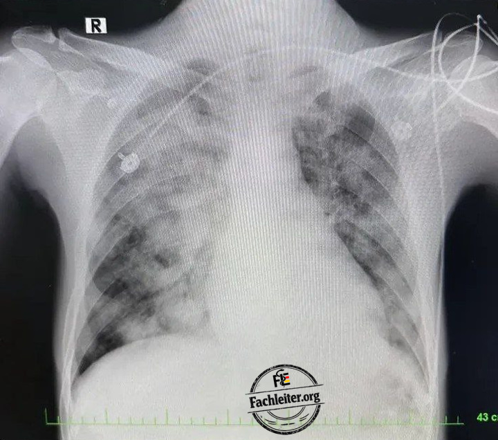
A patient is struggling to breathe. Oxygen saturation is falling. The chest X‑ray shows opacities. Is this already acute respiratory distress syndrome (ARDS)?
The answer cannot be derived from a single symptom or image. Crucial are the time course, the cause, the oxygenation and the question whether the imaging findings explain the respiratory disturbance. A pneumonia can trigger ARDS. A sepsis from the abdomen can as well. A pulmonary embolism, by contrast, often causes severe dyspnea without ARDS being present.
What does ARDS mean?
The Acute Respiratory Distress Syndrome (ARDS) is an acute, inflammation‑mediated lung injury. Because the alveolocapillary barrier is disturbed, fluid enters the lung tissue and the alveoli. Gas exchange deteriorates.
The respiratory impairment develops within one week after a known trigger or after the onset or worsening of respiratory symptoms. Imaging shows bilateral opacities that are not fully explained by pleural effusions, atelectasis or other localized findings. Heart failure or fluid overload must not be the principal explanation for the respiratory disturbance. Not every patient subsequently develops pulmonary fibrosis.
The Horovitz ratio
The Horovitz ratio describes the relationship between the arterial oxygen partial pressure (PaO₂) from the blood gas analysis and the fraction of inspired oxygen (FiO₂) actually inhaled:
PaO₂/FiO₂ = PaO₂ in mmHg ÷ FiO₂ as a decimal number
On room air FiO₂ is about 0,21. If the patient is receiving oxygen therapy, use the FiO₂ at the time of the blood gas analysis instead. If it is set, for example, to 0,50, divide the PaO₂ by 0,50.
Example: PaO₂ 75 mmHg with FiO₂ 0,50 → 75 ÷ 0,50 = 150 mmHg.
According to the Berlin definition, ARDS is assessed at a ratio of ≤ 300 mmHg under appropriate positive airway pressure: mild 201–300, moderate 101–200, severe ≤ 100 mmHg. The more recent global definition takes into account, under certain conditions, patients on high‑flow oxygen therapy. A low ratio alone is not sufficient for the diagnosis of ARDS.
Case 1: Influenza A pneumonia with development of ARDS
Case and history
A 50‑year‑old man, 178 cm tall and 82 kg, presents with four days of fever, dry cough, headache and myalgia. Since the morning he has also been short of breath at rest. His wife had a confirmed influenza A infection the previous week.
He has known arterial hypertension. He takes amlodipine 5 mg p.o. once daily. No other chronic medications or drug allergies are known. There is no chronic heart or lung disease.
Initial examination, laboratory and radiography
The patient is awake and oriented but, because of dyspnea, speaks only in short phrases. Bilateral basal crackles are audible.
| Finding | Value |
|---|---|
| Temperature | 39,0 °C |
| Blood pressure | 116/70 mmHg |
| Heart rate | 114/min |
| Respiratory rate | 31/min |
| SpO₂ on room air | 87 % |
| PaO₂ on room air | 56 mmHg |
| Leukocytes | 8,9 G/l |
| CRP | 108 mg/l |
| Procalcitonin | 0,12 ng/ml |
| Creatinine | 0,9 mg/dl |
PCR from a nasopharyngeal swab detects influenza A. The chest X‑ray initially shows patchy opacities in both lower lung zones. There is no large pleural effusion. Blood cultures and a respiratory sample are taken. A bacterial coinfection cannot be excluded on admission.
Initial treatment – medical orders
-
Start oxygen via Venturi mask with FiO₂ 0,40. Continuously monitor SpO₂ and work of breathing and repeat the arterial blood gas analysis while on oxygen.
-
Start oseltamivir 75 mg p.o. every 12 hours. Treatment is initiated immediately despite symptom onset four days earlier because the patient is hospitalized with severe influenza pneumonia. Initially five days of treatment are planned; duration will be reassessed in case of severe course.
-
Until a bacterial coinfection is excluded, start ceftriaxone 2 g i.v. every 24 hours and azithromycin 500 mg i.v. every 24 hours.
-
Treat the patient on a monitoring unit. Closely monitor respiratory rate, oxygen requirement, blood pressure and level of consciousness.
Deterioration and repeat diagnostics
After about 30 hours the oxygen requirement increases. The patient is breathing 38 times per minute, requires CPAP and appears increasingly exhausted.
The deterioration can occur despite oseltamivir already being given: the influenza has triggered inflammatory lung injury that does not resolve immediately with the start of antiviral therapy. A bacterial coinfection, fluid overload and heart failure are additionally re‑evaluated.
The follow‑up chest X‑ray now shows extensive, partially confluent opacities in both lungs. Echocardiography shows no left ventricular failure that would primarily explain the respiratory disturbance. There are no large pleural effusions.
On CPAP with PEEP 8 cmH₂O and FiO₂ 0,50 the arterial PaO₂ is 75 mmHg:
Horovitz ratio: 75 ÷ 0,50 = 150 mmHg.
The acute course, the bilateral opacities and the hypoxemia are consistent with moderate ARDS. Blood cultures to date are negative. After intubation required because of exhaustion, tracheal secretions are additionally sent for microbiological testing.
Subsequent therapy – medical orders
-
Intubate and apply lung‑protective ventilation.
-
Set initial tidal volume to 440 ml. At 178 cm height the predicted body weight is about 73 kg; 440 ml corresponds to roughly 6 ml/kg predicted body weight. Keep plateau pressure below 30 cmH₂O. Adjust PEEP and FiO₂ based on repeated blood gases and circulatory response.
-
Continue oseltamivir 75 mg every 12 hours via the nasogastric tube. Reassess therapy duration after five days based on the clinical course.
-
Continue ceftriaxone and azithromycin initially until the repeat diagnostics are evaluated. Blood and tracheal secretion cultures remain negative; further clinical assessment also yields no evidence of bacterial pneumonia. After this reassessment both antibiotics are discontinued.
-
Perform prone positioning if significant hypoxemia persists despite optimized ventilation.
Clear cause: The proven influenza A infection caused a viral pneumonia. The inflammation subsequently injured both lungs and led to moderate ARDS. An additional bacterial pneumonia was not detected in this case.
Case 2: Perforated diverticulitis with septic shock and ARDS
Case and history
A 67-year-old man, 175 cm tall and 75 kg, is admitted for increasing left lower abdominal pain and fever. His wife reports that he has been confused for several hours and has produced very little urine. He has no cough or sputum.
He has type 2 diabetes mellitus and arterial hypertension. He takes Metformin 1.000 mg p.o. twice daily and Ramipril 5 mg p.o. once daily. No drug allergies are known. Diverticulosis was noted on a previous colonoscopy.
Initial examination, laboratory and radiography
The patient is disoriented and diaphoretic. The lower abdomen is tender with guarding.
| Findings | Value |
|---|---|
| Temperature | 38,8 °C |
| Blood pressure | 78/44 mmHg |
| Heart rate | 128/min |
| Respiratory rate | 27/min |
| SpO₂ on room air | 94 % |
| Leukocytes | 20,4 G/l |
| Lactate | 5,2 mmol/l |
| Creatinine | 2,1 mg/dl |
The abdominal CT shows a perforated sigmoid diverticulitis with free air and intraabdominal fluid. The initial chest x-ray does not yet show bilateral pulmonary infiltrates. The initially estimated creatinine clearance is about 35 ml/min. The patient is in septic shock with acute renal dysfunction.
Initial treatment – medical orders
-
After immediate collection of blood cultures, start Piperacillin/Tazobactam 4 g/0,5 g i.v. every 8 hours. The dosing takes into account the currently estimated creatinine clearance of 35 ml/min; renal function and dose will be reassessed daily.
-
Give balanced crystalloid infusion solution initially in 500-ml boluses i.v. After each bolus reassess blood pressure, peripheral perfusion, respiratory findings and volume response. For 75 kg the guideline-based initial reference amount of 30 ml/kg is 2.250 ml within three hours; ongoing administration is guided by repeated assessment.
-
If hypotension persists, start Noradrenaline i.v. as an infusion pump at 0,05 µg/kg/min. For 75 kg this corresponds to 3,75 µg/min. Titrate according to blood pressure and organ perfusion; the initial target is a mean arterial pressure of approximately 65 mmHg.
-
Arrange urgent operative source control of the abdomen. Repeatedly monitor lactate, creatinine and urine output.
-
Pause Metformin and Ramipril while septic shock and acute renal dysfunction persist.
The perforated bowel segment is surgically treated and the abdominal cavity is decontaminated. Source control is an essential part of sepsis therapy; antibiotics alone do not eliminate the perforation.
Deterioration and repeat diagnostics
Several hours after the operation the oxygen demand increases markedly. The new chest x-ray shows extensive bilateral infiltrates. There are no large pleural effusions. Echocardiography shows no left heart failure to primarily explain this finding.
The patient is intubated. Under PEEP 10 cmH₂O and FiO₂ 0,80 the PaO₂ is 64 mmHg:
Horovitz quotient: 64 ÷ 0,80 = 80 mmHg.
This corresponds to severe ARDS. Blood cultures later grow Escherichia coli; susceptibility to Piperacillin/Tazobactam is confirmed. The source of infection was surgically treated, but the systemic inflammatory response already triggered had damaged both lungs.
Subsequent therapy – medical orders
-
Lung-protective ventilation with an initial tidal volume of 420 ml. At 175 cm body height the calculated body weight is about 71 kg; 420 ml correspond to roughly 6 ml/kg. Keep plateau pressure below 30 cmH₂O.
-
Due to severe ARDS, perform prone positioning for more than twelve hours per day under intensive care monitoring.
-
Continue Piperacillin/Tazobactam 4 g/0,5 g i.v. every 8 hours as long as renal function and pathogen susceptibility support this order. Determine creatinine clearance and dosing daily.
-
Titrate Noradrenaline according to circulatory response. Give further fluids only after reassessing volume needs. Monitor lactate, urine output and fluid balance.
Clear cause: A perforated sigmoid diverticulitis triggered septic shock. The resulting systemic inflammatory response subsequently caused bilateral lung injury and severe ARDS.
Case 3: Aspiration of gastric contents with ARDS
Case and history
A 42-year-old man, 180 cm tall and 78 kg, is admitted after a generalized seizure. His partner observed that he vomited during the subsequent impaired consciousness. He then began to cough heavily. Before the seizure he had neither fever nor respiratory complaints.
He has known epilepsy. Levetiracetam 1.000 mg p.o. twice daily is prescribed; he missed several doses. No other chronic medications or drug allergies are known. There is no heart or lung disease.
Initial examination, laboratory and radiography
The patient is rousable again and can protect his airway. He coughs persistently. Crackles are audible at the right base.
| Findings | Value |
|---|---|
| Temperature | 37,4 °C |
| Blood pressure | 134/82 mmHg |
| Heart rate | 108/min |
| Respiratory rate | 30/min |
| SpO₂ on room air | 88 % |
| Leukocytes | 12,5 G/l |
| CRP | 18 mg/l |
| Creatinine | 0,9 mg/dl |
The chest x-ray shows a right-predominant opacity in dependent lung regions. The immediate onset after vomiting during impaired consciousness suggests aspiration of gastric contents with chemical lung injury.
Initial treatment – medical orders
-
Control the airway, suction visible material as needed and give oxygen. Closely monitor level of consciousness, SpO₂ and work of breathing.
-
Administer Levetiracetam 1.000 mg reliably every 12 hours. As long as the patient cannot safely swallow, give it intravenously.
-
Do not start antibiotics. The observed course and the initially absent clear signs of infection suggest a chemical aspiration pneumonitis. Reassess temperature, secretions, inflammatory markers and the clinical course.
Deterioration and repeat diagnostics
After 18 hours the respiratory rate increases to 36/min; the patient requires increasing oxygen and becomes exhausted. The follow-up x-ray now shows patchy infiltrates in both lungs. He still has no fever; reassessment yields no convincing evidence of an additional bacterial pneumonia.
The patient is intubated. Under PEEP 8 cmH₂O and FiO₂ 0,60 the PaO₂ is 90 mmHg:
Horovitz quotient: 90 ÷ 0,60 = 150 mmHg.
The course and the new bilateral findings are consistent with moderate ARDS following aspiration.
Subsequent therapy – medical orders
-
Lung-protective ventilation with an initial tidal volume of 450 ml. At 180 cm body height the calculated body weight is about 75 kg; 450 ml correspond to 6 ml/kg. Keep plateau pressure below 30 cmH₂O.
-
Adjust PEEP and FiO₂ based on blood gas analyses and circulatory response. Reassess airway secretions, temperature and imaging findings over time.
-
Continue levetiracetam 1.000 mg i.v. every 12 hours as long as reliable oral intake is not possible.
-
Do not prescribe antibiotics unless the clinical course suggests a bacterial infection. ARDS alone does not prove bacterial pneumonia.
Clear cause: The seizure resulted in impaired consciousness with aspiration of gastric contents. The chemical lung injury progressed to both lungs and caused a moderate ARDS.
Case 4: Pulmonary embolism with hypoxemia – not ARDS
Case and history
A 49-year-old woman, weighing 70 kg, suddenly develops dyspnea, pleuritic chest pain and palpitations. Her left leg has been swollen since the previous day.
After a lower leg fracture two weeks ago the leg was immobilized; since then she has been poorly mobile. There is no known cardiac or pulmonary disease or prior thromboses. She takes no long-term medications and has no known drug allergies. There is no active bleeding.
Initial examination, laboratory tests and radiography
| Finding | Value |
|---|---|
| Blood pressure | 118/72 mmHg |
| Heart rate | 124/min |
| Respiratory rate | 29/min |
| SpO₂ on room air | 90 % |
| PaO₂ on room air | 60 mmHg |
| Creatinine | 0,8 mg/dl |
The left leg is unilaterally swollen. The ECG shows sinus tachycardia. The chest x-ray shows no new bilateral opacities. CT pulmonary angiography demonstrates a bilateral pulmonary embolism. The initial echocardiography shows no marked right heart strain.
On room air the Horovitz ratio is:
60 ÷ 0,21 ≈ 286 mmHg.
The value indicates an oxygenation disturbance. It does not establish ARDS here, because the CT demonstrates another cause and the bilateral lung opacities required for ARDS are absent.
Initial treatment – physician orders
-
Start apixaban 10 mg p.o. twice daily for seven days. Then continue apixaban 5 mg p.o. twice daily. The patient can reliably take tablets and, according to the case information, has no contraindication to this treatment.
-
Administer oxygen according to the measured hypoxemia. Monitor SpO₂, blood pressure, heart rate and symptoms in hospital.
-
Monitor for signs of bleeding during anticoagulation. The further duration of treatment will be determined taking the provoking risk factor into account.
Deterioration and repeat diagnostics
A few hours later the heart rate rises to 138/min and oxygen requirement increases. Blood pressure remains stable. Repeat echocardiography now shows an enlarged right ventricle with impaired function; troponin is elevated. The follow-up chest x-ray still shows no new bilateral ARDS opacities.
The findings indicate increasing right heart strain due to the pulmonary embolism. The patient is at risk of developing shock, but at the time of the reassessment she is still hemodynamically stable.
Subsequent therapy – physician orders
-
Continue apixaban at the initiated therapeutic dose.
-
Monitor on an appropriate unit with frequent checks of blood pressure, heart rate, oxygen requirements, level of consciousness and signs of bleeding.
-
Inform the team responsible for possible reperfusion early. Do not perform systemic thrombolysis as long as the patient remains hemodynamically stable. If shock occurs, reperfusion and bleeding risk must be reassessed immediately.
Clear cause: Immobilization of the leg after the fracture promoted a venous thrombosis. This led to a pulmonary embolism that explains the sudden dyspnea, hypoxemia and later right heart strain. In this case there is no ARDS.
ARDS briefly explained
The acute respiratory distress syndrome (ARDS) is an inflammatory lung injury that develops within one week. Fluid enters the alveoli and impairs gas exchange. Typical features are bilateral opacities and marked hypoxemia. The symptoms should not be primarily explained by heart failure or volume overload.
The Horovitz ratio is calculated from the arterial oxygen partial pressure (PaO₂) and the actually inspired fraction of oxygen (FiO₂): PaO₂ ÷ FiO₂. Example: 75 mmHg ÷ 0,50 = 150 mmHg. A value of ≤ 300 mmHg is an ARDS criterion; the value alone does not prove the diagnosis.