
Greeting
Good afternoon, dear Professor Doctor …
I would like to present the patient Ralf Winter to you now.
He is 57 years old, one meter seventy-six tall and weighs approximately 89 kg. The body mass index (BMI) is 28.7 kg/m² and therefore in the overweight range.
Current history
The patient presented because of, since this morning early persistent, suddenly occurring chest tightness.
He complained of stabbing pain radiating to the neck and the left mandible.
As accompanying symptoms the patient reported dyspnea, tachypnea, tachycardia, dizziness, nausea, cold sweating and pallor.
He also described a pronounced fear of dying as well as a feeling of annihilation.
The following symptoms were denied:
fever, pressure tenderness in the thoracic area
panic attack
respiratory- or position-dependent increase in pain
cough, palpitations, emesis, orthopnea, peripheral edema as well as newly onset nocturia
circumference difference between the lower extremities, coagulopathy
He added that he had been experiencing similar complaints for about six weeks – mostly with physical exertion, such as climbing stairs or walking about 150 meters.
He has also noticed postprandial pain that did not respond to pantoprazole.
As a trigger he described that his grandchild had suffered seizures in the morning, which distressed him greatly. During the joint ambulance ride to the hospital the current complaints suddenly occurred.
So far, Mr. Winter reports he did not consult a doctor because he did not take the symptoms seriously.
The autonomic history
The autonomic history is unremarkable, apart from an insomnia with difficulties falling and staying asleep, which the patient attributes to familial and occupational stress. (no evidence of obstructive sleep apnea syndrome)
He also reports loss of appetite with unintended weight loss and a food intolerance with bloating, especially after consumption of legumes.
Past medical history
His past medical history includes the following:
A type 2 diabetes mellitus has existed for eight years and is well controlled metabolically using a glucose sensor in the upper arm. Diabetes-associated complications have not occurred to date.
|
No diabetes |
Prediabetes |
Diabetes |
|
|
Fasting blood glucose |
< 100 mg/dl |
100–125 mg/dl |
≥ 126 mg/dl |
|
Random blood glucose |
– |
– |
≥ 200 mg/dl |
|
HbA1c |
< 5,7 % |
5,7–< 6,5 % |
≥ 6,5 % |
|
OGTT (2 h value) |
< 140 mg/dl |
140–199 mg/dl |
≥ 200 mg/dl |
An arterial hypertension has also been present for eight years. Due to irregular medication intake there are blood pressure fluctuations. The diagnosis was established using a 24-hour blood pressure measurement that recorded blood pressure every 20 minutes.
|
systolic (mmHg) |
diastolic (mmHg) |
Treatment |
|
|
Normal blood pressure |
< 120 |
< 70 |
– |
|
Elevated blood pressure |
120–139 |
70–89 |
+ |
For 3 years there has also been a benign prostatic hyperplasia (main complaint: nocturia), which is treated medically.
A few years ago the patient suffered a syncope. He was then monitored inpatient in the intensive care unit and received intravenous fluid therapy.
For some time the patient has had exertional pain in the right shoulder. About a year ago an orthopedist diagnosed a bursitis subacromialis. With overuse pain and limited motion recur. Rest of the shoulder was recommended but is not always possible due to work.
Previous surgeries
At the age of 50 a stool test was performed, after which the family doctor initiated a colonoscopic evaluation. Seven years ago a colonoscopy was performed in which a benign polyp (adenoma) was removed.
Two years ago the patient fell while ice skating and sustained a fracture of the right Humerus to. The fracture was surgically treated using plate osteosynthesis treated. Postoperatively he received analgesic therapy with ibuprofen. The metal plate was removed about six months later.
Note: If a statement can be objectively confirmed by a medical examination – for example by visible scars – the indicative is appropriate. (No physical examination takes place in the medical language exam.)
Gynecology
————–
Medications
The patient is currently taking the following medications as prescribed:
Ramipril 5 milligrams in the evening
Metoprolol 95 milligrams at midday
Hydrochlorothiazide 12.5 milligrams in the morning
Metformin 500 milligrams, 1-0-1
Tamsulosin 0.4 milligrams
Dutasteride 0.5 milligrams
Ibuprofen 400 mg as needed
Lefax– chewable tablets as needed
Note: If a statement can be objectively confirmed by a medical observation – for example by a prescription – the indicative is appropriate.
Allergies
He is allergic to rubber gloves; he gets an itchy rash on his hands.
He also reacts to metamizole, i.e. Novalgin, with urticaria and shortness of breath – at the time he even had to be monitored in the intensive care unit.
Exposures
The patient has been a non-smoker for three years. Previously he had smoked about half a pack of cigarettes per day for approximately 20 years, which corresponds to about 10 pack-years equivalents. On medical advice he quit smoking completely.
He reports drinking one to two glasses of beer in the evenings to help him fall asleep.
The patient reported not using drugs.
Social history
He is self-employed and owns a butcher’s shop.
After separating from his wife, he lives alone in an apartment on the first floor.
He has three children – a 20-year-old son and two daughters aged 26 and 29. All are healthy.
Physical activity: –
Family history
In the family history the patient reported that his father died at age 65 of a brain tumor and his mother died at age 69 of a pulmonary embolism .
His nine-year-old granddaughter underwent surgery for an aortic valve malformation.
Suspected and differential diagnosis
In my view the history, especially the character of the pain accompanied by dyspnea, most likely indicates an acute coronary syndrome.
Risk factors for an acute coronary syndrome (ACS) in this patient include age, former smoker, lack of physical activity, arterial hypertension, diabetes mellitus, and overweight.
An ECG and determination of troponin levels can confirm or exclude a myocardial infarction.
Against a pericarditis argues the absence of fever, preceding infections, and pleuritic (respiration-dependent) pain.
In an aortic dissection unstable hemodynamics and blood pressure differences between the upper extremities are to be expected.
A panic attack seems unlikely because the symptoms also occur during physical exertion and not exclusively in stressful situations. A panic attack seems unlikely because the symptoms also occur during physical exertion and not exclusively in stressful situations. In addition, a cardiac cause must always be ruled out.
Against a pulmonary embolism argues the absence of risk factors such as immobilization or surgery as well as the lack of cough and hemoptysis.
A Prinzmetal angina (vasospastic angina pectoris) is also considered in the differential diagnosis.
Diagnostic work-up
If myocardial infarction is suspected, the patient must be admitted immediately and the necessary measures initiated, as every second counts.
physical examination
Following the history, a thorough physical examination should be performed. This begins with recording the vital signs, including heart rate, blood pressure, respiratory rate, oxygen saturation and temperature.
(The physical examination alone is not sufficient to diagnose an acute coronary syndrome (ACS).)
First comes inspection, where we look at respiratory movements, edema, pallor, jugular venous distention and other abnormalities.
This is followed by palpation to check for local findings such as tenderness and to assess peripheral pulses (e.g., A. radialis or A. tibialis posterior).
After that, percussion is performed,
to obtain clues to possible lung disease or hepatosplenomegaly.
Finally, auscultation is performed to detect pathological breath sounds and abnormal heart sounds. Pathological auscultatory findings such as crackles, wheeze, a pericardial friction rub, or a heart murmur consistent with aortic regurgitation (murmur of aortic regurgitation) may be noted.
In heart failure, for example, we may hear basal crackles over the lungs – an indication of pulmonary congestion – as well as a third heart sound (gallop rhythm), which speaks for diastolic dysfunction.
The cardiac apex is usually located in the 5th intercostal space at the left midclavicular line. Palpation of the apical impulse provides information about heart size and contractile force. A displaced or broadened palpable apical impulse can indicate left heart failure. Additionally, signs of jugular venous congestion as well as a positive hepatojugular
Since the female patient has both arterial hypertension and diabetes mellitus, initial indications of complications such as retinopathy, neuropathy, or peripheral arterial occlusive disease should also be checked as part of the basic diagnostics.
Electrocardiogram
An electrocardiogram (ECG) should be performed within ten minutes of first contact. Repeats should be performed every five to ten minutes.
An ECG is performed by placing electrodes on the skin of the chest, arms, and legs to record the electrical activity of the heart.
Typical findings are ST-segment elevations, which indicate an ST-elevation myocardial infarction (STEMI), or ST-segment depressions and T-wave inversions, which can occur with a non-ST-elevation myocardial infarction (NSTEMI) or unstable angina pectoris. In addition, pathological Q waves may occur, indicating a previous myocardial necrosis. Cardiac arrhythmias such as ventricular extrasystoles, atrial fibrillation or Tachycardias can be observed in the context of an ACS. A Left bundle branch block, especially if newly occurring, can be an indication of an acute myocardial infarction and must be taken into account accordingly.
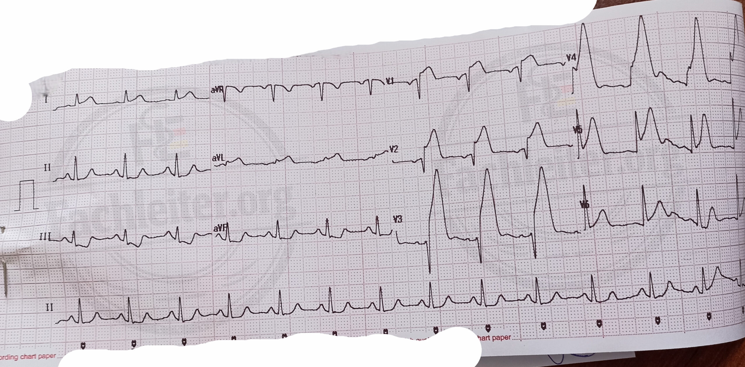
|
Infarction |
Leads |
Artery |
|
Anterior wall infarction (anterior)/ septal infarction |
V1 – V4 |
Ramus interventricularis anterior (RIVA / LAD) |
|
Lateral wall infarction (lateral) |
I, aVL |
Ramus circumflexus (LCx) |
|
Inferior |
II, III, aVF |
Mostly right coronary artery (ACD/ RCA) |
|
aVL and I (reciprocal) |
Laboratory tests
If an acute coronary syndrome is suspected, a small blood count, C-reactive protein, electrolytes, lipid profile, renal function tests, TSH (thyroid-stimulating hormone), long-term blood sugar (HbA1c), fasting blood glucose, PT, PTT, INR, troponin I, creatine kinase–myocardial band (CK-MB) as well as B-type natriuretic peptide should be determined. natriuretic peptide can be measured.
Anatomic diagnosis
Chest X-ray
The patient removes jewelry and clothing in the chest area. He stands or sits facing the X-ray plate. During the exposure he is asked to take a deep breath and hold it briefly. Among other things, the cardiac silhouette (e.g., in cardiac hypertrophy), the vascular markings (signs of pulmonary congestion or congestion) as well as possible signs of pulmonary edema are assessed.
Echocardiography:
Echocardiography is an imaging procedure for assessing cardiac structure and function, wall motion and valve motion using ultrasound. For transthoracic echocardiography (TTE) no special preparation is required. The patient usually lies in the left lateral position while the probe is placed on the chest wall to visualize different planes of the heart. Among other things, the ejection fraction (EF) can be measured, an important parameter for assessing left ventricular pump function.
An echocardiogram, also called a cardiac ultrasound, shows whether the patient has a reduced ejection fraction (less than 40%), a moderately reduced ejection fraction (between 40 and 50%), or a preserved ejection fraction.
(For transesophageal echocardiography (TEE) the patient must fast for about six hours. Prior to the examination, informed consent and, if necessary, sedation are provided. During TEE an ultrasound probe is introduced via the esophagus to obtain detailed images of the heart, especially the valves and atria.)
Treatment
Lifestyle modification
• Weight reduction
• regular physical activity
• Reduction of salt intake
General acute measures in ACS:
• Calming/reassurance
• Monitoring: ECG monitoring, blood pressure, heart rate, oxygen saturation
• Oxygen administration: Only if SpO2 < 90% or clinical signs of hypoxia
• Analgesia: Morphine
• Pharmacological basic therapy: Acetylsalicylic acid (ASA, 162 to 325 mg, followed by 81 mg daily, lifelong), P2Y12 inhibitors (e.g., clopidogrel or ticagrelor, up to one year), anticoagulation (e.g., heparin), statin, PPI or H2 blocker, beta blocker (has already been taken by this patient for 8 years)
Coronary angiography:
Coronary angiography with primary percutaneous coronary intervention (PCI) should be performed within 120 minutes of first contact.
(If PCI is not available within this time, fibrinolysis should be considered within 10 minutes of diagnosis.) Fibrinolysis should be considered within 10 minutes after diagnosis.)
In a high-risk non–ST-elevation myocardial infarction – characterized by persistent chest pain, hemodynamic instability, dynamic changes in troponin values or ECG changes – coronary angiography should be performed within 24 hours.
Angiography is a minimally invasive procedure to visualize and, if necessary, treat arteries using X-rays and contrast agent. After disinfecting the puncture site, the contrast agent is injected via a larger artery such as the femoral or radial artery. X-ray images are then taken to visualize blood flow as well as possible thromboses or stenoses. In certain cases this procedure is replaced by duplex sonography, as it is noninvasive and does not require contrast. The procedure usually takes 30 to 60 minutes and requires no elaborate preparation. If necessary, a therapeutic measure can be performed simultaneously, for example using a balloon catheter or a stent to treat vascular narrowing.
Because contrast is used in angiography, we should ask about renal insufficiency, metformin use, possible contrast allergies, and hydration status.
Coronary bypass surgery (CABG)
Surgical treatment for acute coronary syndrome (ACS) is performed in certain cases – especially with multiple affected coronary arteries, left main stenosis or failed PCI
Coronary bypass surgery (CABG) means diverting blood flow using vascular grafts (e.g., saphenous vein, internal mammary artery)
Complications
• Pump failure (heart failure)
• Arrhythmias
• recurrent ischemias and reinfarctions
Pathophysiology and forms of acute coronary syndrome (ACS)
Angina pectoris
Angina pectoris occurs when the heart receives too little oxygen.
This happens when the oxygen demand of the myocardium – for example due to physical exertion or stress – increases, but the narrowed coronary arteries cannot deliver enough blood.
This creates an imbalance between oxygen supply and demand.
Typical are pressure or tightness sensations in the chest, sometimes radiating to the arm, neck or jaw.
The most common cause of stable angina pectoris is atherosclerosis of the coronary arteries, i.e., vascular narrowing due to deposits.
Typical angina pectoris:
• Retrosternal pressure or tightness
• Triggered by physical exertion or emotional stress
• Improves with rest or after nitroglycerin administration
Acute coronary syndrome (ACS)
The term acute coronary syndrome refers to an acute reduction in blood flow to the heart muscle, which can present as unstable angina pectoris or myocardial infarction (NSTEMI or STEMI).
In most cases, ACS is caused by rupture of an atherosclerotic plaque in a coronary artery.
Platelets adhere, a thrombus forms, and blood flow is reduced or completely blocked.
Unstable angina pectoris (UA)
Unstable angina pectoris is characterized by:
• new-onset chest pain (for less than 2 months),
• longer duration (over 20 minutes),
• increased intensity, or
• symptoms at rest.
Perfusion is reduced but still sufficient to prevent necrosis of myocardial cells.
Therefore cardiac enzymes (CK-MB, troponin) are normal.
Non–ST-elevation myocardial infarction (NSTEMI)
In NSTEMI cardiac enzymes (CK-MB, troponin) are elevated,
but no ST elevations are seen on the ECG.
Here there is a partial or transient narrowing of the coronary artery.
The injury usually affects only the inner layer of the myocardium (subendocardial).
ST-elevation myocardial infarction (STEMI)
A STEMI is present when cardiac enzymes are elevated and ST elevations or a new left bundle branch block are visible on the ECG.
The cause is a complete and persistent occlusion of a coronary artery.
This causes myocardial cell death, resulting in a transmural infarction.
Extra
Names:
Gertrud Wolf, Kraus Krämer, Brigitta Müller, Markus Scholz, Josef Schreiner, Hobert Kuhler, Anna Pulver, Veronika Voß, Hanns Zierzicht, Carla Dräger, Josif Rinnir
Occupations:
The patient was a pastry chef by profession (owner). She and her husband had taken over the pastry shop from her in-laws. The pastry shop was on the ground floor.
Tax consultant
Interior designer specializing in old building renovation
Former teacher
Details:
She wanted to pick up her grandchild from kindergarten and the pain started while walking. She called her daughter-in-law, who picked up the patient on the way and brought her to the hospital.
The customer cancelled a large order because they no longer needed the products. This upset the patient. Because he is self-employed and works as an electromechanic, this contract was very important to him to support his company with 5 employees. Therefore the typical ACS symptoms appeared suddenly.
why the patient continues to suffer from difficulty falling asleep despite the improvement in his granddaughter’s health.
In my chest I have the feeling as if a large car tire is pressing on it.
In her childhood the patient fell from a climbing frame and sustained a forearm fracture.
He received nitroglycerin/a spray in the ambulance.
These occurred after a phone call in which a customer cancelled an important project
A 78-year-old retiree went for a walk in the forest with his dog in the morning.
When the dog suddenly ran off, he ran after it.
He developed an acute retrosternal pain and shortness of breath.
He called his daughter-in-law, who immediately brought him to the hospital.
Past medical history:
Compression therapy for DVT, previously heparin for 3 months
Constipation, treated with psyllium
She has had undiagnosed urinary incontinence for a few months. She described it as urinary weakness and urgency.
Furthermore, the patient underwent a transvaginal uterine myomectomy.
Hypercholesterolemia
Status post left rib fracture from a bicycle accident 13 years ago, treated conservatively.
Traumatic brain injury (TBI) after a car accident at age 14
Status post hysterectomy due to postpartum hemorrhage
Status post tendon tear at the right thenar 10 years ago
Known anemia, regularly treated with parenteral iron administration every three months
Status post bilateral cataract surgery two years ago
After a gastroscopy in 2023 a Helicobacter pylori infection was detected and subsequently treated with eradication therapy.
Status post osteosynthesis after an ankle fracture (work accident) 5 years ago; plates and screws have since been removed.
Status post orchiectomy due to a testicular carcinoma at age 26.
20 years ago the patient had a deep vein thrombosis (DVT). At that time she was taking oral contraceptives and had recently taken a long-distance flight. She subsequently stopped taking the contraceptives.
Allergies:
Hazel pollen allergy: rhinoconjunctivitis
Medications:
Lisinopril 10 mg
Melatonin spray, as needed
Lorano® (antihistamine)
Family history:
a grandchild who is suffering from scarlet fever.
congenital lung disease in his child, primary ciliary dyskinesia or congenital pulmonary airway malformation
Father: died about 2 years ago of peritonitis (perforated appendicitis) or had a foot amputated
Mother: 80 years old, living with TIA
Mother: colon carcinoma, currently undergoing chemotherapy
Father: died of myocardial infarction
The father died of a cardiac arrhythmia; he had a pacemaker.
The mother died of renal insufficiency.
Sister: pulmonary embolism.
My brother, who is five years younger than me, has heart problems, probably coronary artery disease (CAD). He is supposed to have bypass surgery, but he is very afraid of dying during the procedure.
Substance use:
Alcohol: Radler
Party drugs in adolescence
Pipe (smoking)
Questions
1. Which sports would be appropriate for this patient?
The choice of sports should be individualized. In particular, enjoyment of the activity plays an important role to promote long-term motivation.
Recommended activities are mainly low-impact endurance sports such as walking, cycling and swimming.
2. What should you do if the patient does not want to stay in the hospital?
If a patient refuses hospital admission even though it is medically indicated, I should first provide comprehensive information about the diagnosis, the possible risks and consequences of refusal, and the recommended therapeutic measures.
I would try to take his concerns seriously and address his fears or doubts in a calm conversation.
If the patient still refuses, I have him sign written documentation of risk counseling and a discharge against medical advice – provided he is competent to consent.
3. What types of ankle fractures are there?
Weber A: fracture below the syndesmosis (infrasyndesmotic) – usually stable.
Weber B: fracture at the level of the syndesmosis (transsyndesmotic) – stability depends on the condition of the syndesmosis.
Weber C: fracture above the syndesmosis (suprasyndesmotic) – usually unstable, often with accompanying syndesmosis rupture.
4. How much alcohol is too much daily?
For men: no more than 24 g pure alcohol/day
For women: no more than 12 g pure alcohol/day
Approximately 10–12 g of pure alcohol are contained in 0.25 l of beer, 0.1 l of sparkling wine, 0.1 l of wine
5. Which diagnoses would be considered in a younger patient with similar symptoms?
In a younger patient with symptoms of an acute coronary syndrome, cardiomyopathies, pericarditis, myocarditis, congenital heart defects and hormonal causes such as hyperthyroidism are primarily to be considered. Substance abuse (e.g., alcohol or drugs) could also be a possible trigger.
6. What are the advantages of switching from Marcumar to Eliquis?
Eliquis (apixaban) – like other direct oral anticoagulants – offers compared with Marcumar (phenprocoumon) the advantage that no regular INR monitoring is required, there are fewer food interactions and the overall risk of bleeding, particularly intracranial, is lower.
7. Why can the troponin rise be low in a STEMI despite a poor prognosis?
Troponin is a marker of myocardial necrosis but does not directly reflect prognosis.
In some patients with ST-elevation myocardial infarction, the troponin increase may be relatively small despite a worse prognosis – particularly due to the timing of blood sampling, no‑reflow phenomena and hemodynamic complications such as cardiogenic shock or malignant arrhythmias.
Medical orders for ACS (STEMI/NSTEMI)
• Early cardiology notification/alert catheterization readiness
• Monitoring: 3-/12-lead ECG immediately, continuous telemetry ECG, blood pressure, SpO₂. First ECG <10 min after arrival; if complaints are unclear, repeat serially.
• Defibrillator
• Resuscitation team / emergency team
• 2 large-bore IV accesses, blood sampling (laboratory incl. hs‑troponin according to 0/1 h algorithm, CBC, electrolytes, fasting glucose, renal values, coagulation, lipids), blood group/antibody screen
• O₂ if SpO₂ < 90% or signs of hypoxemia/dyspnea.
• Intubation readiness
• Analgesia/antiemesis: morphine IV; antiemetic e.g., ondansetron
• Nitrates: nitro spray/nitroglycerin IV for persistent pain/hypertension, not in hypotension, not in suspected RV infarction.
• Keep fasting
• Aspirin 150–300 mg orally (chewable) or 75–250 mg IV as loading dose.
• P2Y12 inhibitor: ticagrelor 180 mg orally (standard) or prasugrel 60 mg (if PCI planned and no contraindication), clopidogrel 600 mg if the others are not suitable (e.g., anticoagulation/bleeding/drug interaction).
• Anticoagulation: UF heparin or enoxaparin/fondaparinux depending on strategy (if planned primary PCI → UF heparin preferred).
• High‑dose statin: atorvastatin 80 mg orally (as early as possible)
• Beta‑blocker (e.g., metoprolol) for hypertension/tachycardia without signs of acute heart failure, hypotension, AV block or bronchospasm.
• ACE inhibitor/ARNI within 24 h with stable hemodynamics, especially in anterior MI/LVEF↓
• PPI
Notes
BMI
Body mass index (BMI)
Definition: BMI is a simple ratio measure to classify body weight. It is calculated by dividing body weight in kilograms by the square of height in meters.
Clinical classification: With the given measurements (1.76 m, 89 kg) the BMI is 28.7 kg/m², which falls into the overweight category.
early
Manual review required: Uncertain assignment: The term ‘early’ refers to time of day (‘in the morning’), while the note ‘for 3 hours’ indicates a duration. The meaning is therefore unclear or not unambiguously assignable to the term.
Mandible
Mandible (lower jaw)
Definition: The mandible is the movable bone of the lower jaw and forms the lower half of the face.
Dyspnea
Dyspnea (shortness of breath)
Term explanation: Dyspnea denotes the subjective sensation of breathlessness or difficulty breathing.
Tachypnea
Tachypnea
Brief description:
Accelerated respiratory movements with increased respiratory rate.
Tachycardia
Tachycardia
Brief description:
Increased heart rate, often subjectively perceived as palpitations.
Clinical significance:
Tachycardia occurs with physical exertion, stress, anxiety, volume depletion, or cardiovascular disease.
Nausea
Nausea
Brief description:
Subjective sensation of nausea with possible tendency to vomit.
Clinical significance:
Nausea can be caused by gastrointestinal disorders, medication side effects, vestibular disturbances, or central/autonomic reactions to anxiety.
Pallor
Pallor
Brief description:
Noticeable reduction of the normal skin coloration (pallor).
Causes and significance:
Pallor can result from a lowered hemoglobin level (anemia) or peripheral vasoconstriction due to sympathetic activation; it is also seen in hypovolemia or shock.
Fear of death
Fear of death
Pronounced, acute fear of dying. Affected individuals often describe an intense sense of life threat and helplessness; the symptom frequently occurs together with autonomic reactions such as shortness of breath, rapid breathing, palpitations, dizziness, nausea, cold sweat, and pallor.
Tenderness to pressure
Tenderness to pressure
Denotes a localized pain elicited by finger pressure, for example on the chest wall. Such pressure tenderness may indicate an inflammatory change of the costochondral junctions (costochondritis), but it is not specific and can also occur with other chest wall problems. In the present history it was explicitly stated, however, that no pressure tenderness in the thoracic area was present.
respiratory-
Indication of pleuritis or pneumonia
position-dependent
Indication of pericarditis
Cough
Cough
Definition and function: Cough is a reflex mechanism to remove secretions, particles, or foreign bodies from the airways.
Clinical relevance: Persistent or productive cough can indicate a respiratory infection such as pneumonia, but it is not specific. The absence of cough reduces the likelihood of a typical symptomatic pneumonia, but does not rule it out, especially in older or immunocompromised patients.
Palpitations
Palpitations
Term explanation: Palpitations describe the subjective perception of the heartbeat, e.g. as strong pounding, fluttering, or skipping.
Causes and significance: Causes range from benign extrasystoles and anxiety to clinically relevant arrhythmias, electrolyte disturbances, or endocrine disorders. Occurring palpitations can indicate an arrhythmia requiring treatment.
Emesis
Emesis (denied in the history)
Definition: Emesis denotes the active vomiting of gastric contents through spasmodic contractions of the abdomen and diaphragm.
Orthopnea
Orthopnea
Definition: Orthopnea denotes the onset or worsening of breathlessness when lying down compared with the upright position.
Pathophysiology and significance: Orthopnea commonly arises from increased venous return and pulmonary congestion in left-sided heart failure and is considered an indicator of cardiac decompensation. It differs from paroxysmal nocturnal dyspnea and other position-dependent breathing complaints.
Edema and newly onset nocturia
Brief finding: Peripheral edema and newly onset nocturia
Assessment: The simultaneous presence of leg edema and newly onset nocturia should primarily suggest cardiac decompensation, since in heart failure fluid is mobilized back into the circulation at night. Isolated nocturia, however, can be caused by benign prostatic hyperplasia or evening intake of diuretics. Benign prostatic hyperplasia typically does not explain pronounced peripheral edema, which is why renal or hepatic causes should also be considered when both symptoms occur together.
Difference in circumference between lower extremities, coagulopathy
Indications of DVT (deep vein thrombosis)
Insomnia
Brief finding: Insomnia
Description: The patient reports difficulties falling and staying asleep, which he attributes to stress in the family and work domains. Otherwise there are no abnormalities in the autonomic history; indications of obstructive sleep apnea syndrome were denied.
Meteorism
Brief finding: Meteorism
Description: The patient complains of increased flatulence and reports a food intolerance that particularly after consumption of legumes leads to pronounced meteorism. Additionally there is loss of appetite with unintentional weight loss.
Legumes
Legumes
Examples and properties
Legumes include lentils, chickpeas, peas, and beans. These foods are high in fiber and contain specific carbohydrates (e.g. oligosaccharides) that can lead to increased gas production in sensitive individuals.
Clinical context
With a food intolerance after eating legumes, bloating and meteorism often occur; this can promote loss of appetite and associated weight loss if affected individuals restrict their intake.
Type 2 diabetes mellitus
Type 2 diabetes mellitus
Core feature
Type 2 diabetes is a chronic metabolic disease primarily characterized by reduced sensitivity of target tissues to insulin (insulin resistance) and a progressive impairment of insulin secretion over time.
Distinction from other forms
In type 1 diabetes there is autoimmune destruction of the insulin-producing beta cells leading to absolute insulin deficiency. Gestational diabetes arises from hormonally mediated, temporary insulin resistance during pregnancy.
Complications
Diabetes-associated complications
Important complications
Retinopathy: Metabolic disorder–related damage to the retina that can impair vision.
Nephropathy: Diabetes-related kidney damage with loss of renal function and typically early onset of albuminuria.
Neuropathy: Damage to peripheral and/or autonomic nerves with sensory deficits, pain, or autonomic dysfunction.
Prostatic hyperplasia
Definition:
Benign prostatic hyperplasia (BPH) is a noncancerous enlargement of the prostate gland that mainly occurs in older men.
Pathophysiology:
Age-related hormonal changes play an important role; in particular the conversion of testosterone to dihydrotestosterone (DHT) contributes to proliferation of prostatic cells.
Clinical signs:
Typical complaints affect the lower urinary tract: increased urinary frequency, decreased urinary stream, nocturia, a feeling of residual urine, and in severe cases acute urinary retention.
Complications:
If untreated, residual urine, recurrent urinary tract infections, bladder stone formation, and secondary kidney damage can occur.
Treatment options:
Therapy is guided by symptoms and severity. Options range from medical treatment (e.g. alpha blockers or 5‑alpha‑reductase inhibitors) through minimally invasive procedures including laser-based techniques to surgical interventions such as transurethral resection of the prostate (TURP).
Prognosis:
Symptoms can usually be well controlled with appropriate therapy; timely treatment reduces the risk of the complications mentioned.
Syncope
Short definition:
A syncope is a brief, spontaneous loss of consciousness due to a temporary reduction of cerebral blood flow.
Common causes:
Reflex triggers (vasovagal syncope), blood pressure drop on standing (orthostatic syncope), and cardiac causes such as arrhythmias or structural diseases (e.g., high-grade aortic stenosis) can provoke a syncope.
Clinical presentation and diagnostics:
The circumstances before the event (e.g., emotional stress), accompanying symptoms, and cardiological assessment are important to narrow down the cause.
Acute management:
If syncope is suspected, monitoring and evaluation are performed; if there is clinically significant volume depletion, fluid administration may be necessary. Specific further treatment depends on the underlying cause.
Bursitis
Definition:
A bursitis is an inflammation of a bursa, which acts as a fluid-filled cushion reducing friction between tendons/muscles and bony prominences.
Occurrence and causes:
Bursitis commonly occurs at the shoulder, elbow, hip, and knee and is usually caused by overuse, repeated microtrauma, or less commonly by infectious or systemic inflammatory processes.
Symptoms:
Typical findings are local pain, swelling, and restricted range of motion in the affected area; in subacromial bursitis, raising the arm is particularly painful.
Treatment:
Conservative measures such as activity modification, physical therapy, and pain-relieving/anti-inflammatory treatments are often effective; for persistent symptoms, interventional options can be considered.
Stool test
Types of tests:
For fecal tests to detect hidden blood in stool, immunological tests (iFOBT) that specifically detect human hemoglobin are distinguished from guaiac-based tests (gFOBT), which were used historically.
Purpose:
These tests serve as screening tools to detect gastrointestinal bleeding sources early and to decide whether colonoscopic evaluation is necessary.
Screening practice:
In many programs stool testing begins at a certain age; a positive stool test usually leads to a recommendation for colonoscopy for evaluation and possible removal of lesions.
coloscopic
Colonoscopy
Description: A colonoscopy is an endoscopic examination of the large intestine in which a flexible instrument visualizes the colon and often the terminal ileum. Visible mucosal abnormalities can be directly assessed, tissue samples taken, and polyps removed immediately.
Indications and procedure: It is used for positive stool screening results, unclear gastrointestinal symptoms, or for surveillance. After a normal result, individuals at average risk are often advised to wait a longer interval before the next exam.
Polyps
Colorectal polyps
Definition and significance: Polyps are mucosal protrusions in the colon. Many are benign, but some can develop into malignancy over years, especially if large or if they have certain histological features.
Clinical aspects: Polyps often remain asymptomatic; occasionally there is blood in the stool or changes in bowel habits. The recommended action is usually endoscopic removal during colonoscopy to reduce the risk of malignant transformation. With early detection and treatment, the prognosis is generally favorable.
Humerus
Humerus (upper arm bone)
Anatomy and function: The humerus is the long bone of the upper arm connecting shoulder and elbow. Proximal and distal joint segments enable force transmission and arm mobility.
Clinical relevance: Humeral fractures can be treated conservatively or surgically depending on fracture pattern and circumstances. Injuries to this bone are common in falls and affect mobility and arm function.
Plate osteosynthesis
Plate osteosynthesis
Procedure: In plate osteosynthesis a metal plate is fixed to the bone with screws to stabilize fracture fragments and maintain correct alignment during healing.
Indications and follow-up: This surgical method is used for certain fracture types when stability and precise repositioning are required. Possible complications include infection, implant irritation, delayed bone healing, or failure of the osteosynthesis. While some implants remain permanently, removal may be considered in individual cases if symptoms or complications occur.
Ramipril
Drug class
ACE inhibitor.
Effect
Inhibits angiotensin-converting enzyme, reducing formation of angiotensin II and causing vasodilation.
Indications
Used for hypertension and as supportive treatment in chronic heart failure.
Important risks and side effects
Common adverse effects include dizziness, headache, and a dry irritating cough. Laboratory-wise, potassium levels may increase. Very rarely there is a risk of angioedema.
Metoprolol
Drug class
Selective beta‑1 blocker (beta blocker).
Effect
Primarily blocks beta‑1 adrenergic receptors in the heart, reducing heart rate and contractility and thereby decreasing cardiac workload.
Indications
Used for hypertension, angina pectoris, rate control in certain arrhythmias, and commonly in long-term therapy after myocardial infarction as well as in stable heart failure.
Important risks and side effects
May cause slow heart rate, low blood pressure, fatigue, and reduced exercise capacity. In patients with reactive airway disease bronchospasm can occur; in diabetes, hypoglycemia warning signs may be masked. Abrupt discontinuation can cause rebound symptoms.
Hydrochlorothiazide
Drug class
Thiazide diuretic.
Effect
Reduces sodium reabsorption in the distal tubule of the kidney, leading to increased excretion of water and sodium.
Indications
Used to treat hypertension and to reduce fluid accumulation (edema).
Important risks and side effects
Common effects include electrolyte disturbances such as decreased potassium levels, dizziness, and fatigue; gastrointestinal complaints may also occur.
Tamsulosin
Group:
Alpha‑1 adrenoceptor blocker (selective for alpha‑1A receptors of the prostate and urinary tract).
Mechanism of action:
Tamsulosin relaxes smooth muscle of the prostate and bladder neck by blocking alpha‑1 adrenergic receptors, thereby improving urinary flow.
Use:
Tamsulosin is used to relieve symptomatic complaints of benign prostatic hyperplasia, especially difficulty voiding and residual urine sensation.
Important risks and notes:
Common adverse effects include circulatory reactions such as dizziness and orthostatic hypotension and ejaculatory disorders. Nasal congestion and headache may also occur. Interactions with other antihypertensives and PDE‑5 inhibitors can further lower blood pressure; therapy should therefore be monitored.
Dutasteride
Group:
5‑alpha‑reductase inhibitor.
Mechanism of action:
Dutasteride inhibits the enzyme 5‑alpha‑reductase, which converts testosterone into the more potent dihydrotestosterone (DHT); this lowers DHT concentration in prostate tissue.
Indication and effect:
Dutasteride is used to treat benign prostatic hyperplasia by reducing prostate volume and improving urinary symptoms. Effective results typically develop only after several months of treatment.
Important risks and notes:
Sexual side effects such as decreased libido and erectile dysfunction and, less commonly, breast changes may occur. Dutasteride affects PSA levels and can complicate their interpretation. The medication must not be handled by pregnant women because it can affect male fetal genital development.
Lefax
Active ingredient:
Simethicone (contained as the active ingredient in Lefax products).
Mode of action:
Simethicone reduces the surface tension of gas‑filled foams in the gastrointestinal tract, causing gas bubbles to coalesce so gas can be more easily expelled. The effect is local in the GI tract with negligible systemic absorption.
Use:
Simethicone is used for symptomatic treatment of fullness, bloating, and excessive gas.
Side effects and notes:
Side effects are rare; allergic reactions can occur in isolated cases. Due to its local action, systemic effects are unlikely.
Pack‑years
Pack years
Definition: A pack year is a unit used to estimate cumulative tobacco exposure. It is calculated by dividing the number of cigarettes smoked per day by 20 and multiplying by the years of smoking.
Significance: This value is used to assess the risk for smoking-associated diseases.
Example in the present case: With a consumption of about 10 cigarettes per day over 20 years, (10/20) × 20 = 10 pack years; the patient has not smoked for three years.
Butcher’s shop
Butcher’s shop
General description: A butcher’s shop is a retail business that offers fresh meat and sausage products and processes them on the premises.
Typical activities: Selling meat and sausage products, butchering and further processing of meat; in some establishments, slaughtering or additional refinement steps also take place.
Pulmonary embolism
Pulmonary embolism
Definition and pathogenesis: A pulmonary embolism is the occlusion of a pulmonary artery, usually by a thromboembolus that often originates from the deep leg veins.
Risk factors: Common triggers or predisposing factors include immobility, surgery, malignant diseases, pregnancy and other thrombophilia-increasing factors (smoking can also increase the risk).
Clinical presentation: Typical symptoms are sudden shortness of breath, pleuritic chest pain, increased heart and respiratory rate; cough or hemoptysis, dizziness and syncope may occur; severe emboli lead to blood pressure drop and circulatory failure.
Therapy: Treatment is primarily based on anticoagulation measures; in life-threatening cases, depending on findings and condition, thrombolysis or interventional/surgical procedures may be considered.
Pericarditis
Pericarditis
Clinical picture: Pericarditis often causes sharp chest pain that decreases when leaning forward or sitting and worsens when lying down or with deep inspiration; fever is common but not obligatory.
Diagnostics: Key information is provided by history and physical examination; additionally, ECG changes (e.g., diffuse ST elevations with possible PR depressions), echocardiography and inflammation-related laboratory values (e.g., CRP, leukocytes) are important.
Aortic dissection
Clinical picture
An aortic dissection typically begins with sudden, very severe pain that reaches maximum intensity immediately and often radiates to the back or between the shoulder blades. Signs of hemodynamic instability may accompany it.
Key findings
A conspicuous blood pressure difference between the arms or a pulseless limb can indicate a dissection. ECG changes are performed to differentiate a concurrently possible acute coronary syndrome.
Diagnostics
Evaluation includes measuring blood pressure in both arms, physical examination and ECG as well as laboratory parameters (e.g., D‑Dimer as a supportive but not excluding test). Imaging is decisive: CT angiography of the aorta is often the primary modality; alternatively, MRI or transesophageal echocardiography can be used.
Pulmonary embolism
Definition and etiology
A pulmonary embolism arises from the acute occlusion of a pulmonary artery, usually by a thrombotic embolus from the deep leg veins. Risk factors include immobility, recent surgery, malignancies and other thrombophilic conditions.
Clinical presentation
Typical symptoms are sudden onset dyspnea, pleuritic chest pain and occasionally cough or hemoptysis. Severe courses can show cardiopulmonary instability.
Diagnostics
Clinical risk assessment (e.g., Wells score) together with a D‑Dimer measurement serves as the initial evaluation. The imaging modality of choice for diagnostic confirmation is CT pulmonary angiography; echocardiography and blood gas analysis can provide additional information on hemodynamic burden.
Blood pressure
Device and application
For noninvasive blood pressure measurement, an inflatable cuff, a pump mechanism for inflation and a pressure indicator (manometer) are usually used; for manual measurement, a stethoscope complements auscultation. There are also fully automatic systems that determine pressure electronically.
Significance in the examination process
Blood pressure measurement is a fundamental vital sign and is routinely recorded together with heart rate, respiratory rate, oxygen saturation and temperature.
Respiratory rate
Term and measurement
Respiratory rate indicates the number of breaths per minute and is a basic vital parameter.
Clinical classification
In adults, the normal respiratory rate is approximately between 12 and 20 breaths per minute. Rapid breathing (tachypnea) begins above about 20/min, slowed breathing (bradypnea) at values below about 12/min.
Note on measurement
The rate should be recorded at rest and over a defined period, as activity and stress can influence the values.
Oxygen saturation
Measurement method
Peripheral oxygen saturation (SpO2) is measured noninvasively with a pulse oximeter, usually at the finger, earlobe or toe.
Informative value and limitations
The device estimates the proportion of hemoglobin bound with oxygen. Measurements can be affected by poor perfusion, motion artifacts, nail polish or certain gases; therefore interpretation in the clinical context is required.
Temperature
Measurement sites and comparison
Rectal temperature most closely approximates core body temperature and is often considered the reference. Oral (mouth) measurements are usually slightly lower than rectal temperature; typical estimates assume about 0.3–0.5 °C lower oral values. Axillary measurements are generally even lower, often by about 0.5 °C compared with rectal temperature.
Practical note
Measurement technique, patient condition and device influence the values; therefore the same measurement method should be used for follow-up checks.
Edema
Definition
A pitting edema is an accumulation of interstitial fluid in which pressure leaves a temporary indentation.
Clinical significance
Pitting edema often indicates a systemic fluid excess. Possible causes include reduced cardiac function or renal dysfunction; liver diseases and other causes can also underlie it. Not every form of edema is pitting, so precise characterization is clinically important.
Pallor
Pallor — significance on inspection
On external examination, pale skin or mucous membrane color is noticeable. Pallor can indicate a reduced number of red blood cells (anemia) or reduced skin perfusion, for example due to poor cardiac output or peripheral vasoconstriction. The observation should be evaluated in the context of vital signs and other findings.
Jugular venous distension
Jugular venous distension — clinical implication
Visible prominence of the neck veins signals increased central venous pressure. Marked jugular venous distension can occur in right heart failure or other conditions with congested venous return and provides important clues about volume and cardiac function status.
percussed
Percussion — examination method and interpretation
Percussion is a diagnostic tapping test in which a finger-shaped pleximeter is placed on the body surface and briefly tapped with another finger (plector) to produce sounds. Based on the sounds produced (e.g., resonant, tympanitic or dull), one can infer the underlying air, fluid or tissue relationship; a dull sound can indicate fluid collections or dense tissue.
Signs of jugular venous distension
Examination for jugular venous distension: The torso is elevated by 30–45°, the head slightly turned to the opposite side. With lateral illumination, the pulsations of the right internal jugular vein are assessed. If its highest visible point lies more than 3–4 cm vertically above the sternal angle, this indicates increased right atrial pressure. The venous pulse is normally not palpable and changes with respiration and body position. Jugular venous distension can, for example, indicate right heart failure or volume overload.
hepatojugular
Hepatojugular reflux
The test is performed by applying steady pressure to the right upper abdominal area while observing the neck veins. If the jugular venous pressure remains elevated for a sustained period (classically about >10 seconds), the finding is considered positive. A positive hepatojugular reflux indicates impaired right heart function and can occur in right heart failure.
Blood count
Blood count
A small blood count is obtained to detect the presence of anemia or other relevant changes in blood cell numbers. This information can influence diagnostics and further management in suspected acute coronary syndrome.
Electrolytes
Electrolytes
Measurement of electrolytes (e.g., sodium, potassium) serves to assess electrolyte balance. Electrolyte disturbances can be both a consequence of underlying diseases and influenced by antihypertensive medications and are important for therapy decisions in cardiovascular diseases.
Lipid profile
Lipid profile
The lipid profile typically includes measurement of total cholesterol, LDL cholesterol, HDL cholesterol and triglycerides. These values are used for cardiovascular risk assessment and planning of primary or secondary prevention measures.
Troponin
Short description
Troponins are cardiac structural proteins whose increased concentration in the blood indicates damage to the heart muscle.
Clinical significance
In acute coronary syndrome, troponin measurements are central to diagnosis; modern troponin‑I assays are often preferred for early detection of myocardial injury.
Limitations
Certain comorbidities, particularly chronic renal insufficiency, can alter serial troponin values and complicate interpretation.
CK-MB
Short description
CK‑MB is an isoform of creatine kinase found in the heart muscle.
Clinical significance
Measurement of CK‑MB can be helpful to detect a recurrent myocardial infarction event, as its concentrations behave temporally differently from troponin values.
Limitations
CK‑MB is less specific than troponin and can be affected by skeletal muscle injury.
B-Type
Short description
B‑type natriuretic peptide (BNP) is a hormonal marker released in response to pressure or volume overload of the ventricles.
Clinical significance
Elevated BNP concentrations indicate cardiac stress or heart failure and support assessment of cardiac function.
Limitations
BNP values can also be influenced by other diseases, such as renal insufficiency or pulmonary disorders, and must be interpreted in the clinical context.
pulmonary congestion
Definition
Pulmonary congestion denotes the predominance of increased hydrostatic pressures in the pulmonary vessels with consequent accumulation of fluid in the vascular bed and interstitium.
Causes and findings
A common cause is left heart failure with backward pressure into the pulmonary veins; radiologically, increased vascular markings, perihilar vascular congestion and, where applicable, interstitial or alveolar pulmonary edema are often seen.
Clinical relevance
Pulmonary congestion is an indication of cardiac decompensation and requires evaluation and appropriate therapy of cardiac function.
Ejection fraction
Ejection fraction
The ejection fraction (EF) indicates the percentage of the blood volume ejected from the left ventricle with each contraction. It is a key parameter for assessing systolic pump function of the heart and is frequently determined by transthoracic echocardiography. A reduced EF suggests impaired left ventricular output, while a normal EF does not exclude other forms of cardiac dysfunction.
Weight reduction
Weight reduction
A reduction in body weight can be therapeutically beneficial. Key measures include an adapted, balanced diet and behavioral changes for sustainable weight loss. Implementation should be planned individually and supervised by a physician or nutrition professional.
Salt intake
Salt intake
Reducing salt intake is often part of measures to prevent and treat cardiovascular diseases. An important point is awareness of hidden salt in processed foods such as bread, cold cuts, cheese or convenience products. Practically useful measures include choosing fresh, unprocessed foods and reading product labels to reduce intake.
Acetylsalicylic acid
Acetylsalicylic acid (ASA)
Group: Nonsteroidal anti‑inflammatory drug with platelet aggregation‑inhibiting properties.
Effect: Inhibits cyclooxygenase enzymes, thereby reducing prostaglandin synthesis; furthermore, irreversible inhibition of platelet COX leads to a lasting impairment of platelet aggregation.
Use: Analgesic, antipyretic and anti‑inflammatory; low doses are used for secondary prevention of cardiovascular events to reduce thrombotic events.
Important risks: Common side effects are gastrointestinal complaints up to ulcers, increased bleeding tendency; hypersensitivity reactions may also occur.
Clopidogrel
Clopidogrel
Group: P2Y12 receptor antagonist (thienopyridine) to inhibit platelet aggregation.
Effect: Irreversible blockade of the platelet P2Y12 ADP receptor, thereby reducing ADP‑mediated aggregation.
Use: Part of dual antiplatelet therapy after acute coronary syndrome and/or percutaneous coronary intervention; usual practice includes a single loading dose followed by a daily maintenance dose.
Important risks: Increased bleeding risk, rare hematological adverse effects; clinical efficacy can be affected by pharmacogenetic factors and interactions (e.g., CYP2C19 inhibitors).
Heparin
Heparin
Group: Anticoagulant; subdivided into unfractionated heparin (UFH) and low‑molecular‑weight heparins (LMWH).
Effect: Potentiates the effect of antithrombin III, leading to inhibition of thrombin (factor IIa) and factor Xa (with LMWH predominantly inhibiting factor Xa).
Use: Used for prevention and treatment of thromboembolic events; various preparations and routes of administration are available.
Important risks: Bleeding risk and, as a relevant complication, heparin‑induced thrombocytopenia (HIT). Note: Enoxaparin is a low‑molecular‑weight heparin; fondaparinux is not a heparin but a synthetic anti‑Xa pentasaccharide and is used as an alternative anticoagulant.
Fibrinolysis
Definition and purpose:
Fibrinolysis refers to the pharmacological promotion of fibrin breakdown to dissolve blood clots. In clinical practice, recombinant plasminogen activators such as alteplase and tenecteplase are used for this purpose.
Mechanism of action:
These agents accelerate the conversion of inactive plasminogen to active plasmin. Plasmin cleaves fibrin fibers in the thrombus, allowing restoration of the vessel lumen.
Application aspect:
In acute vessel occlusion (e.g., STEMI), fibrinolytic therapy is a treatment option when timely percutaneous coronary intervention is not available; the decision for lysis therapy should therefore be made quickly within the recommended time windows.
Major risks:
The most common and clinically important complication is bleeding of all severities, especially gastrointestinal bleeding and intracranial hemorrhage, which can be life‑threatening. Individual bleeding risk must be weighed before use.
PatientSalutation: Mr./Ms. Senior Physician ……….., I would like to present Mr. Ralf Winter to you. |
Date02/07/2025 |
| Age57 years | Weight89 kg | Height176 cm |
Allergies (keywords)
Contact allergy to rubber gloves (itchy rash)
Allergy to metamizole with generalized exanthem and shortness of breath → intensive care monitoring required
Substances / drugs (keywords)
Former smoker (10 pack-years), abstinent for 3 years
1–2 glasses of beer in the evening
Drug use: denied
Social history (keywords)
The patient lives alone in a first-floor apartment after separating from his wife. He is a self-employed butcher.
He has three healthy children (20, 26 and 29 years old).
Family history (keywords)
Father: died at age 65 of a cerebral tumor
Mother: died at 69 of pulmonary embolism
Granddaughter: aortic valve surgery in childhood
Medications (keywords)
Ramipril 5 mg in the evening
Metoprolol 95 mg at noon
Hydrochlorothiazide 12.5 mg in the morning
Metformin 500 mg 1-0-1
Tamsulosin 0.4 mg
Dutasteride 0.5 mg
Ibuprofen 400 mg as needed
Lefax chewable tablets as needed
History (full sentences)
The patient presented because of suddenly occurring chest tightness that started this morning.
He complains of stabbing pain radiating to the neck and the left mandible.
Accompanying symptoms reported were dyspnea, tachypnea, tachycardia, dizziness, nausea, cold sweating, pallor, fear of dying and a feeling of doom.
The following symptoms were denied: fever, localized chest tenderness, panic attacks, respiration- or position-dependent increase in pain, cough, palpitations, vomiting and signs of heart failure or embolism.
He reported thoracic pain for about six weeks consistent with angina pectoris, occurring both with exertion and postprandially.
As a trigger he cited worries about his grandchild, who had a seizure today. While accompanying the child in the ambulance, the complaints suddenly began.
The autonomic history is unremarkable, apart from insomnia with difficulties falling and staying asleep, which the patient attributes to family and job stress.
He also reports loss of appetite with unintended weight loss and a food intolerance with meteorism, particularly after consuming legumes.
Known Pre-existing conditionsFor eight years there has been well-controlled type 2 diabetes mellitus and arterial hypertension with occasional blood pressure fluctuations.
For three years there has been medically treated benign prostatic hyperplasia.
Several years ago a syncope occurred after a family conflict.
There is also exertion-related right shoulder pain with known subacromial bursitis.
At age 50 a colonoscopy was performed with removal of a benign adenoma.
Two years ago the patient sustained a right humerus fracture while ice skating, which was surgically fixed with a plate and removed after six months.
Suspected diagnosis – differential diagnosis (full sentences)
The history of thoracic pain and dyspnea most likely indicates an acute coronary syndrome.
Risk factors are age, former smoker status, lack of exercise, arterial hypertension, diabetes mellitus and overweight.
Against pericarditis speaks the absence of fever, infections and respiration-dependent pain.
An aortic dissection appears unlikely due to stable hemodynamics and the lack of a blood pressure difference between the upper extremities.
Further procedure – therapy (full sentences)
Hospital admission for further cardiological evaluation is indicated.
Planned are an ECG and echocardiography, measurement of cardiac enzymes and further laboratory tests.
The patient will be monitored on a telemetry ward.
A cardiology consultation will be obtained for further diagnostics and therapy recommendations, in particular to assess the indication for coronary angiography.
An optimization of antihypertensive therapy is also planned.
Signature
First name Last name
Sincerely
1. Age
2. BMI
3. Main complaint
4. similar complaints
1. Over 40 years
2. Overweight or obesity
3. thoracic tightness with stabbing chest pain, sudden onset, progressive since then
4. similar complaints for several weeks, with greater physical exertion, not of current intensity/ duration
5. Location
6. Radiation
7. Stressor
8. Accompanying symptoms
5. Behind the sternum
6. Arm/shoulder (left), neck, lower jaw
7. distressing news (e.g. injury of the grandchild) or physical exertion
8. Dyspnea, tachypnea, tachycardia, dizziness, nausea, cold sweats, pallor, fear of dying
9. Important negatives
10. Risk factors
9. no increase of pain with breathing or position, cough, peripheral edema, signs of DVT, fever
10. Physical inactivity, ex-smoker or smoker, type 2 diabetes mellitus, arterial hypertension, dyslipidemia, positive family history
11. Physical examination
Vital signs
Left heart failure: dyspnea, chest pain, fatigue, orthopnea
Right heart failure: leg edema, nocturia, jugular venous distension and positive hepatojugular reflux
Auscultation: crackles, wheezing, a pericardial friction rub or a heart murmur consistent with aortic valve insufficiency
12. Differential diagnosis
Pericarditis: pain ↓ when sitting/leaning forward, ↑ when lying down/inspiration; + fever
Aortic dissection: significant blood pressure difference between the arms
Panic attack: exclusion of organic causes
Pulmonary embolism
13. Laboratory
complete blood count, C-reactive protein, electrolytes, lipid panel, renal function tests, TSH, long-term blood sugar (HbA1c), fasting blood glucose, troponin I, creatine kinase–MB (CK-MB) and B-type natriuretic peptide
14. Further management
Electrocardiogram (ECG): ST-segment elevations
ST-segment depressions and T-wave inversions
Left bundle branch block and cardiac arrhythmias
Chest X-ray: cardiomegaly or pulmonary congestion
Echocardiography: determination of ejection fraction (EF)
15. Lifestyle modification
15. Weight reduction, regular physical activity, reduction of dietary salt intake, smoking cessation, control of hypertension and diabetes
16. Acute measures
Oxygen therapy: only if SpO₂ < 90% or clinical signs of hypoxia
Morphine, acetylsalicylic acid, clopidogrel, anticoagulation, heparin, statin, PPI or H2 blocker, beta blocker
17. Coronary angiography
within 120 min after first contact for ST-elevation
with X-rays + contrast agent
Access: A. femoralis or A. radialis
Therapy: balloon catheter or stent
Now imagine: How would you typically report about an ACS?
The patient presented due to suddenly occurring, increasing thoracic tightness and stabbing retrosternal pain since this morning. The pain radiates to the neck and the left mandible.
Accompanying symptoms: dyspnea, tachypnea, tachycardia, dizziness, nausea, cold sweats, pallor, fear of dying
General term:
1. Shortness of breath, breathlessness
2. Pressure/tightness in the chest
3. Chest pain behind the sternum
4. Shortness of breath when lying down (needing to sleep with pillows)
5. Swollen legs
6. Tiredness, exhaustion
7. No energy, lack of drive
8. Rapid breathing
Technical term
1. Dyspnea
2. Thoracic tightness
3. Retrosternal pain
4. Orthopnea
5. Leg edema
6. Fatigue
7. Lack of motivation
8. Tachypnea
General term:
9. Dry cough
10. Vertigo, lightheadedness
11. Profuse sweating
12. Nocturia
13. Apneas during sleep
14. Loss of appetite
15. Constipation
Technical term:
9. Irritative cough
10. Dizziness
11. Hyperhidrosis
12. Nocturia
13. Sleep apnea
14. Inappetence
15. Obstipation
General term:
16. Sleep disturbances
17. Fainting, brief loss of consciousness
18. Nausea
19. Vomiting
20. Racing heart
21. Chest tightness, chest pain, heart pain
22. Heart palpitations
Technical term:
16. Insomnia
17. Syncope
18. Nausea
19. Emesis
20. Tachycardia
21. Angina pectoris
22. Palpitation
Definition:
1. Unstable angina pectoris (UA)
2. Non-ST-elevation myocardial infarction
3. ST-elevation myocardial infarction
1. Blood flow still sufficient → prevents myocardial necrosis
2. Incomplete/intermittent occlusion → myocardial necrosis, usually limited subendocardially
3. Complete, persistent thrombotic occlusion → death of myocardial cells
ECG: localization
fachleiterinfo@gmail.com
instagram.com/fachleiterinfo
|
Infarct |
Lead |
Artery |
|
Anterior wall infarct (ant)/ septal infarct |
V1 – V4 |
Anterior interventricular branch |
|
Lateral wall infarction (Lateral) |
I, aVL |
Circumflex branch (LCx) |
|
Inferior |
II, III, aVF |
Most commonly the artery |
|
aVL and I |
To run an AI simulation, download the PDF with the prompt, then upload it to an AI application such as ChatGPT.
Page 1 · 1–20 / 96
DOCTOR: Hello,
My name is Doctor Kühlmann.
I am working as a resident physician on the ward today.
I would like to take a medical history with you.
If you did not understand something or have questions, please let me know.
First I need some personal information, then we can continue.
What is your name?
PATIENT: My name is Ralf Winter.
DOCTOR: Did I write it correctly?
PATIENT: Oh no, Winter with a W.
DOCTOR: Sorry, I confused it with English.
First I would like to know whether you can tolerate the pain or discomfort reasonably well.
PATIENT: The discomfort is really distressing.
DOCTOR: I will clarify immediately which treatment is best for you.
How old are you, Mr. Winter?
PATIENT: I am fifty-seven years old.
DOCTOR: When exactly were you born?
PATIENT: My birthday is on the 17th of November 1967.
DOCTOR: How tall are you?
PATIENT: I am 1.76 meters tall.
DOCTOR: How much do you weigh?
PATIENT: I weigh about 89 kilograms.
DOCTOR: What is the name of your primary care physician?
PATIENT: My primary care physician is Doctor Karla Scholz.
DOCTOR: What brings you to us today?
PATIENT: Since this morning I have had this tightness in my chest.
It feels like strong pressure, almost as if an elephant were lying on my chest.
DOCTOR: Are you also having difficulty breathing?
PATIENT: Yes, I have the feeling I am not getting enough air.
DOCTOR: Do you also have chest pain?
PATIENT: Yes, I also have severe pain directly behind the sternum.
DOCTOR: Are the pains constant or do they occur in phases?
They are present all the time.
DOCTOR: Did the pains start abruptly or did they develop slowly?
PATIENT: The pains started very suddenly and have become stronger over time.
DOCTOR: How would you describe the pain?
Is it more dull, pulling, burning or cramp-like?
PATIENT: It feels like someone is pricking or pressing in briefly.
DOCTOR: Does the pain radiate anywhere?
PATIENT: Yes, I also feel the pain in my lower jaw and neck.
DOCTOR: On which side of the lower jaw do you feel the pain?
PATIENT: mainly in the left lower jaw.
DOCTOR: How severe is your pain on a scale of 1 to 10?
1 would be very mild, and 10 unbearable.
Once when you are sitting quietly, and once when you exert yourself?
PATIENT: At the moment I would say about an eight.
But I noticed that I’ve had similar pains for about six weeks – whenever I exert myself more, for example when I climb stairs or carry something heavy.
Back then it wasn’t as bad as now, more like a four or five.
DOCTOR: Mr. Winter, since an emergency is suspected, we have to end the conversation at this point.
We will admit you to the hospital first and perform an electrocardiogram.
We will take care of that immediately.
PATIENT: All right, doctor.
DOCTOR: What were you doing when the pain first occurred?
PATIENT: My grandson Lukas had seizures this morning. That stressed me out a lot, and I went to the hospital with him by ambulance.
During the ride I suddenly got these pains – and also a real panic that I might die.
DOCTOR: That sounds like a very stressful situation.
How is Lukas doing now?
PATIENT: He had to stay in the hospital and received treatment there. Fortunately he’s already feeling somewhat better.
DOCTOR: How many stairs can you climb without a break? Or how much can you lift approximately when the pains occur?
PATIENT: For example, I recently had complaints when I carried a crate weighing about twenty kilos – or when I climb two flights of stairs or perhaps walk about one hundred fifty meters briskly.
DOCTOR: Have you tried anything against it?
PATIENT: No, the complaints have always disappeared on their own so far – completely without medication.
I just needed some rest.
Page 2 · 21–40 / 96
DOCTOR: Have you noticed any pressure sensitivity in the chest area?
PATIENT: If I press on it or touch it, it doesn’t hurt extra.
DOCTOR: Have you recently had the feeling of suddenly becoming very anxious?
PATIENT: No, I didn’t have such anxieties.
But I’ve been under a lot of stress since this morning.
DOCTOR: I understand. Does the intensity of the pain change when coughing or when inhaling and exhaling?
PATIENT: No, they remain the same strength.
DOCTOR: Does the pain intensity change with a certain body position, for example when you bend forward?
PATIENT: No, that makes no difference.
DOCTOR: Have you noticed any changes in breathing or coughing in addition?
PATIENT: Yes, I noticed that I breathe a bit faster than usual.
DOCTOR: Have you noticed palpitations, pounding of the heart or an irregular heartbeat?
PATIENT: Yes, I feel like my heart is beating faster than usual.
DOCTOR: Have you had fainting spells or the feeling that things go black before your eyes?
PATIENT: No, I didn’t actually faint, but I was dizzy.
DOCTOR: When does the dizziness occur particularly for you?
PATIENT: The dizziness occurs especially when I stand up suddenly – for example, when the doorbell rings and I get up quickly from the sofa, things go black before my eyes.
DOCTOR: Have you noticed nausea or vomiting?
PATIENT: Yes, I feel nauseous, but I haven’t vomited.
DOCTOR: Have you been sweating more as well?
PATIENT: Yes, I’m sweating quite heavily.
DOCTOR: Have you noticed that you get short of breath when lying flat?
Do you need to prop your upper body up higher to breathe better?
PATIENT: No, I haven’t noticed that.
I can lie flat normally.
DOCTOR: Have you noticed recently that your legs or ankles are swelling – for example, that your shoes fit tighter than usual?
PATIENT: No, I haven’t noticed anything like that.
DOCTOR: Have you noticed that your skin has become paler than usual?
PATIENT: I often look very pale – I notice it myself and others do too.
For example, recently I walked up the stairs with my colleague, and he then said that my skin and my lips had become very pale.
We then took a short break.
DOCTOR: Do you have to get up more often at night to urinate?
PATIENT: Three years ago I regularly had to wake up at night to urinate. So I went to a urologist.
He suggested two options: either medication or surgery. Because I’m afraid of surgery, I’ve been taking capsules since then.
DOCTOR: What are the capsules called?
PATIENT: They are called Tamsulosin.
DOCTOR: Are you also taking Finasteride or Dutasterid?
PATIENT: Yes, I take Dutasterid.
DOCTOR: How effective was the treatment?
PATIENT: To be honest, I’m very satisfied with it.
DOCTOR: Have you noticed that one leg is thicker than the other?
PATIENT: No, I haven’t noticed any swelling or difference in the circumference of my legs.
DOCTOR: Do you have any known coagulation disorders?
PATIENT: No, I have not been diagnosed with any clotting problems so far.
DOCTOR: Have you noticed breathing pauses during sleep or has anyone told you that you snore?
PATIENT: No, I’m not aware of that.
Page 3 · 41–60 / 96
DOCTOR: Do you feel like everything around you is collapsing or overwhelming?
PATIENT: Yes, I have a feeling of being crushed.
DOCTOR: Why haven’t you sought medical help in the last six weeks?
PATIENT: You know, doctor, at the beginning I thought that something like this can happen at my age – I’m not a young man anymore.
So at first I didn’t take the complaints seriously.
DOCTOR: Age alone doesn’t have to mean that you have to put up with symptoms.
Are you accompanied by someone?
PATIENT: Yes, my son will be here soon.
He is on his way here.
DOCTOR: Do you have fever, night sweats or chills?
PATIENT: No, I don’t have any of that.
DOCTOR: How is your appetite?
PATIENT: I eat less than usual.
DOCTOR: Why?
Since when is that the case?
PATIENT: You know, doctor, in recent weeks the complaints also occurred after eating.
So I eat less – that works somewhat.
DOCTOR: Have you tried a medication like omeprazole or famotidine – something for stomach acid?
PATIENT: Yes, I took Pantoprazol for a few days, but it didn’t help much.
DOCTOR: What dose and at what time did you take the Pantoprazol?
PATIENT: I took 20 milligrams – each time about 30 minutes before breakfast.
DOCTOR: Have you had gastrointestinal problems in the past?
PATIENT: Nothing special. Seven years ago I had a colonoscopy where a benign polyp was removed.
DOCTOR: Why was the colonoscopy performed back then?
PATIENT: At 50 I first did a stool test, and afterwards my GP referred me for a colonoscopy.
DOCTOR: Did everything go well back then?
PATIENT: Yes, there were no complications.
DOCTOR: How do you eat – vegan, vegetarian or do you eat everything?
PATIENT: I generally eat everything, normally.
But I’ve had problems with bloating for a long time – especially after legumes, for example when I eat falafel.
For that I sometimes take Lefax chewable tablets, that usually helps quite well.
DOCTOR: Have you recently lost or gained weight unintentionally?
PATIENT: I’m not entirely sure, but I may have lost about one kilogram – probably due to reduced appetite.
DOCTOR: Do you have difficulty falling asleep or staying asleep?
PATIENT: Yes, I have trouble both falling asleep and staying asleep.
DOCTOR: Since when have you been suffering from that?
Why?
PATIENT: I’ve actually been under constant stress, but I’d say at this intensity for about four months.
I am self-employed and run a butcher’s shop with two employees, for whose salaries I am responsible.
About four months ago some of my customers – especially restaurants – canceled their contracts, which has severely affected my business. They only wanted to offer vegetarian options anymore.
Additionally, I am worried about my granddaughter, who had a heart valve operation.
DOCTOR: That sounds like a very stressful situation – both professionally and personally.
I wish you all the best. But as they say: after the rain comes sunshine.
Do you know the name of the heart valve that was operated on in your granddaughter? Was it likely the mitral valve or the aortic valve?
PATIENT: As far as I know, it was the aortic valve that was operated on.
DOCTOR: And how old is she?
PATIENT: Kristin is 9 years old.
DOCTOR: What are you doing about your sleep disturbances?
PATIENT: I drink one to two glasses of beer in the evening so that I can sleep better.
DOCTOR: Although alcohol initially has a calming effect, it is not an appropriate option because it is harmful to health and has a high potential for dependence.
We can gladly discuss this in more detail in a conversation – I am available to you at any time.
PATIENT: Thank you very much, Doctor.
DOCTOR: Have you had constipation or diarrhea recently?
PATIENT: No, not really.
DOCTOR: Do you suffer from chronic diseases such as high blood pressure, diabetes, or elevated blood lipid levels?
PATIENT: I have had high blood pressure and diabetes for 8 years.
Page 4 · 61–80 / 96
DOCTOR: How was the diagnosis made?
PATIENT: That was back then during a preventive check-up with my GP.
They measured my blood pressure – it was 160 over 95.
She then said that was too high and suspected hypertension.
After that she put one of those long-term monitors on me that measures blood pressure every 20 minutes.
DOCTOR: Do you also measure your blood pressure and blood sugar at home?
What are the values?
PATIENT: Yes, I measure my blood pressure at home and also check my blood sugar using a chip in my upper arm.
The values can be read directly from the device.
my blood pressure values fluctuate.
DOCTOR: Do you know what might be causing that?
PATIENT: To be honest, I sometimes forget to take one of the medications.
That unfortunately happens to me often.
DOCTOR: It is important that you take your medications regularly to prevent secondary damage.
A phone reminder or a medication checklist can help you with that.
Are there complications such as kidney problems, visual disturbances, or cardiovascular diseases?
PATIENT: No, as far as I know I do not have such complaints or secondary diseases.
DOCTOR: Have you ever been hospitalized because of too high or too low blood pressure or blood sugar values?
PATIENT: No, so far I have not needed inpatient treatment for that.
DOCTOR: When did you last see an ophthalmologist?
I was at the eye doctor 2 years ago.
DOCTOR: What do you take for high blood pressure and diabetes?
PATIENT: I take a total of four medications:
For blood sugar Metformin,
and for high blood pressure Ramipril, Metoprolol and Hydrochlorothiazide.
DOCTOR: In what dosage and how often do you take the medications?
PATIENT: Metformin 500 milligrams in the morning and evening.
Metoprolol 95 milligrams at midday.
Ramipril 5 milligrams in the evening.
And Hydrochlorothiazide 12.5 milligrams in the morning as well.
DOCTOR: Have you had any unpleasant or serious side effects so far?
PATIENT: At the beginning I had abdominal pain and nausea, which disappeared after a few days.
DOCTOR: Are there any other pre-existing conditions that you are aware of?
PATIENT: Back then I had a severe argument with my wife, we separated – and I felt so bad mentally that in the end I ended up in the intensive care unit.
DOCTOR: What diagnosis was made back then?
And what treatments did you receive?
PATIENT: The diagnosis was a circulatory collapse – probably a fainting spell.
I was then monitored in the intensive care unit and received infusions.
In addition, I went ice skating with my grandson, and I fell – I broke my upper arm in the process.
DOCTOR: When was that?
PATIENT: That was two years ago.
DOCTOR: Which side was affected?
PATIENT: The right side.
DOCTOR: And how was that treated?
PATIENT: It was operated on – with some kind of metal plate, I think.
And at that time I was also given ibuprofen for the pain.
DOCTOR: When was the material removed?
PATIENT: After about six months everything was removed.
DOCTOR: Are there any other complaints or pre-existing conditions you would like to tell me about?
PATIENT: Yes, I’ve had problems with the right shoulder for a while.
About a year ago a bursitis was diagnosed there.
If I overload the shoulder, pain and movement restrictions sometimes occur.
DOCTOR: What treatment was carried out for the bursitis?
PATIENT: The orthopedist advised me to avoid overloading the shoulder as much as possible – but because of my job that is not always avoidable.
For the pain I take ibuprofen, 400 milligrams, twice daily.
DOCTOR: Do you take any other medications?
PATIENT: No, none others.
DOCTOR: Do you have a prescription for your medications?
PATIENT: Yes, I do.
DOCTOR: Are you fully vaccinated, that is, have you received all vaccinations since your childhood?
PATIENT: Yes, I am fully vaccinated.
I also wanted to get a flu shot this year, but I was a bit too late – the vaccine was already sold out everywhere.
Page 5 · 81–96 / 96
DOCTOR: Do you have known allergies, for example to foods, medications, animals or other substances?
PATIENT: I get an itchy skin rash on my hands from rubber gloves,
And with Novalgin I get an itchy rash all over my body and even shortness of breath.
DOCTOR: When did you first notice the allergy to Novalgin?
PATIENT: I noticed the allergy to Novalgin when I was in the hospital for my upper arm fracture.
They administered the medication to me, and afterwards I had to be taken to the intensive care unit.
DOCTOR: Do you smoke?
How many cigarettes do you smoke daily and since when?
PATIENT: I have been a non-smoker for three years.
Before that I smoked about half a pack a day for about 20 years.
DOCTOR: It’s great that you decided to quit smoking.
Can you tell me what motivated you to do that?
PATIENT: My GP also advised me to quit smoking completely.
DOCTOR: Wonderful. Do you have coughing especially at night?
PATIENT: No, I rarely cough at night.
DOCTOR: Has it ever happened that you had symptoms of intoxication due to excessive alcohol consumption?
PATIENT: No, that has never happened so far.
DOCTOR: May I ask whether you have ever used drugs?
Never
DOCTOR: Do you exercise regularly?
What kind and how often?
PATIENT: I don’t do much sports.
DOCTOR: Alright. Exercise is basically important though – even regular walks can help strengthen the heart and circulation.
Who do you live with?
Where do you live?
PATIENT: I live alone in my apartment.
DOCTOR: On which floor do you live?
Is there an elevator in your building?
PATIENT: I live on the first floor, and there is an elevator.
My son lives one floor above me – so on the second floor.
DOCTOR: How many children do you have and are they boys or girls?
PATIENT: I have three children – one son and two daughters.
DOCTOR: How old are your children and are they healthy?
PATIENT: My son is 20 years old, and my daughters are 26 and 29.
They are healthy.
DOCTOR: Are there any important illnesses in your family, for example in your parents, siblings or other close relatives?
PATIENT: My father died at the age of 65 from a tumor in the head, and my mother suddenly died at 69 from a blood clot in the lung.
DOCTOR: My sincere condolences.
Have you traveled recently?
PATIENT: No, I haven’t traveled recently.
DOCTOR: Do you have any pets?
PATIENT: I don’t have any pets.
DOCTOR: We’re finished with the medical history.
Do you have any more questions?
PATIENT: How can something like that develop?
DOCTOR: Please don’t worry too much, Mr. Winter.
It’s probably a circulatory disorder of the heart, meaning a narrowing of the coronary arteries.
We’ll do a few more examinations now, and afterwards we’ll discuss together which treatment is best for you.