
History
The patient reports approximately two hours of recurrent palpitations. The episodes start suddenly, last a few seconds to minutes each, and occur both at rest and with light physical exertion. He describes the symptoms as irregular, at times very rapid palpitations with individual “skipped beats.”
Concomitantly the patient feels weakness, dizziness and increasing unsteadiness when walking. He denies any brief loss of consciousness, falls or comparable complaints in the past.
Chest pain, chest pressure, dyspnea, cough, fever, nausea, vomiting, neurological deficits and leg swelling are denied. There are likewise no indications of an acute infection, recent diarrhea or pronounced fluid loss.
The patient reports having eaten and drunk sufficiently today. He denies alcohol, drugs and increased caffeine consumption. He is not aware of any new medications or changes to his long-term medication.
Past medical history
- Arterial hypertension
- Hypercholesterolemia
Long-term medication
- ASS 100 mg once daily
- Ramipril 5 mg once daily
- Atorvastatin 40 mg once daily
Allergies
No known drug allergies.
Cardiovascular risk factors
The patient has smoked about 15 cigarettes daily for approximately 45 years. His father died at age 68 from a myocardial infarction.
Physical examination
The patient is initially awake and oriented but appears significantly affected, pale and diaphoretic.
- Blood pressure: 100/70 mmHg
- Heart rate: 70/min
- Respiratory rate: 24/min
- Oxygen saturation: 94% on room air
- Temperature: 36.6 °C
- Blood glucose: 186 mg/dl
Cardiovascular system
Heart sounds are clear. The rhythm is regular. Peripheral pulses are palpable.
Lungs
Vesicular breath sounds bilaterally.
Abdomen and extremities
The abdomen is soft and non-tender. There are no lower leg edemas and no clinical signs of deep vein thrombosis.
Further course
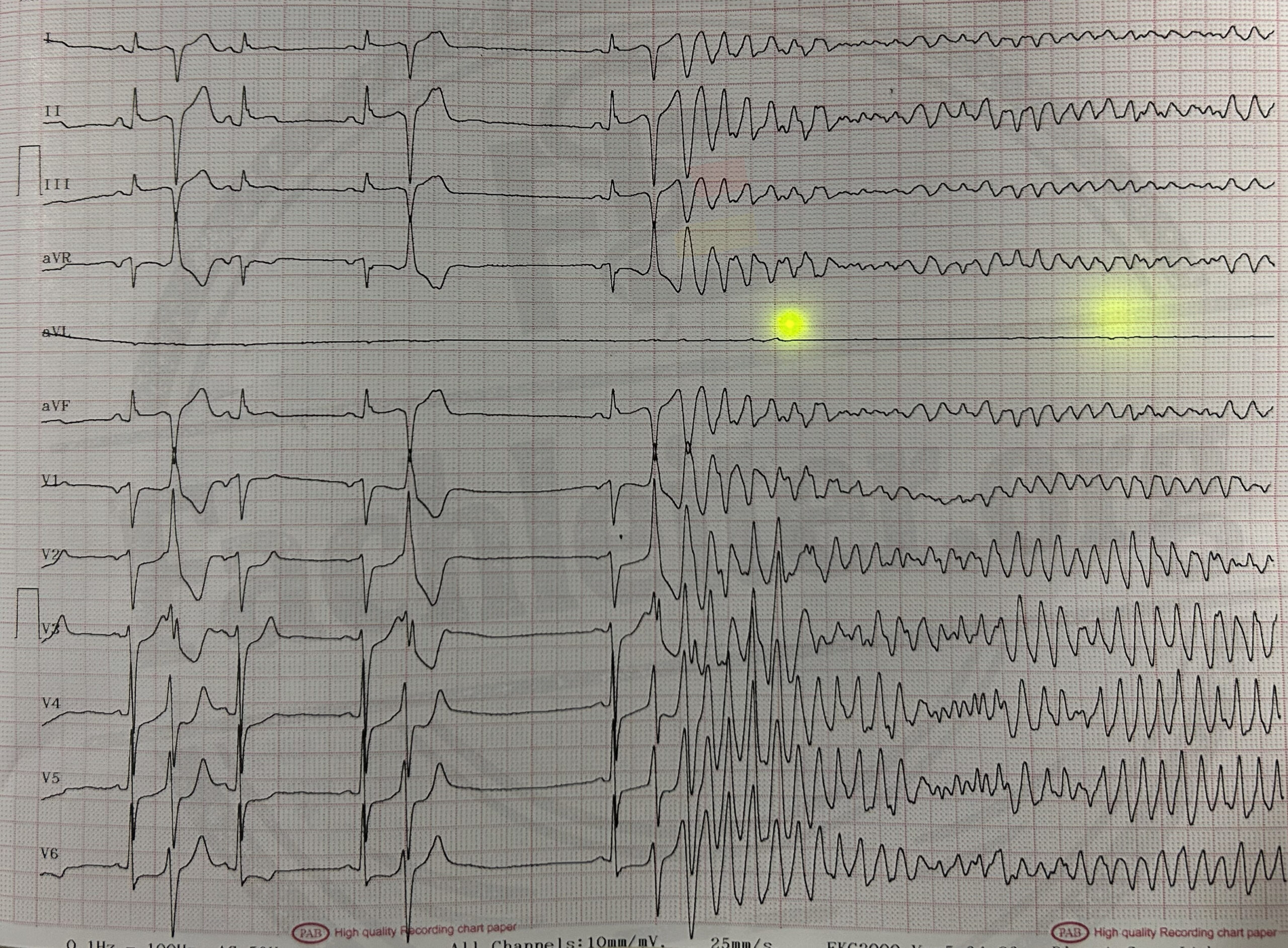
During the ECG recording the following development is observed:
Summary
In the admission ECG there is a rapid polymorphic wide-complex tachycardia with continuously changing amplitude and electrical axis of the QRS complexes. This corresponds to a polymorphic ventricular tachycardia. An R-on-T phenomenon is also visible.
Sudden clinical deterioration
The patient loses consciousness, no longer responds and shows no normal breathing. A central pulse cannot be reliably palpated within at most ten seconds.
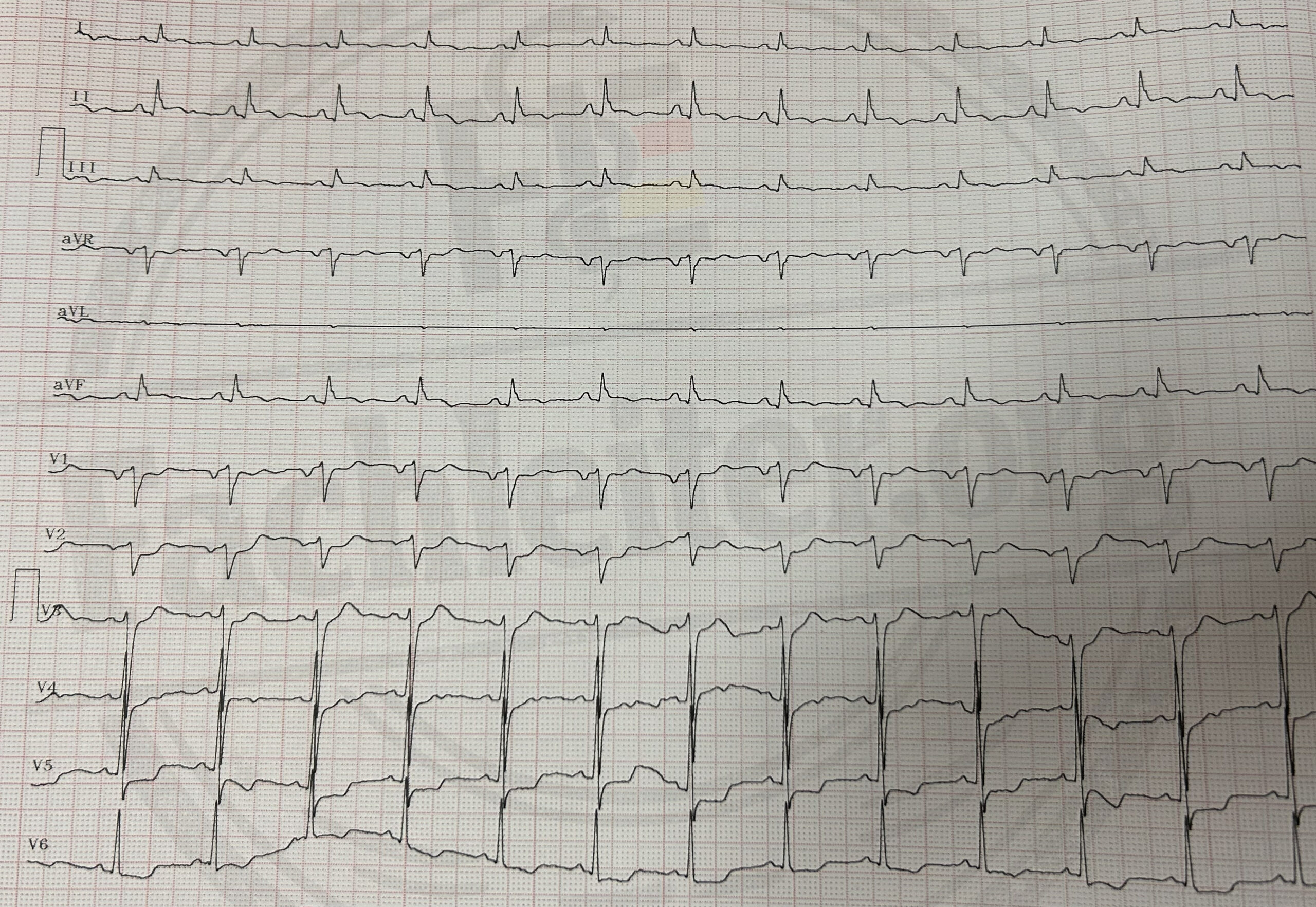
The patient is resuscitated for a total of 20 minutes. At three rhythm checks a shockable rhythm is present, so three biphasic defibrillations of 200 Joules each are delivered. In between a non-shockable rhythm is present. After the third defibrillation spontaneous circulation is restored.
Laboratory
| Parameter | Value | Reference range |
|---|---|---|
| hs-Troponin T | 22 ng/l | < 14 ng/l |
| CK | 178 U/l | < 190 U/l |
| CK-MB | 7,1 µg/l | < 5,0 µg/l |
| Leukocytes | 12,4 /nl | 4,0–10,0 /nl |
| Hemoglobin | 14,2 g/dl | 13,5–17,5 g/dl |
| Platelets | 238 /nl | 150–400 /nl |
| Creatinine | 1,0 mg/dl | 0,7–1,3 mg/dl |
| Sodium | 139 mmol/l | 135–145 mmol/l |
| Potassium | 3,6 mmol/l | 3,5–5,1 mmol/l |
| Magnesium | 0,73 mmol/l | 0,70–1,00 mmol/l |
| Glucose | 192 mg/dl | 70–140 mg/dl |
| CRP | 3,2 mg/l | < 5 mg/l |
| INR | 1,0 | 0,9–1,2 |
| aPTT | 29 s | 25–35 s |
After resuscitation the patient was transferred to the intensive care unit for close monitoring.
Questions
- Explain the step-by-step conduct of resuscitation for this patient.
- Explain the step-by-step conduct of intubation.
- Which medications were likely administered during the resuscitation? Justify your answer.
- Which further measures are required after transfer to the intensive care unit?
- What should you consider in such presenting complaints in a diabetic patient?