
Case description
A 65-year-old female patient presents with an unpleasant sensation between the shoulder blades that has been present for three days.
She denies pressure or tightness in the chest, shortness of breath, palpitations, loss of consciousness, or any relation of the symptoms to physical activity or food intake.
Her history includes a coronary angiography approximately two to three years ago. The patient cannot recall her regular medications.
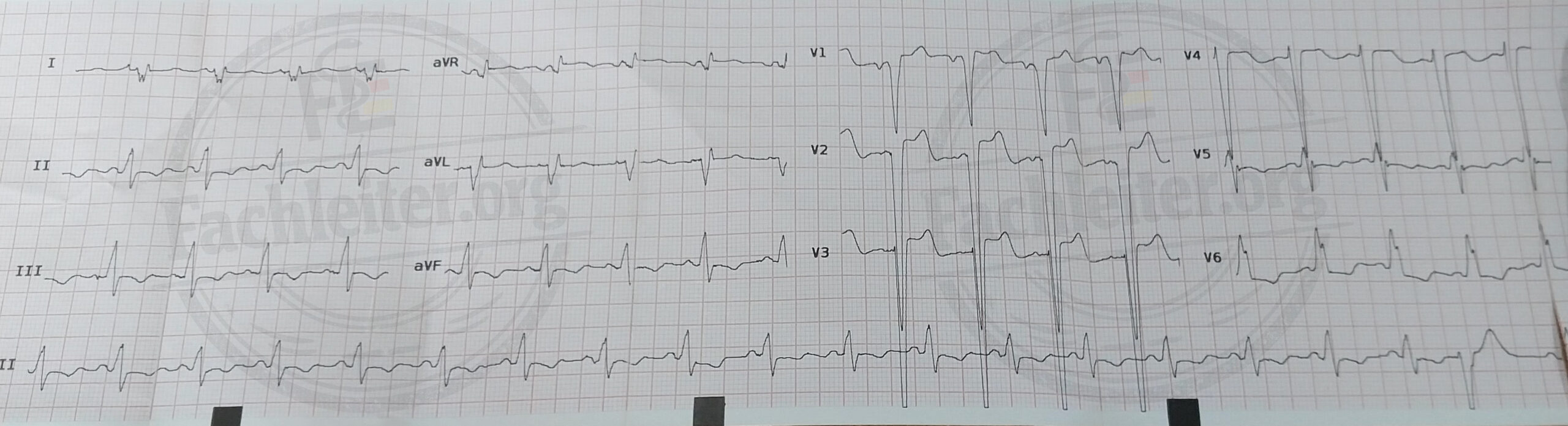
Summary of ECG findings
- Regular sinus rhythm
- Heart rate approx. 95/min
- Right axis
- PR interval within normal limits
- Complete left bundle branch block (LBBB) with widened QRS complex
- Secondary discordant ST- and T-wave changes, consistent with an LBBB
- Signs of left atrial enlargement (LAE)
- No clear ECG evidence of an acute ST-elevation myocardial infarction
- Assessment of ischemic changes and a past myocardial infarction is limited due to the LBBB
Significance
The ECG demonstrates a complete left bundle branch block with the typical secondary repolarization changes. An acute ST-elevation myocardial infarction cannot be reliably detected electrocardiographically. Because an LBBB impairs recognition of ischemic changes, an acute coronary syndrome cannot be confirmed or excluded based on the ECG alone. The symptoms therefore need evaluation in the clinical context and clarification by serial high-sensitivity troponin measurements and, if necessary, further diagnostics (e.g., echocardiography). The left atrial enlargement suggests chronic cardiac strain, for example due to arterial hypertension or structural heart disease.