
Medical history interview
- Greeting and brief introduction
- Personal data
- Current complaints
- Previous episodes
- Autonomic history
- Past medical history
- Operations and hospital stays
- Medications
- Allergies and intolerances
- Gynecological history
- Social history
- Recreational substances and drugs
- Family history
- Summary and explanation
👨⚕️ Good day, my name is Dr. Schneider. I am responsible for you today. Before we begin: How should I address you?
👩 Good day. My name is Sabine Keller.
👨⚕️ Ms. Keller, I would first like to ask you some questions about your complaints and your health. Is that okay with you?
👩 Yes, of course.
👨⚕️ How old are you?
👩 I am 44 years old.
👨⚕️ How tall are you and how much do you weigh?
👩 I am 1,68 Meter tall and weigh approximately 74 Kilogramm.
👨⚕️ Who do you currently live with?
👩 I live with my husband and our daughter.
👨⚕️ Who is your GP?
👩 Dr. Weber. I should have the phone number in my mobile phone.
👨⚕️ What brings you to us today?
👩 My heart suddenly races so strongly. It also feels like it keeps skipping beats.
👨⚕️ When did the complaints start?
👩 About an hour ago.
👨⚕️ Did they start suddenly or more slowly?
👩 Very suddenly. From one moment to the next.
👨⚕️ What were you doing when the palpitations began?
👩 I was at home tidying the kitchen. It wasn’t anything strenuous.
👨⚕️ Has the palpitations been continuous since then?
👩 Yes. Sometimes it’s a bit stronger, but it doesn’t really go away.
👨⚕️ Do you also have pain or a feeling of pressure in the chest?
👩 Yes. It feels like something heavy is lying on my chest.
👨⚕️ Where exactly do you feel the complaints?
👩 Mainly here in the middle and a bit on the left side.
👨⚕️ Do the pains radiate to another part of the body?
👩 Yes, into my left arm.
👨⚕️ How severe is the pain on a scale from zero to ten?
👩 About six out of ten.
👨⚕️ Do the complaints get worse with exertion?
👩 Yes. When I ran from the car to get here, the pressure and the palpitations became significantly worse.
👨⚕️ Do the complaints improve at rest?
👩 A little, but they don’t disappear completely.
👨⚕️ Are the pains related to breathing or a certain movement?
👩 No.
👨⚕️ Can the pain be triggered by pressing on the chest?
👩 No.
👨⚕️ Are you short of breath?
👩 A little. But I can still speak normally.
👨⚕️ Have you felt dizzy or fainted?
👩 I felt a bit dizzy briefly, but I did not faint.
👨⚕️ Have you had nausea, vomiting or unusually heavy sweating?
👩 I feel a bit nauseous and I’m sweating more than usual.
👨⚕️ Have you previously experienced palpitations or skipped beats?
👩 Yes, several times in the past months. It usually lasted only a few minutes and then went away on its own.
👨⚕️ Did you see a doctor about it?
👩 Yes, I saw a cardiologist a few months ago.
👨⚕️ What was found there?
👩 He said that I had extra heartbeats. I should have a Holter monitor and an echocardiogram.
👨⚕️ Were these tests carried out?
👩 No. The symptoms improved at first, so I didn’t pursue it further.
👨⚕️ Was a medication prescribed at that time?
👩 No. The doctor wanted to wait for the further tests first.
👨⚕️ Have you had a fever or an infection recently?
👩 No.
👨⚕️ Have you had unintentional weight loss or severe night sweats?
👩 No.
👨⚕️ Do you suffer from diarrhea, severe tremors, or marked heat intolerance?
👩 No.
👨⚕️ How is your appetite?
👩 Normal.
👨⚕️ Which medical conditions are you known to have?
👩 I have had high blood pressure for about three years.
👨⚕️ Do you have diabetes, high blood lipids, or a thyroid disorder?
👩 My cholesterol was somewhat elevated at the last check. To my knowledge I do not have diabetes or a thyroid disorder.
👨⚕️ Have you ever been diagnosed with coronary artery disease or a heart attack?
👩 No.
👨⚕️ Have you ever had a thrombosis or a pulmonary embolism?
👩 No.
👨⚕️ Have you ever had surgery?
👩 Yes, my appendix was removed when I was 19 years old.
👨⚕️ Were there any complications?
👩 No.
👨⚕️ Were you ever in the hospital for another reason?
👩 No.
👨⚕️ Which medications do you take regularly?
👩 Ramipril, five milligrams in the morning.
👨⚕️ Do you take the medication regularly?
👩 Mostly. Sometimes I forget it on weekends.
👨⚕️ Do you take any other medications, supplements, or herbal remedies?
👩 No.
👨⚕️ Have you taken anything today for the symptoms?
👩 No.
👨⚕️ Do you have allergies or intolerance to certain medications?
👩 As a child I developed a skin rash from penicillin.
👨⚕️ Did you have shortness of breath or swelling with that?
👩 No, only a rash.
👨⚕️ When was your last menstrual period?
👩 About two weeks ago.
👨⚕️ Is pregnancy possible?
👩 No. My husband has been sterilized.
👨⚕️ Have you ever been pregnant?
👩 Yes, once. My daughter is 15 years old.
👨⚕️ Were there complications during the pregnancy or delivery?
👩 No.
👨⚕️ What do you do for a living?
👩 I work as a sales assistant in a supermarket.
👨⚕️ Is the work physically demanding there?
👩 I stand a lot and occasionally have to carry heavy boxes.
👨⚕️ Are you currently experiencing particular work-related or personal stress?
👩 There’s a lot going on at work right now, but not more than usual.
👨⚕️ Do you smoke?
👩 Yes, about ten cigarettes a day.
👨⚕️ How long have you been smoking?
👩 For about 20 years.
👨⚕️ Do you drink alcohol?
👩 Occasionally on weekends one or two glasses of wine.
👨⚕️ Do you take drugs or stimulant substances?
👩 No.
👨⚕️ Do you drink a lot of coffee or energy drinks?
👩 Three to four cups of coffee a day. Today I had two cups. I don’t drink energy drinks.
👨⚕️ Are there cardiovascular diseases in your family?
👩 My father had a heart attack at 52 years of age.
👨⚕️ Are cardiac arrhythmias or sudden deaths known in your family?
👩 My older brother sometimes has cardiac arrhythmias. I don’t know the specifics. As far as I know, there were no sudden deaths.
👨⚕️ Is there diabetes or other significant illnesses in your family?
👩 My mother has diabetes and high blood pressure.
👨⚕️ Ms. Keller, your symptoms and the palpitations indicate a cardiac arrhythmia. Since you additionally have exertion-related chest pain radiating into the left arm, there is suspicion of an acute coronary syndrome.
We will now examine you physically, check your vital signs, perform an ECG immediately and take blood samples. In the meantime we will continuously monitor your heart rhythm. Depending on the findings, you will be transferred to a cardiology center for further treatment.
- Personal data
- Allergies
- Recreational substances and drugs
- Social history
- Family history
- Medications
- Medical history
- Suspected diagnosis and differential diagnoses
- Further management and therapy
Sabine Keller
Age: 44 years
Weight: 74 kg
Height: 168 cm
- Penicillin: itchy rash
- No known shortness of breath or swelling after taking penicillin
- Nicotine: approx. 10 cigarettes daily for 20 years, approx. 10 pack-years
- Alcohol: occasionally 1–2 glasses of wine on weekends
- Coffee: 3–4 cups daily
- Denies illegal drugs and energy drinks
- Married, lives with her husband and her daughter
- Employed as a sales assistant
- Independent in daily activities
- Father: myocardial infarction at the age of 52 years
- Mother: arterial hypertension and diabetes mellitus
- Brother: unspecified cardiac arrhythmia
- No known sudden cardiac deaths in the family
- Ramipril 5 mg 1–0–0, according to the patient occasionally taken irregularly
- No long-term medication with antiplatelet agents or anticoagulants
The 44-year-old patient presented due to a suddenly onset tachycardia with palpitations that had been present for about one hour. She additionally reported retrosternal, left-sided chest pain described as pressure and heaviness radiating to the left arm. The pain intensity was rated 6/10.
The symptoms worsened with physical exertion and only partially improved at rest. She also complained of mild dyspnea, nausea, increased sweating and transient dizziness. Syncope, emesis, and respiratory- or movement-dependent chest pain were denied.
Similar episodes with palpitations had occurred repeatedly in recent months but had lasted only a few minutes each time and ceased spontaneously. Because of these complaints the patient had already undergone cardiological evaluation. Extrasystoles were detected and further diagnostics with Holter ECG and echocardiography were recommended. The patient, however, did not undergo these examinations.
Known pre-existing conditions include arterial hypertension and hypercholesterolemia. Coronary artery disease, diabetes mellitus, thyroid disease and thromboembolic events have not been diagnosed to date.
On physical examination the patient was awake, oriented in all directions and slightly diaphoretic, in reduced general condition. Blood pressure was 148/92 mmHg, heart rate 112/min with irregular cardiac activity, respiratory rate 19/min and oxygen saturation 97% on room air. Body temperature was 36.7 °C.
Auscultation revealed clear heart sounds without pathological murmurs and bilateral vesicular breath sounds without crackles. There was no jugular venous distension and no peripheral edema. The chest pain was not reproducible on palpation.
The 12-lead ECG showed a tachycardic sinus rhythm with a heart rate of approximately 105/min. Numerous monomorphic ventricular extrasystoles consistent with ventricular trigeminy were present. An early ventricular extrasystole fell into the T-wave of the preceding beat, consistent with an R-on-T phenomenon. No persistent ST elevations were present.
Laboratory testing initially showed a slightly elevated hs-Troponin I of 19 ng/l. On repeat measurement after one hour hs-Troponin I rose to 46 ng/l.
With typical angina pectoris symptoms and absent ST elevations on ECG there is a strong suspicion of a non–ST-elevation acute coronary syndrome (NSTE-ACS). Differentiation between unstable angina pectoris and NSTEMI is not yet possible without troponin determination. Additionally, there is frequent monomorphic ventricular extrasystoles with alternating phases of ventricular bigeminy and trigeminy as well as complete compensatory pauses. An R-on-T phenomenon was not observed.
The absence of clinical signs of infection and a preceding infection argue against myocarditis; however, definitive exclusion is not possible without laboratory tests and echocardiography. A coronary spasm appears less likely given the predominantly exertion-related complaints. Structural heart disease must be excluded by echocardiography. Electrolyte disturbances and thyroid dysfunction as possible triggers of PVCs must be evaluated by laboratory testing. The absence of an identifiable trigger argues against a catecholamine-induced arrhythmia.
Examination
Vital signs
| Parameter | Measured value |
|---|---|
| Blood pressure | 148/92 mmHg |
| Heart rate | 112/min, irregular |
| Respiratory rate | 19/min |
| Oxygen saturation | 97 % on room air |
| Body temperature | 36,7 °C |
| Blood glucose | 108 mg/dl or 6,0 mmol/l |
| GCS | 15 points |
| Pain intensity | 6/10 |
Physical examination
General condition
Alert, fully oriented female patient in a slightly reduced general condition. She appears anxious and is diaphoretic. No cyanosis, no dyspnea at rest.
Cardiovascular system
- Heart sounds clear and well delineated
- Tachycardic heartbeat
- Irregular rhythm due to numerous extrasystoles
- No pathological heart murmurs
- Peripheral pulses palpable bilaterally
- Capillary refill time under two seconds
- No jugular venous distension
- No peripheral edema
Lungs
- Vesicular breath sounds bilaterally
- No crackles
- No wheeze or rhonchi
- Bilaterally resonant percussion
Abdomen
- Soft and non-tender
- No guarding
- Normal bowel sounds
Chest
- No tenderness on palpation
- Symptoms not reproducible by palpation or movement
Neurological examination
- Alert and fully oriented
- Pupils isocoric and reactive to light
- No pareses or sensory disturbances
- Speech unremarkable
ECG
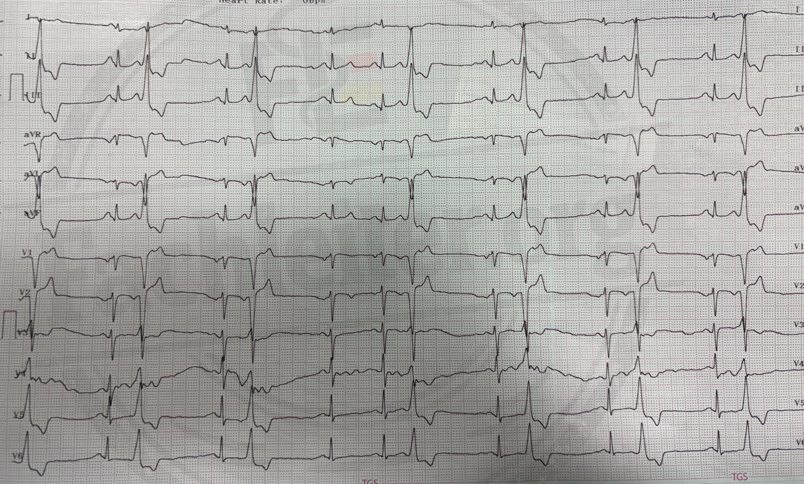
ECG summary
The 12-lead ECG showed a sinus rhythm of normal frequency with a heart rate of approximately 75/min. There were frequent monomorphic ventricular extrasystoles with alternating phases of ventricular bigeminy and trigeminy. The PVCs exhibited wide, deformed and prematurely occurring QRS complexes without immediately preceding P waves. Complete compensatory pauses followed the PVCs. No R-on-T phenomenon or persistent ST elevations were present.
Initial measures after the ECG
Case variant
Laboratory values
| Laboratory parameters | Initial | Reference range |
|---|---|---|
| Hemoglobin | 13,4 g/dl | 12,0–16,0 g/dl |
| Leukocytes | 9,2/nl | 4,0–10,0/nl |
| Platelets | 276/nl | 150–400/nl |
| Sodium | 139 mmol/l | 135–145 mmol/l |
| Potassium | 4,2 mmol/l | 3,5–5,1 mmol/l |
| Magnesium | 0,86 mmol/l | 0,70–1,05 mmol/l |
| Calcium | 2,31 mmol/l | 2,15–2,55 mmol/l |
| Creatinine | 0,78 mg/dl | 0,50–0,90 mg/dl |
| eGFR | 95 ml/min/1,73 m² | > 60 ml/min/1,73 m² |
| Glucose | 112 mg/dl | 70–140 mg/dl |
| CRP | 2,1 mg/l | < 5 mg/l |
| TSH | 1,62 mIU/l | 0,27–4,20 mIU/l |
| CK | 126 U/l | < 170 U/l |
| CK-MB | 3,1 ng/ml | < 4,9 ng/ml |
| hs-Troponin I | 19 ng/l | < 16 ng/l |
| INR | 1,0 | 0,9–1,2 |
| aPTT | 29 s | 25–37 s |
| β-hCG in serum | < 1 IU/l | < 5 IU/l: negative |
Further management without an in-house cardiology department
After admission the patient was continuously monitored on the Chest Pain Unit. On the follow-up ECG there were still frequent monomorphic ventricular extrasystoles with alternating phases of ventricular bigeminy and trigeminy. No sustained ventricular tachycardia or ST elevations occurred.
The initial hs-Troponin I was 19 ng/l and rose to 46 ng/l on repeat measurement after one hour. In conjunction with the typical angina pectoris symptoms, the diagnosis of NSTEMI was confirmed.
Transthoracic echocardiography showed a mildly reduced left ventricular ejection fraction of 50 % with a subtle regional wall motion abnormality. There were no hemodynamically relevant valvular lesions.
Anticoagulation with unfractionated heparin was continued. Coronary angiography was then performed. This revealed a high-grade, approximately 90-percent, thrombotic coronary stenosis as the infarct lesion. Percutaneous coronary intervention with balloon dilatation and implantation of a drug-eluting stent was performed immediately. Final angiographic control showed normal blood flow without relevant residual stenosis.
After PCI the patient additionally received Ticagrelor 180 mg as a loading dose, then 90 mg twice daily. Treatment with acetylsalicylic acid 100 mg once daily and atorvastatin 80 mg once daily was continued.
In the further course the patient was symptom-free and hemodynamically stable. The frequency of ventricular extrasystoles decreased markedly after revascularization. Specific antiarrhythmic therapy was not required. Prior to discharge a Holter ECG and a cardiology follow-up were arranged. The patient was also counseled on smoking cessation and on rigorous control of her cardiovascular risk factors.
Course at the cardiology center
After admission the patient was continuously monitored on the Chest Pain Unit. On the follow-up ECG there were still frequent monomorphic ventricular extrasystoles with alternating phases of ventricular bigeminy and trigeminy. No sustained ventricular tachycardia or ST elevations occurred.
The initial hs-Troponin I was 19 ng/l and rose to 46 ng/l on repeat measurement after one hour. In conjunction with the typical angina pectoris symptoms, the diagnosis of NSTEMI was confirmed.
Transthoracic echocardiography showed a mildly reduced left ventricular ejection fraction of 50 % with a subtle regional wall motion abnormality. There were no hemodynamically relevant valvular lesions.
Anticoagulation with unfractionated heparin was continued. Coronary angiography was then performed. This revealed a high-grade, approximately 90-percent, thrombotic coronary stenosis as the infarct lesion. Percutaneous coronary intervention with balloon dilatation and implantation of a drug-eluting stent was performed immediately. Final angiographic control showed normal blood flow without relevant residual stenosis.
After PCI the patient additionally received Ticagrelor 180 mg as a loading dose, then 90 mg twice daily. Treatment with acetylsalicylic acid 100 mg once daily and atorvastatin 80 mg once daily was continued.
In the further course the patient was symptom-free and hemodynamically stable. The frequency of ventricular extrasystoles decreased markedly after revascularization. Specific antiarrhythmic therapy was not required. Prior to discharge a Holter ECG and a cardiology follow-up were arranged. The patient was also counseled on smoking cessation and on rigorous control of her cardiovascular risk factors.
Laboratory
| Parameter | First blood draw | Follow-up after 2 hours | Example reference range |
|---|---|---|---|
| Hemoglobin | 13,4 g/dl | – | 12,0–16,0 g/dl |
| Leukocytes | 9,2/nl | – | 4,0–10,0/nl |
| Platelets | 276/nl | – | 150–400/nl |
| Sodium | 139 mmol/l | – | 135–145 mmol/l |
| Potassium | 4,2 mmol/l | – | 3,5–5,1 mmol/l |
| Magnesium | 0,86 mmol/l | – | 0,70–1,05 mmol/l |
| Calcium | 2,31 mmol/l | – | 2,15–2,55 mmol/l |
| Creatinine | 0,78 mg/dl | – | 0,50–0,90 mg/dl |
| eGFR | 95 ml/min/1,73 m² | – | > 60 ml/min/1,73 m² |
| Glucose | 112 mg/dl | – | 70–140 mg/dl |
| CRP | 2,1 mg/l | – | < 5 mg/l |
| TSH | 1,62 mIU/l | – | 0,27–4,20 mIU/l |
| β-hCG in serum | < 1 IU/l | – | < 5 IU/l: negative |
| hs-Troponin I | 5 ng/l | 6 ng/l | assay-dependent; here for example < 16 ng/l |
Ms. Keller reported at admission continued retrosternal pressure radiating to the left arm. She remains on the Chest Pain Unit under continuous rhythm and hemodynamic monitoring. During symptoms a repeat 12-lead ECG is immediately recorded; further troponin checks are performed taking into account the early onset of symptoms. The effect of the already administered nitroglycerin and the blood pressure are also reassessed.
Persistent typical pain despite negative troponin values is, in this case, a reason for an urgent cardiology decision regarding coronary angiography during the inpatient stay. If symptoms persist or increase, this is performed promptly. If a relevant infarct lesion is found, PCI can be performed immediately. Unstable angina pectoris remains possible until further clarification.
| Parameter | First blood draw | Follow-up after 2 hours | Example reference range |
|---|---|---|---|
| Hemoglobin | 13,4 g/dl | – | 12,0–16,0 g/dl |
| Leukocytes | 9,2/nl | – | 4,0–10,0/nl |
| Platelets | 276/nl | – | 150–400/nl |
| Sodium | 139 mmol/l | – | 135–145 mmol/l |
| Potassium | 4,2 mmol/l | – | 3,5–5,1 mmol/l |
| Magnesium | 0,86 mmol/l | – | 0,70–1,05 mmol/l |
| Calcium | 2,31 mmol/l | – | 2,15–2,55 mmol/l |
| Creatinine | 0,78 mg/dl | – | 0,50–0,90 mg/dl |
| eGFR | 95 ml/min/1,73 m² | – | > 60 ml/min/1,73 m² |
| Glucose | 112 mg/dl | – | 70–140 mg/dl |
| CRP | 2,1 mg/l | – | < 5 mg/l |
| TSH | 1,62 mIU/l | – | 0,27–4,20 mIU/l |
| β-hCG in serum | < 1 IU/l | – | < 5 IU/l: negative |
| hs-Troponin I | 5 ng/l | 6 ng/l | assay-dependent; here, for example, < 16 ng/l |
On admission, Ms. Keller reports that the chest pressure has significantly decreased. She is hemodynamically stable; the follow-up ECG still shows PVCs, but no new ischemic changes. Rhythm monitoring is initially continued. Because the symptoms began only shortly before the first blood draw, the ECG and hs-Troponin will be reassessed according to the examination protocol used. An echocardiography assesses left ventricular function and possible regional wall motion abnormalities.
If Ms. Keller remains symptom-free and the ECG and troponin remain unremarkable during further observation, the risk for coronary heart disease will be assessed by inpatient cardiology. Coronary angiography is not automatically an immediate measure in this scenario: the cardiology team decides based on the overall findings whether it is necessary during the admission or whether another coronary diagnostic procedure is performed first. The frequent PVCs will subsequently be further evaluated with a long-term ECG.
q