
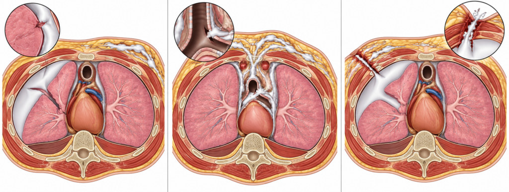
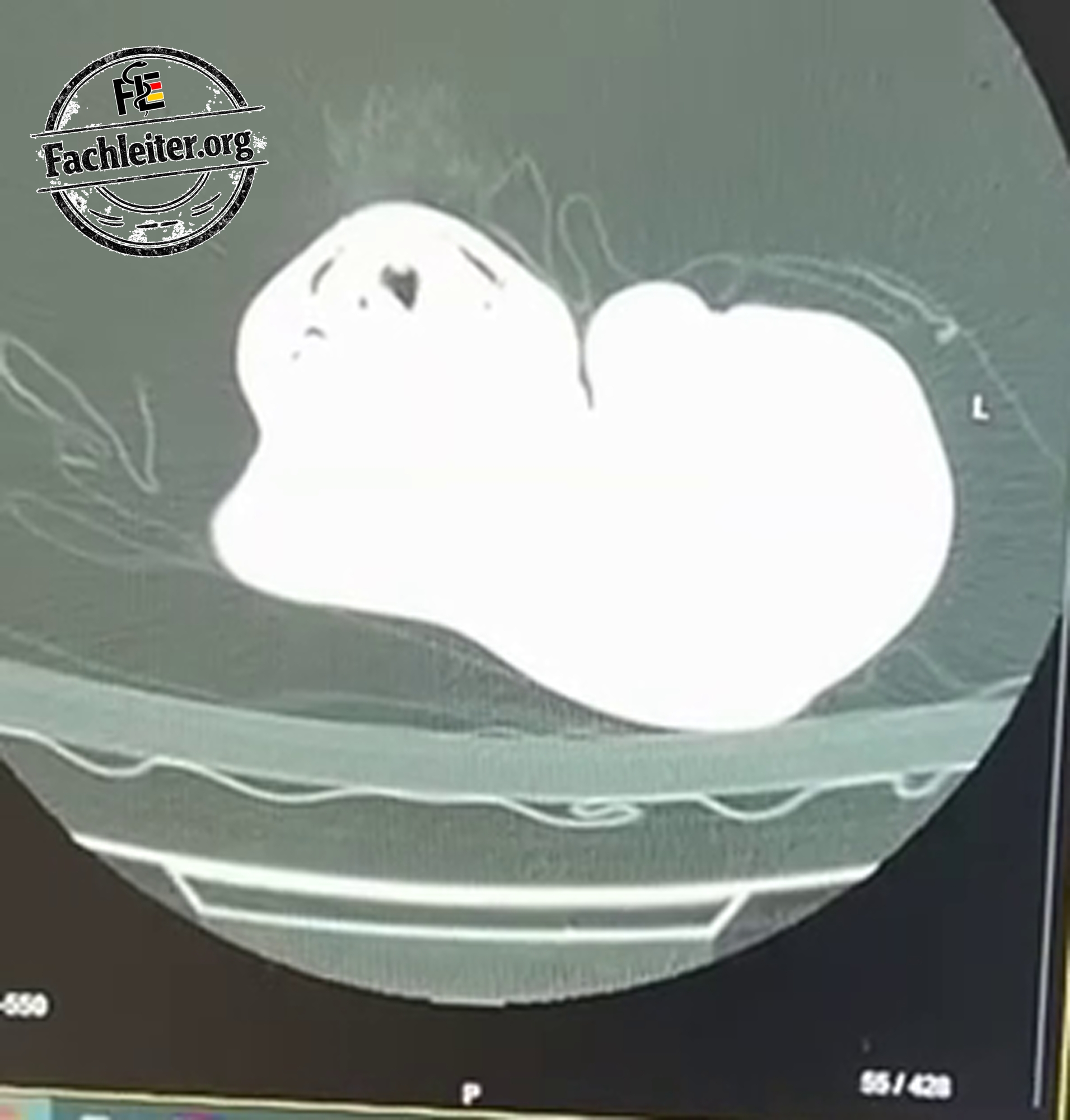
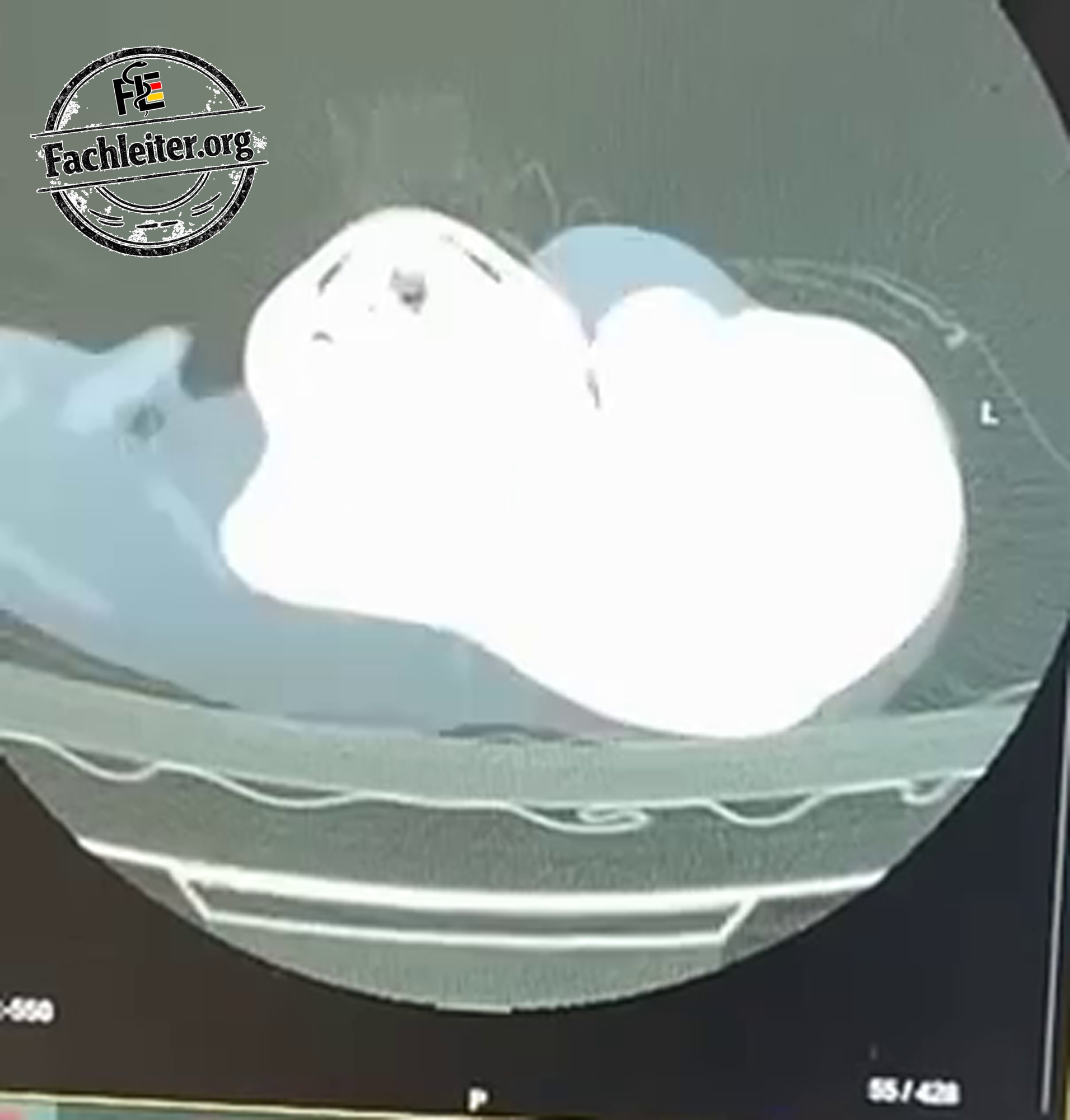
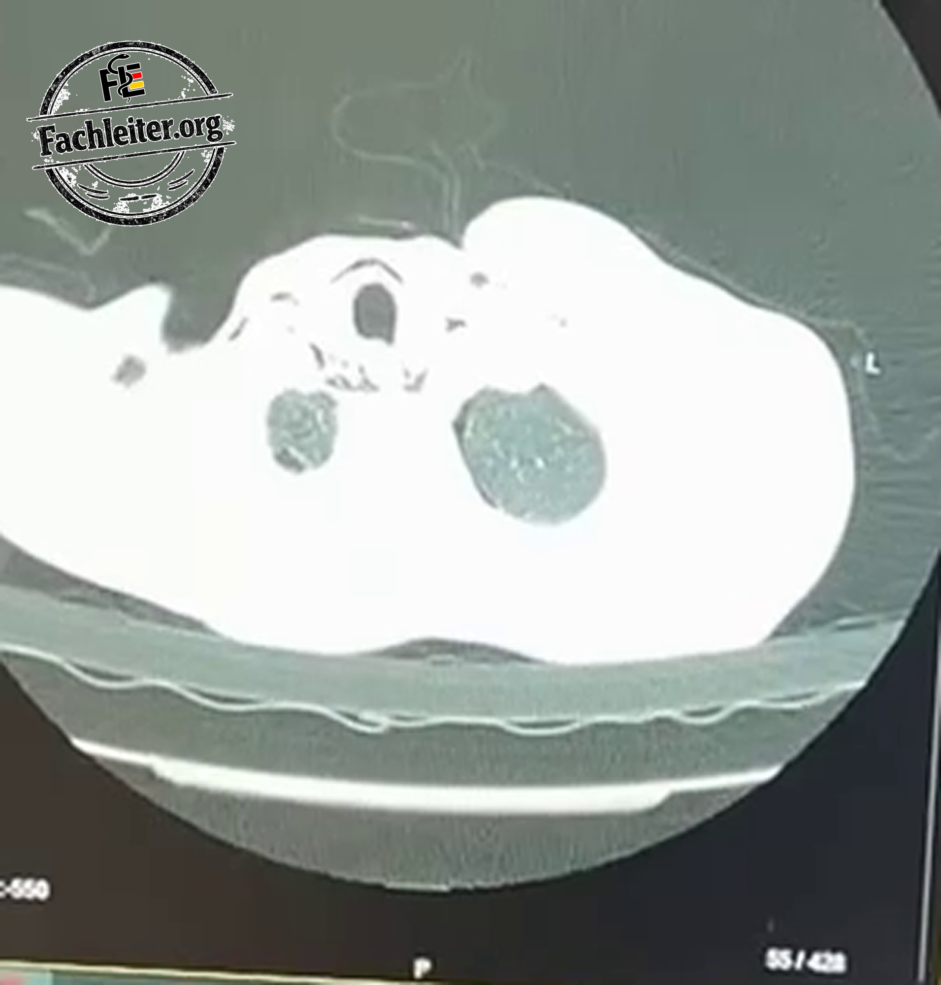
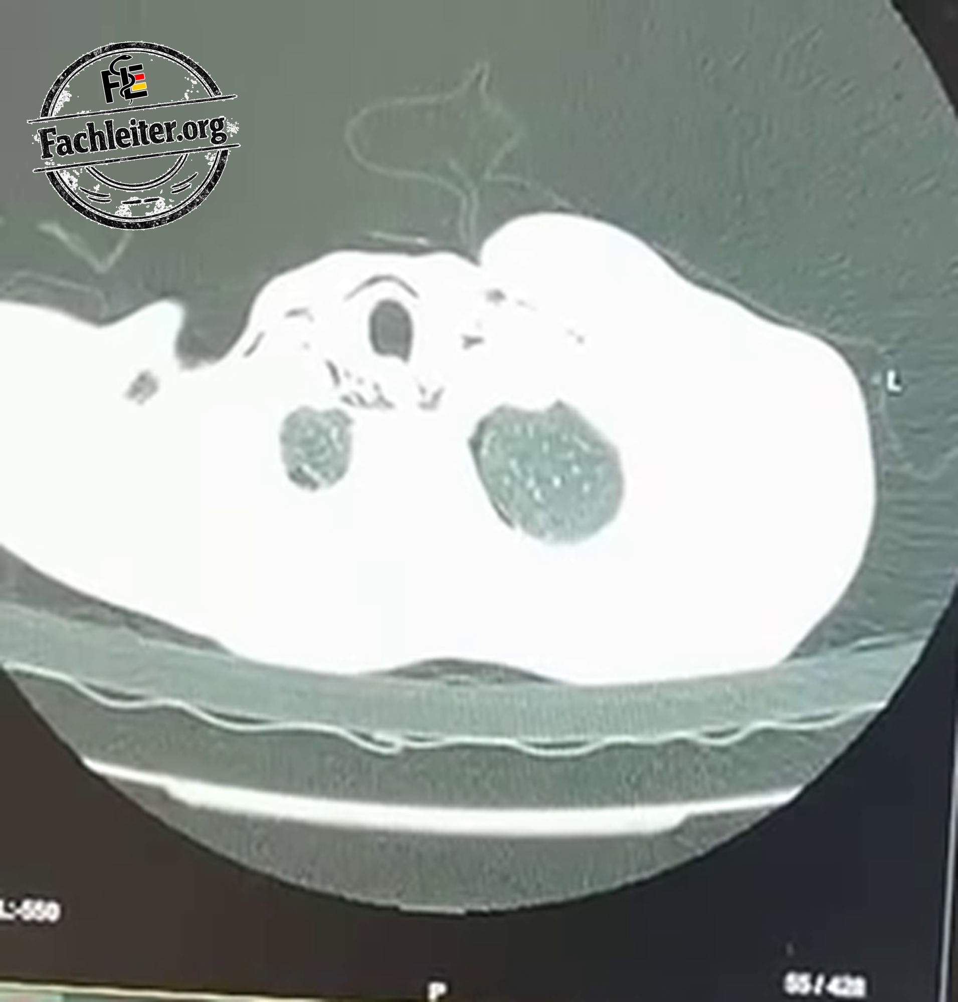
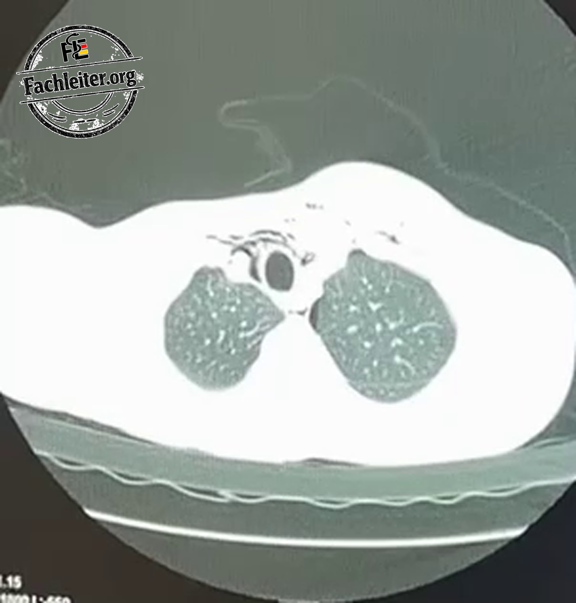
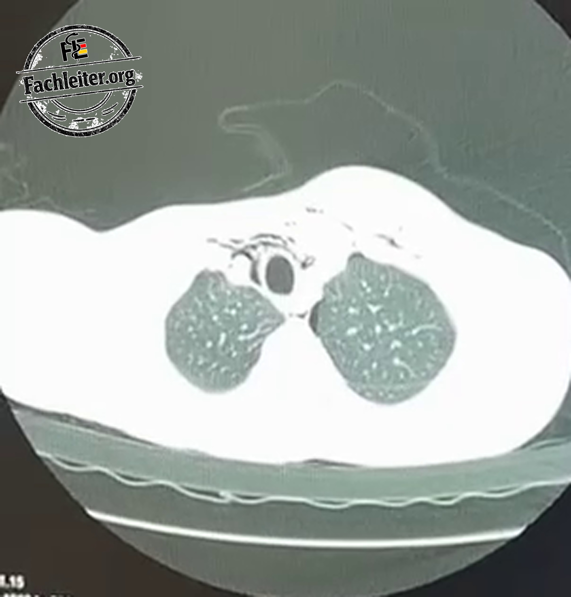
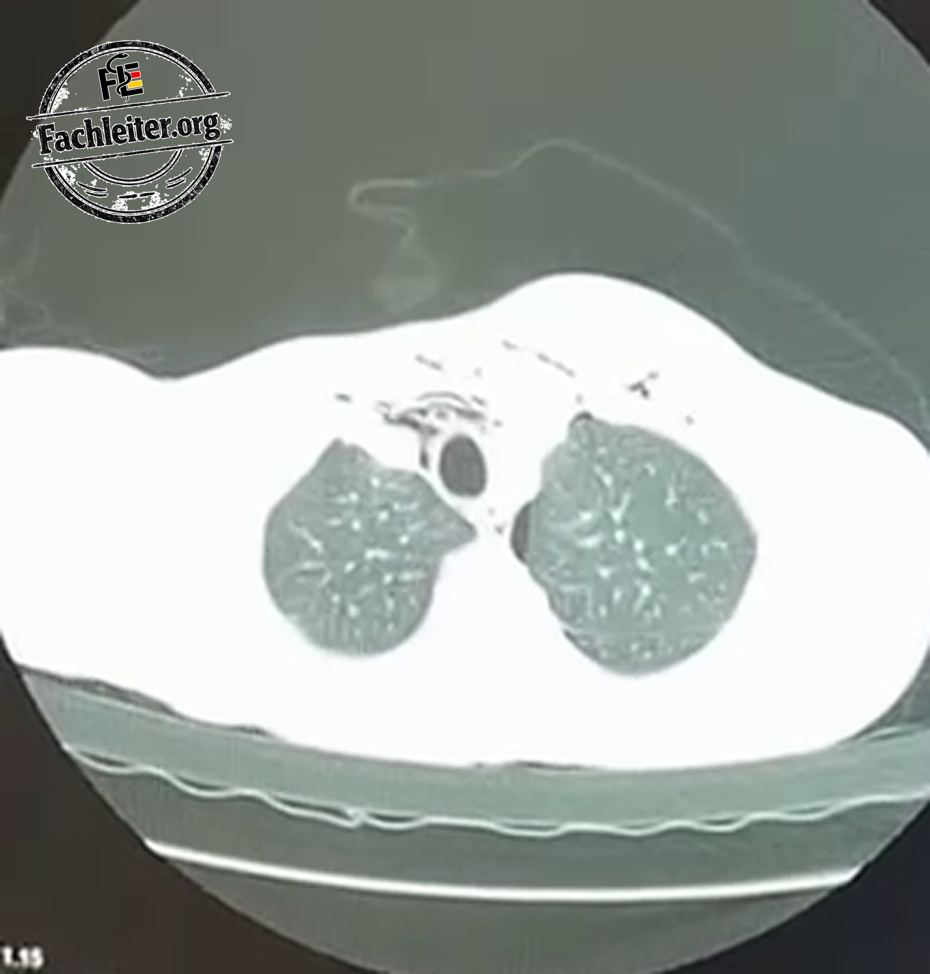
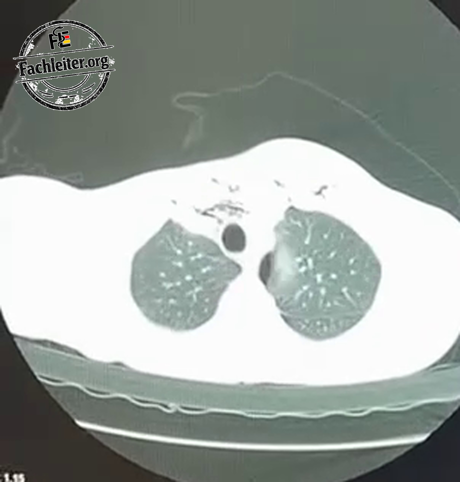
Describe what you see in the three images from left to right.
The illustration shows three possible routes by which air can spread after an injury to the chest. On the left, air escapes from an injured lung and reaches the subcutaneous tissue via the chest wall. In the middle, air leaks from an injured airway and spreads into the mediastinum and along tissue planes up to under the skin. On the right, air enters the pleural space through an open chest wall wound; the lung on that side is collapsed. In all three images air in the subcutaneous tissue is also depicted.
Subcutaneous emphysema
Definition
Subcutaneous emphysema is an accumulation of air in the subcutaneous tissue. This can cause swelling of the chest, neck or face. On gentle palpation the skin often feels crackling.
How does it occur?
Air can escape from an injured lung, the airways or the pleural space and spread along tissue planes under the skin. Possible causes include a fall with rib fracture, a stab wound, a medical procedure, or an air leak from an already placed chest drain.
Subcutaneous emphysema is therefore a finding whose cause must be determined. Especially if the swelling increases rapidly, one must search for an ongoing air leak and further injuries.
Symptoms and physical examination
Affected people often notice swelling or an unusual sense of pressure in the chest or neck. The swelling can extend to the face. The characteristic crackling on palpation is referred to as cutaneous crepitus.
During the examination respiration, oxygen saturation, pulse and blood pressure are checked. The physician examines the extent of the swelling and at the same time looks for signs of a pneumothorax: shortness of breath, chest pain or diminished breath sounds on one side. After an accident the chest wall and any additional injuries are also examined.
What is seen on the X-ray?
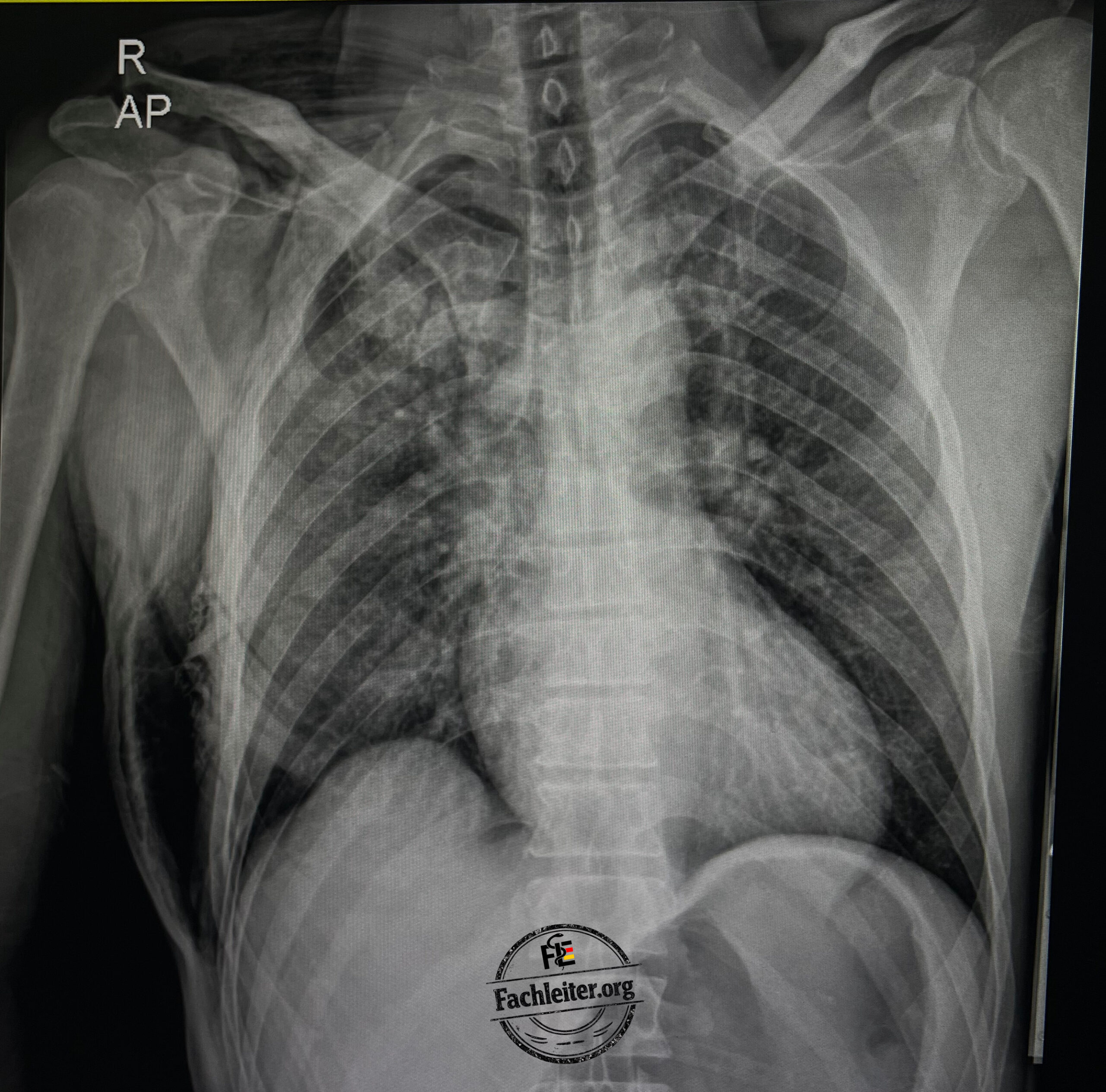
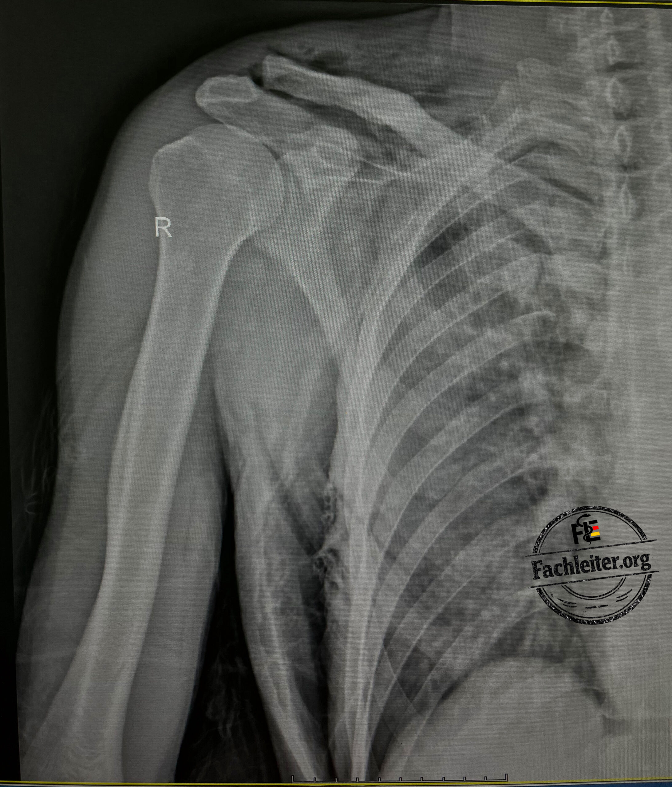
Subcutaneous emphysema can usually be detected during the physical exam. The affected area is swollen; with gentle palpation the skin feels characteristically crackling.
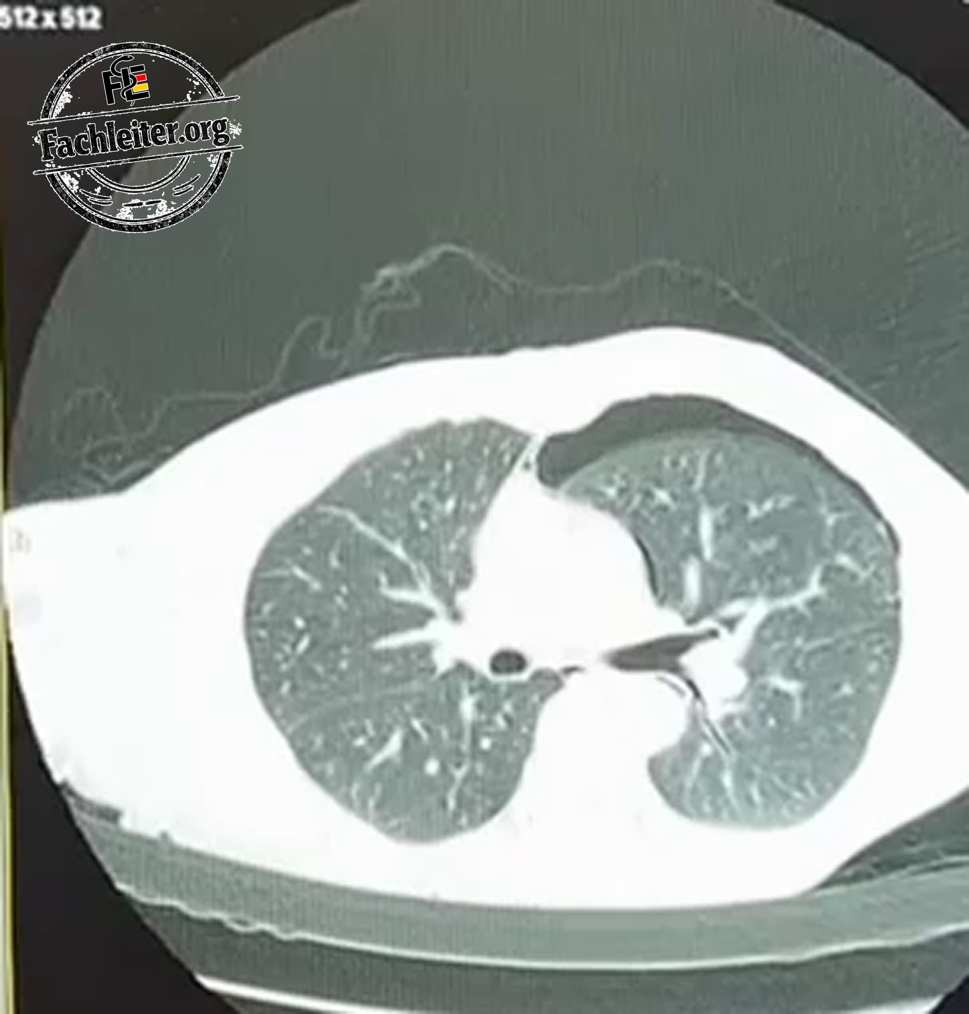
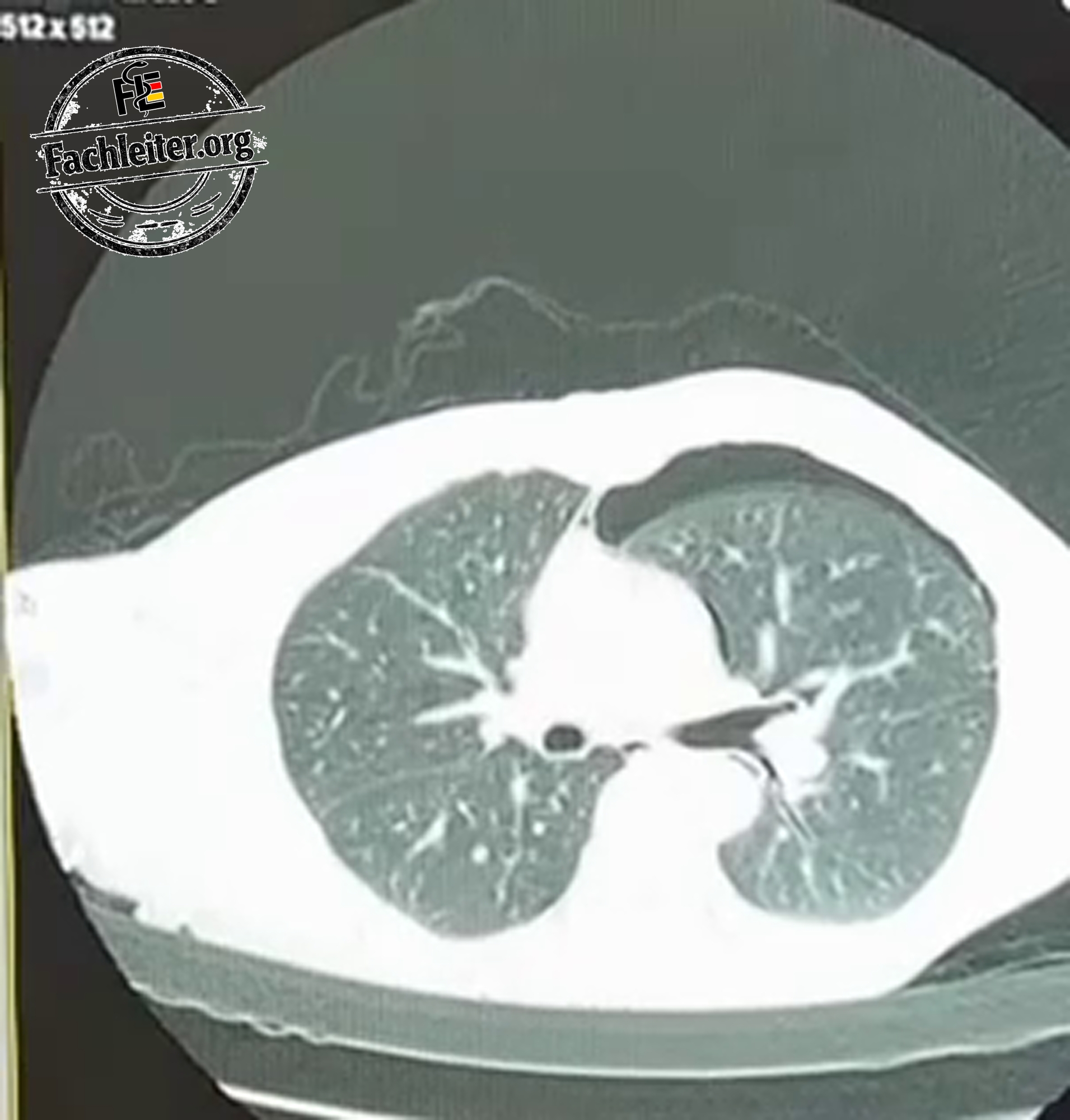
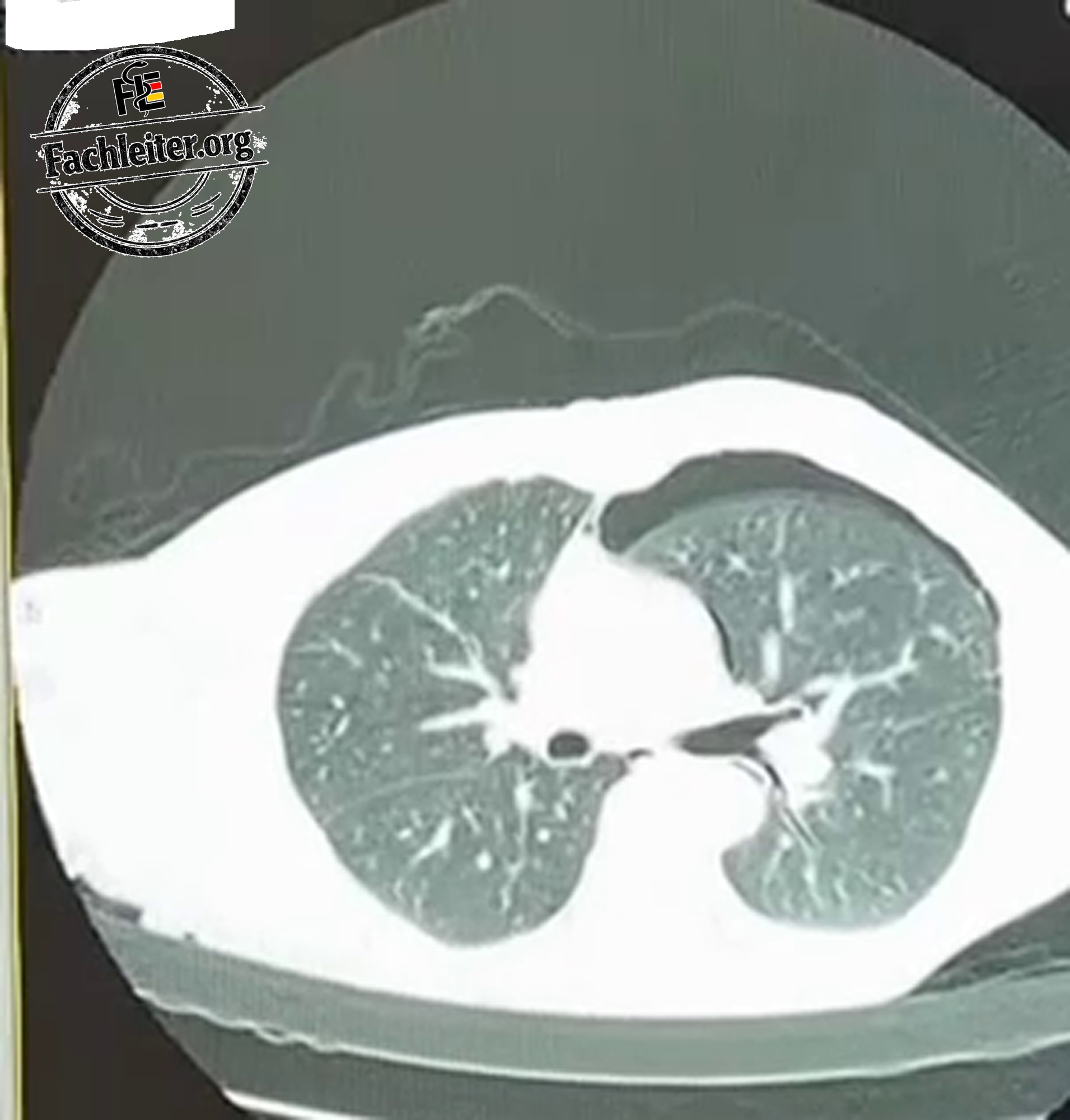
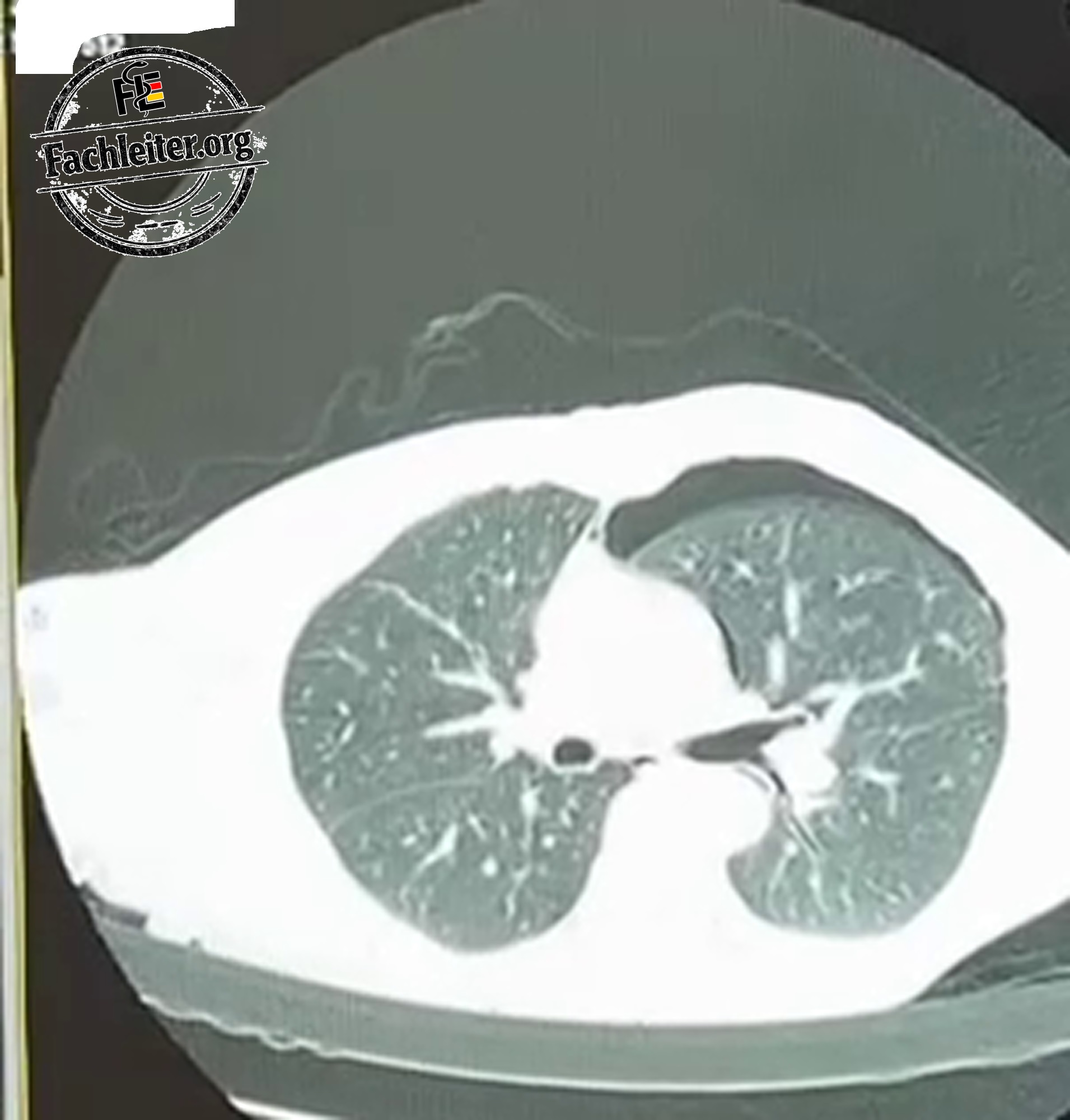
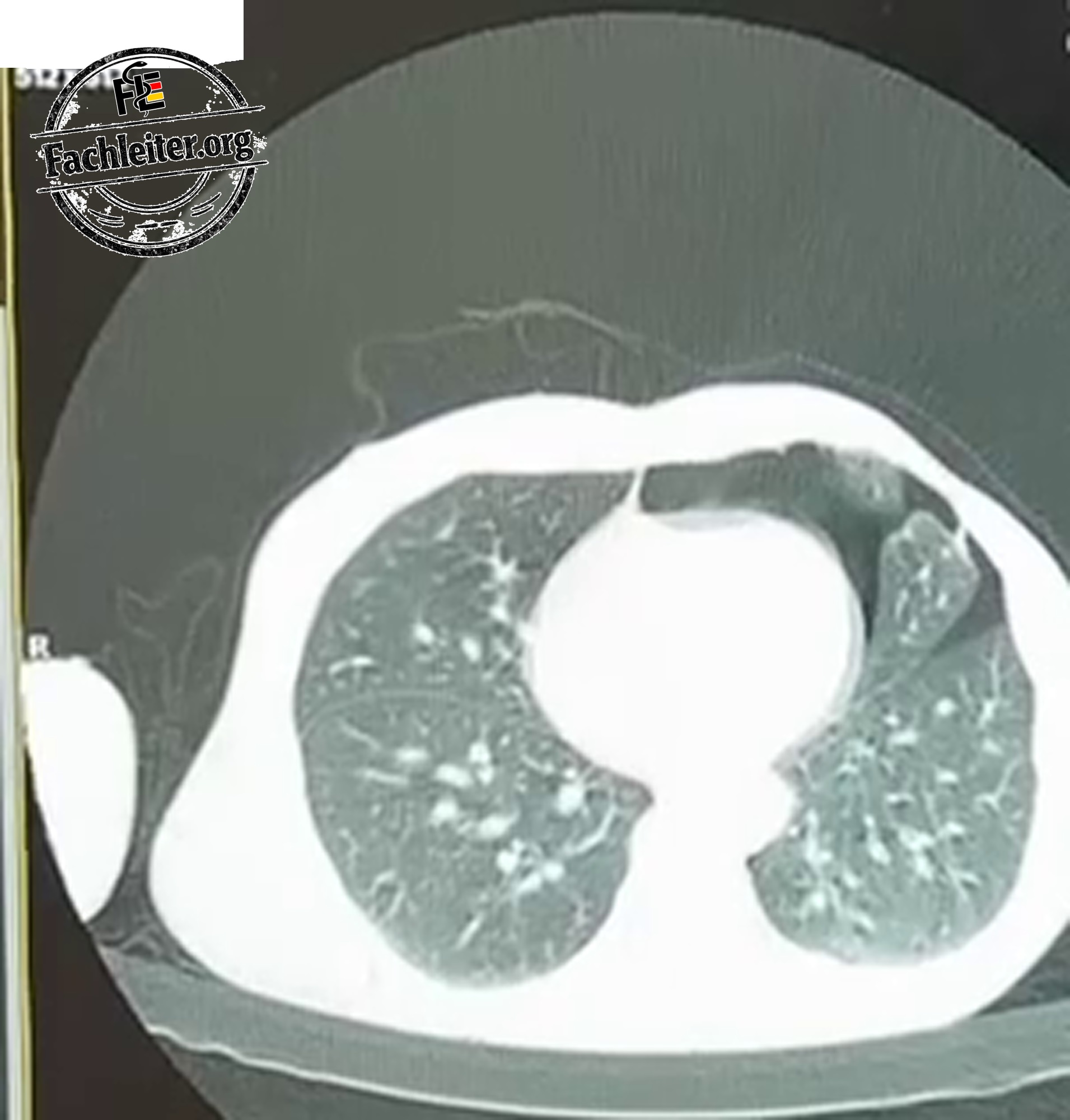
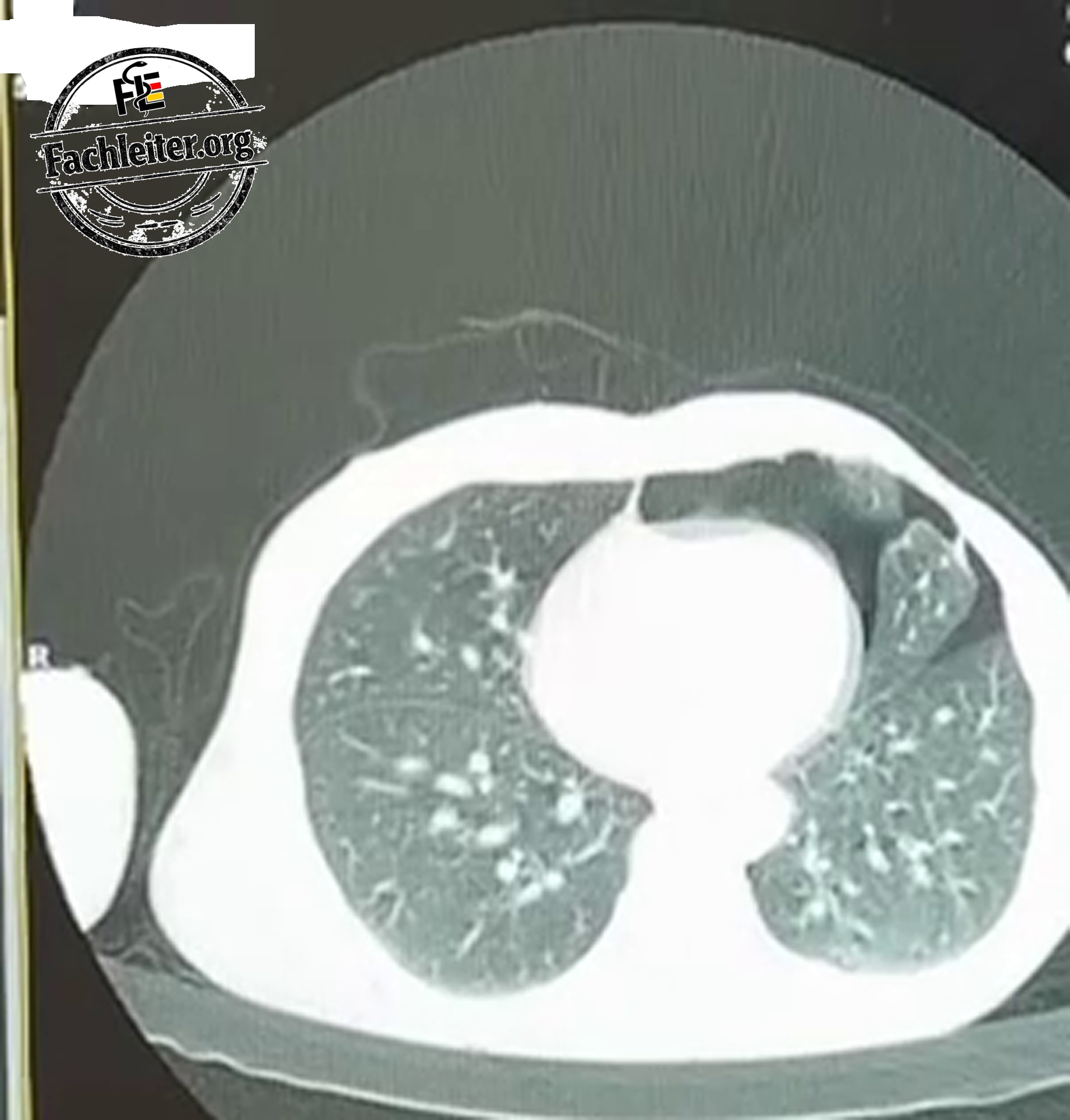
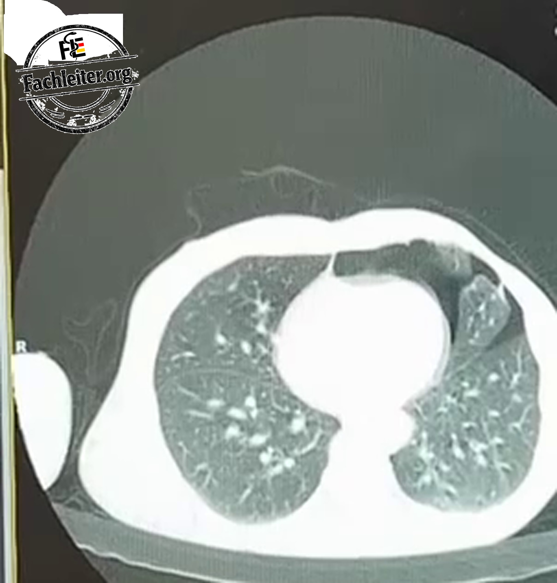
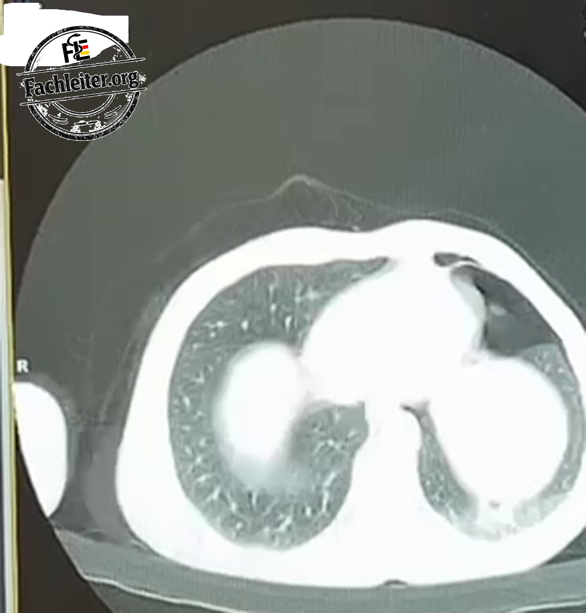
On the X-ray air appears as a dark area in the soft tissues. It can be visible between the tissue layers of the chest wall or in the neck. A large air collection can make assessment of the underlying structures more difficult.
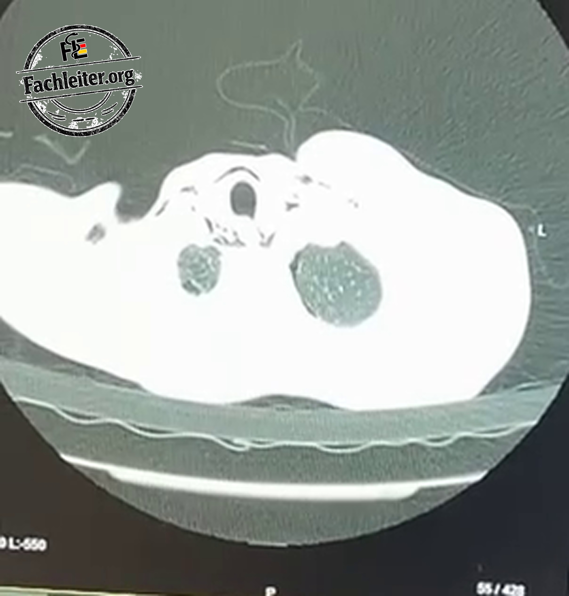
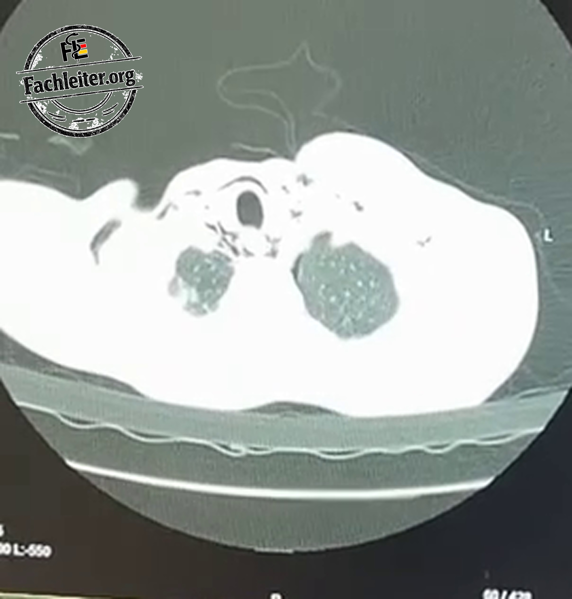
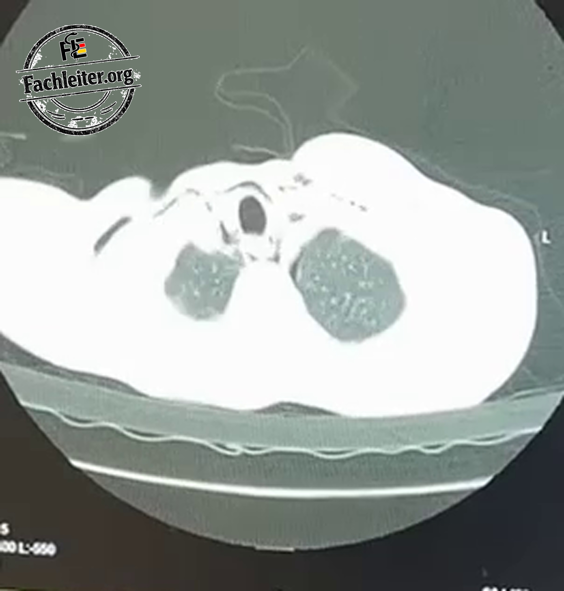
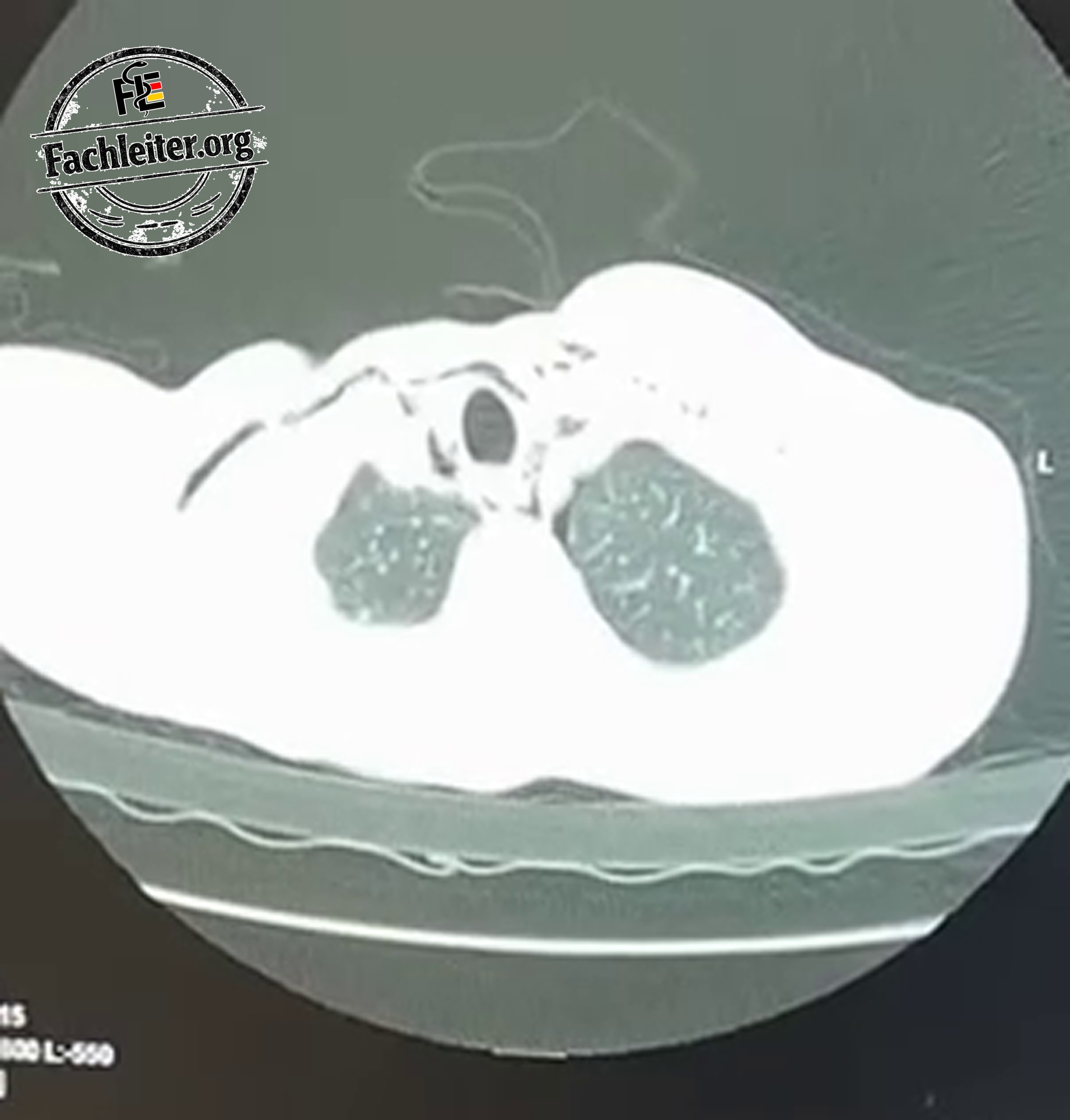
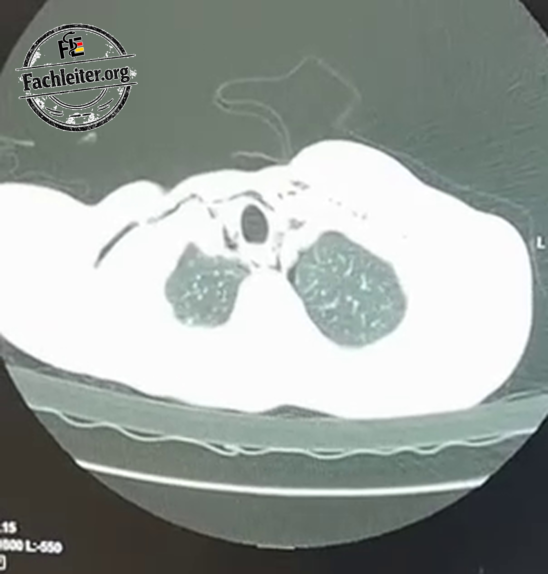
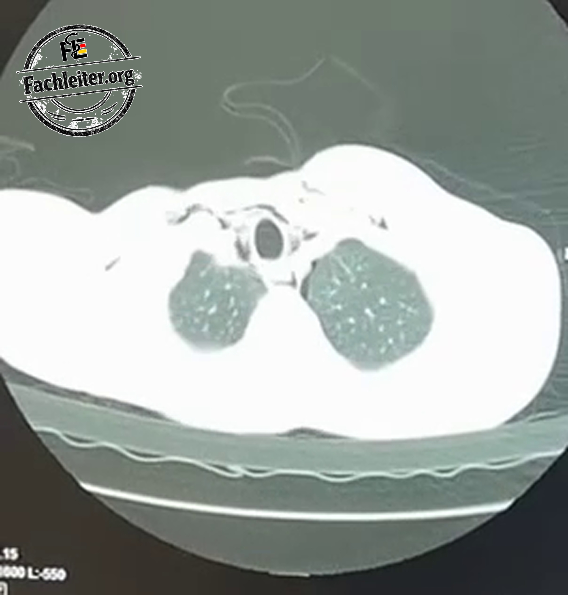
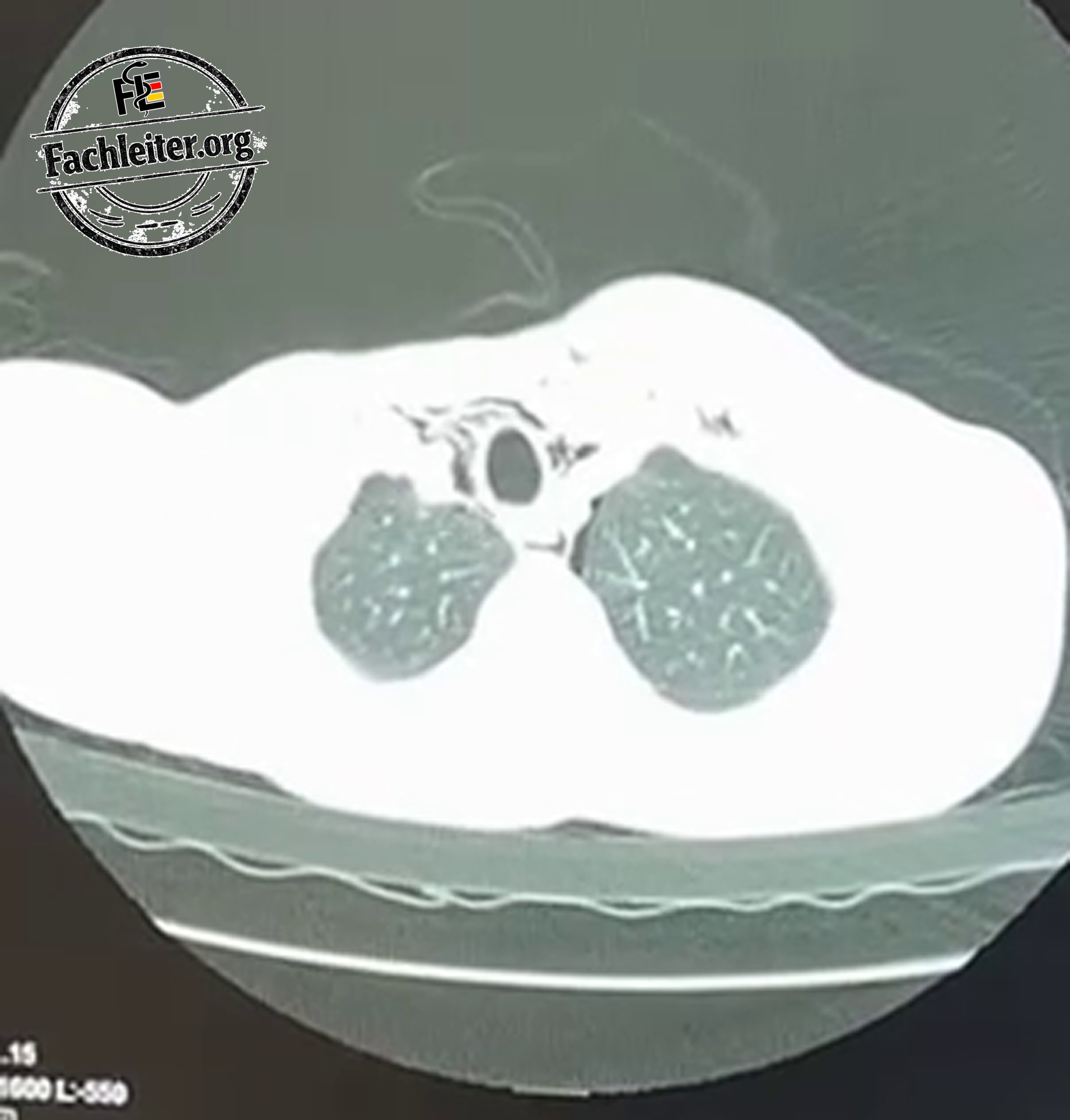
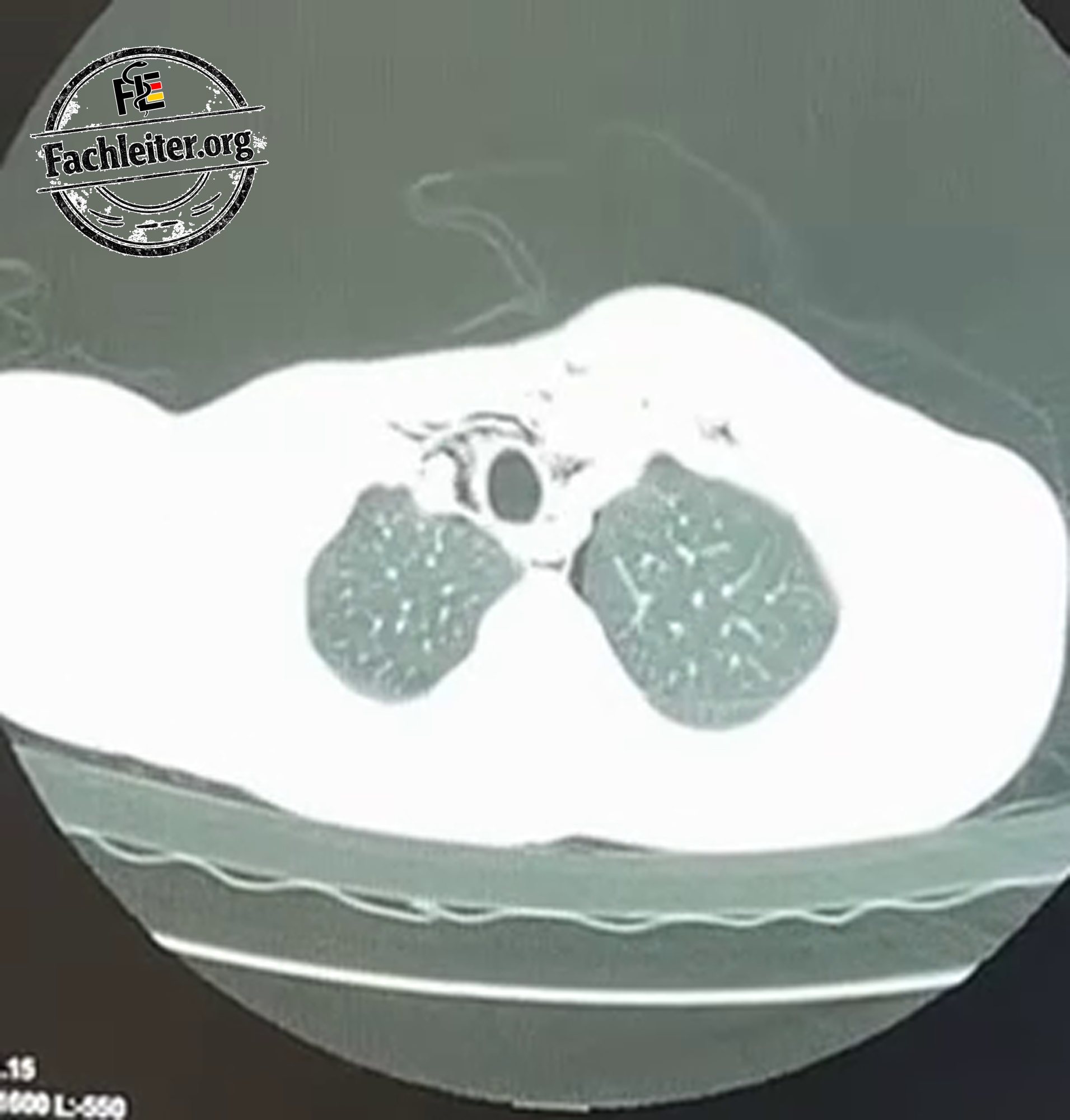
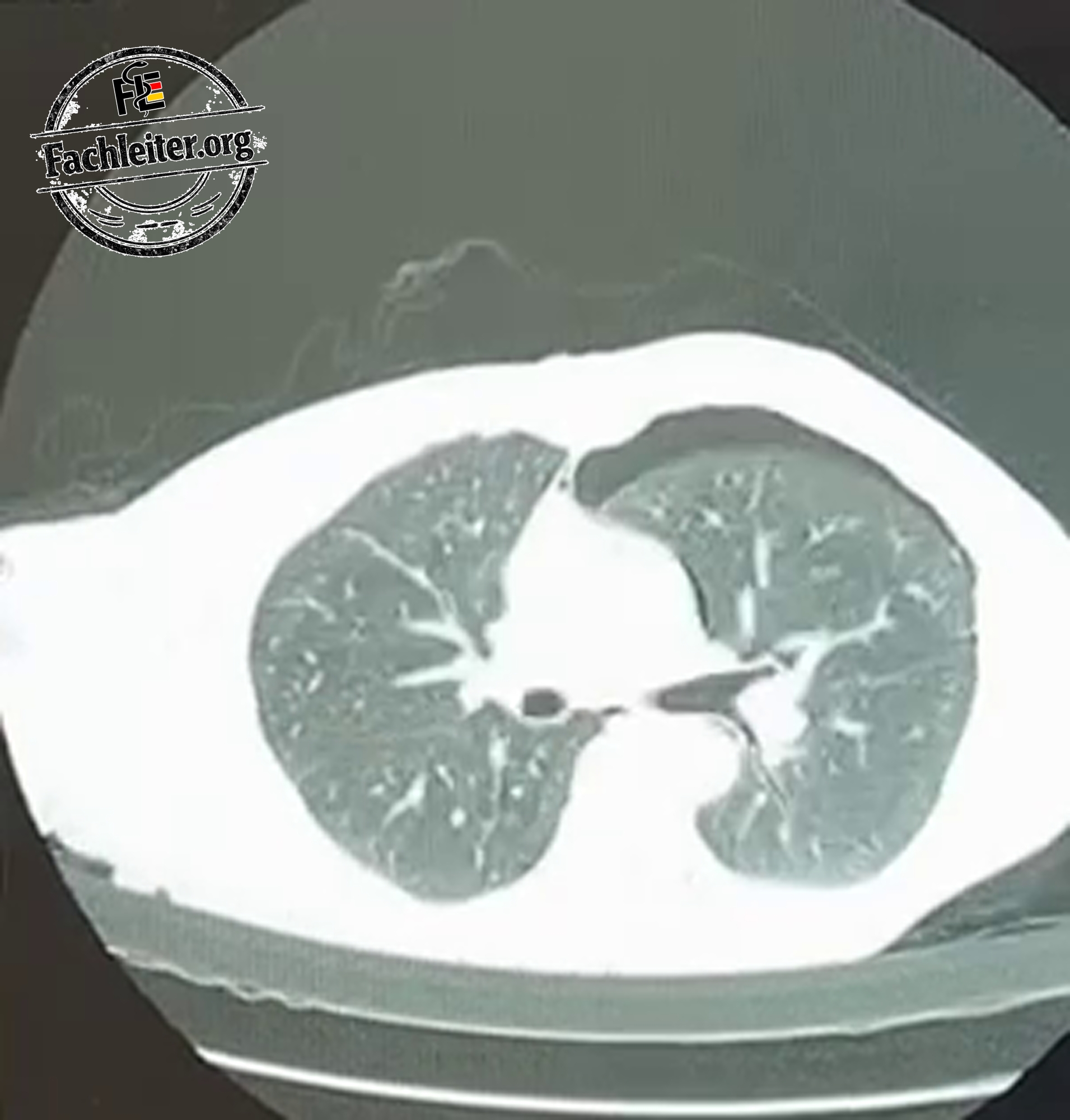
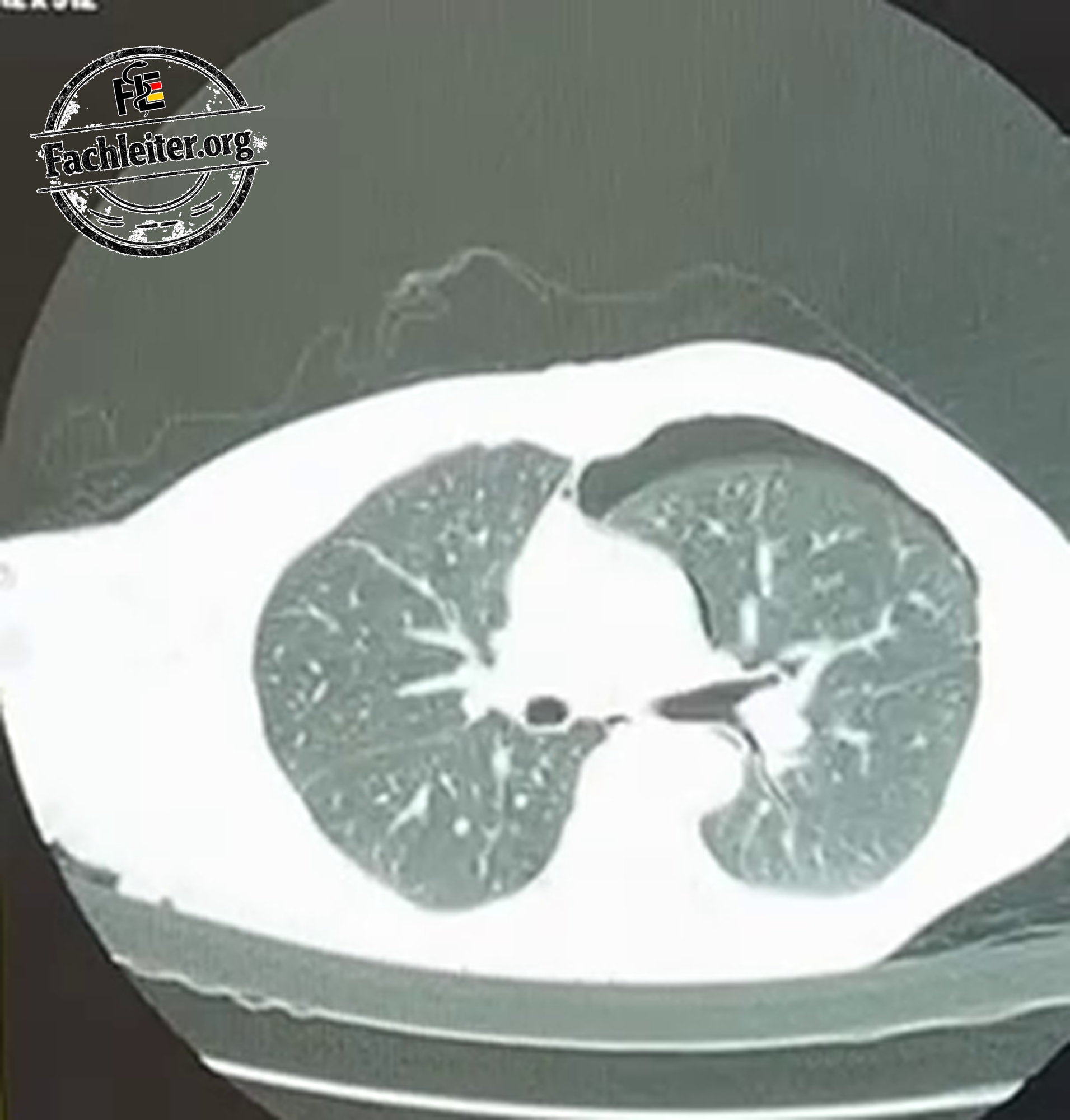
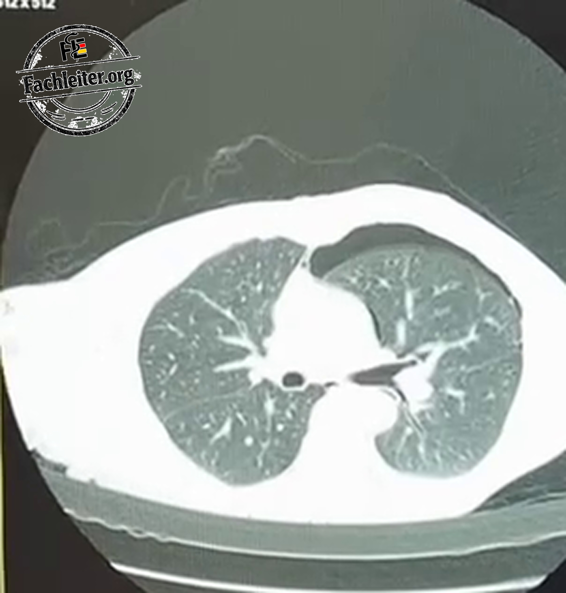
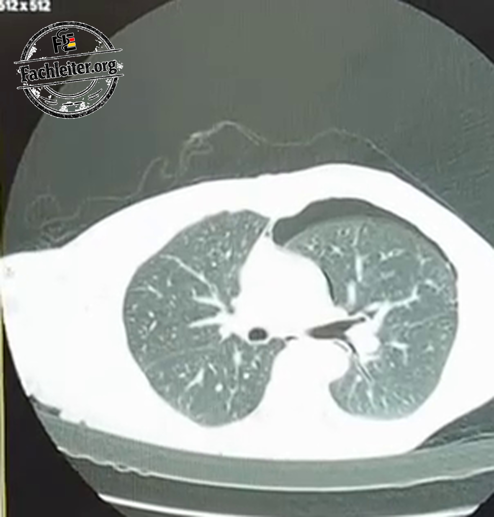
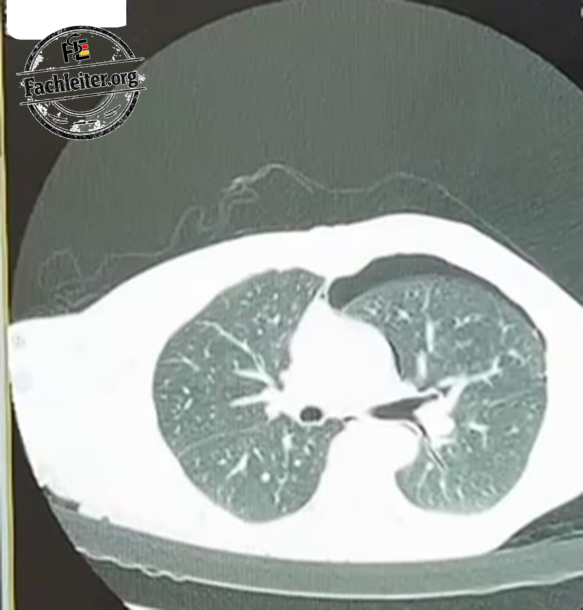
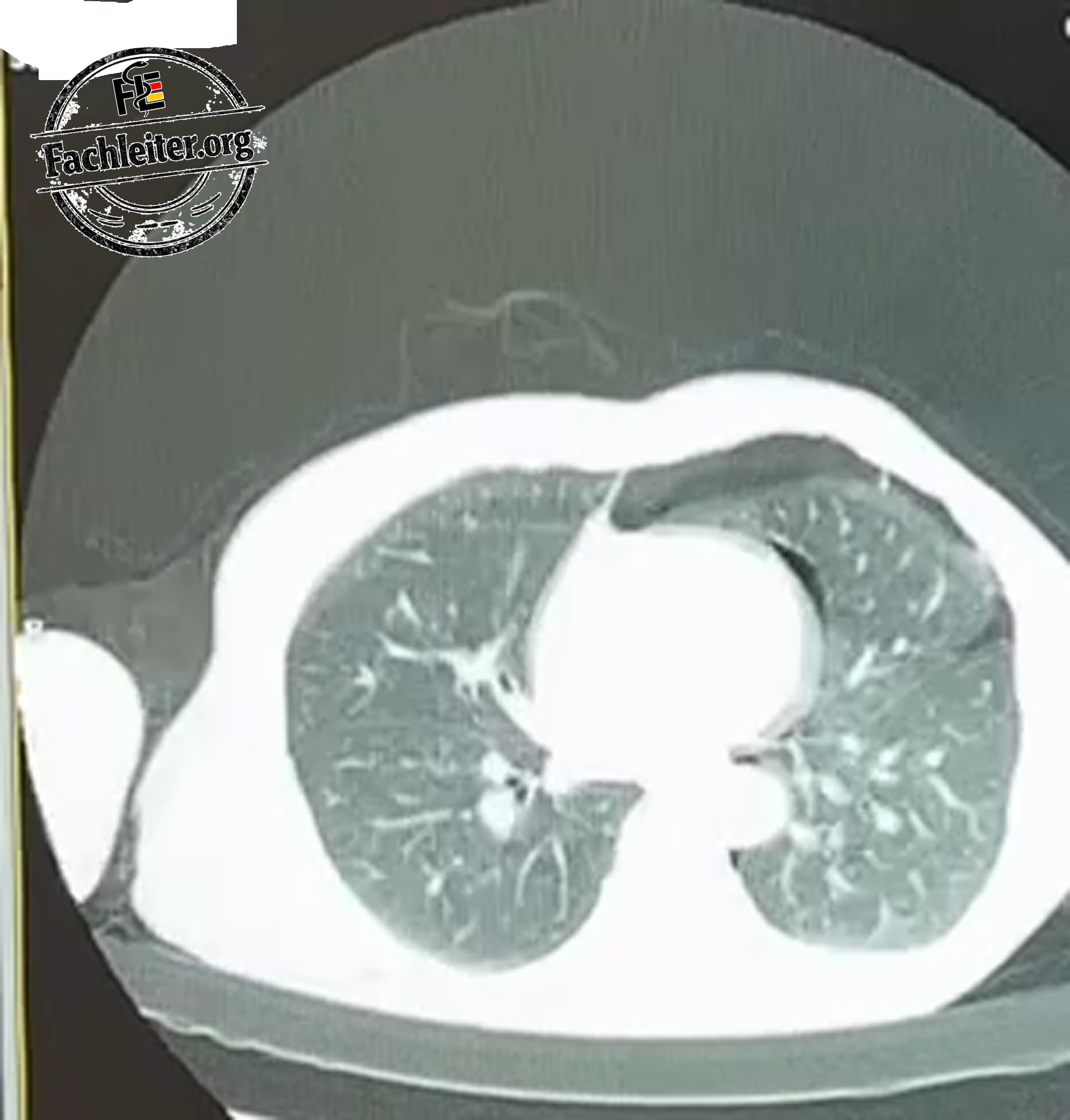
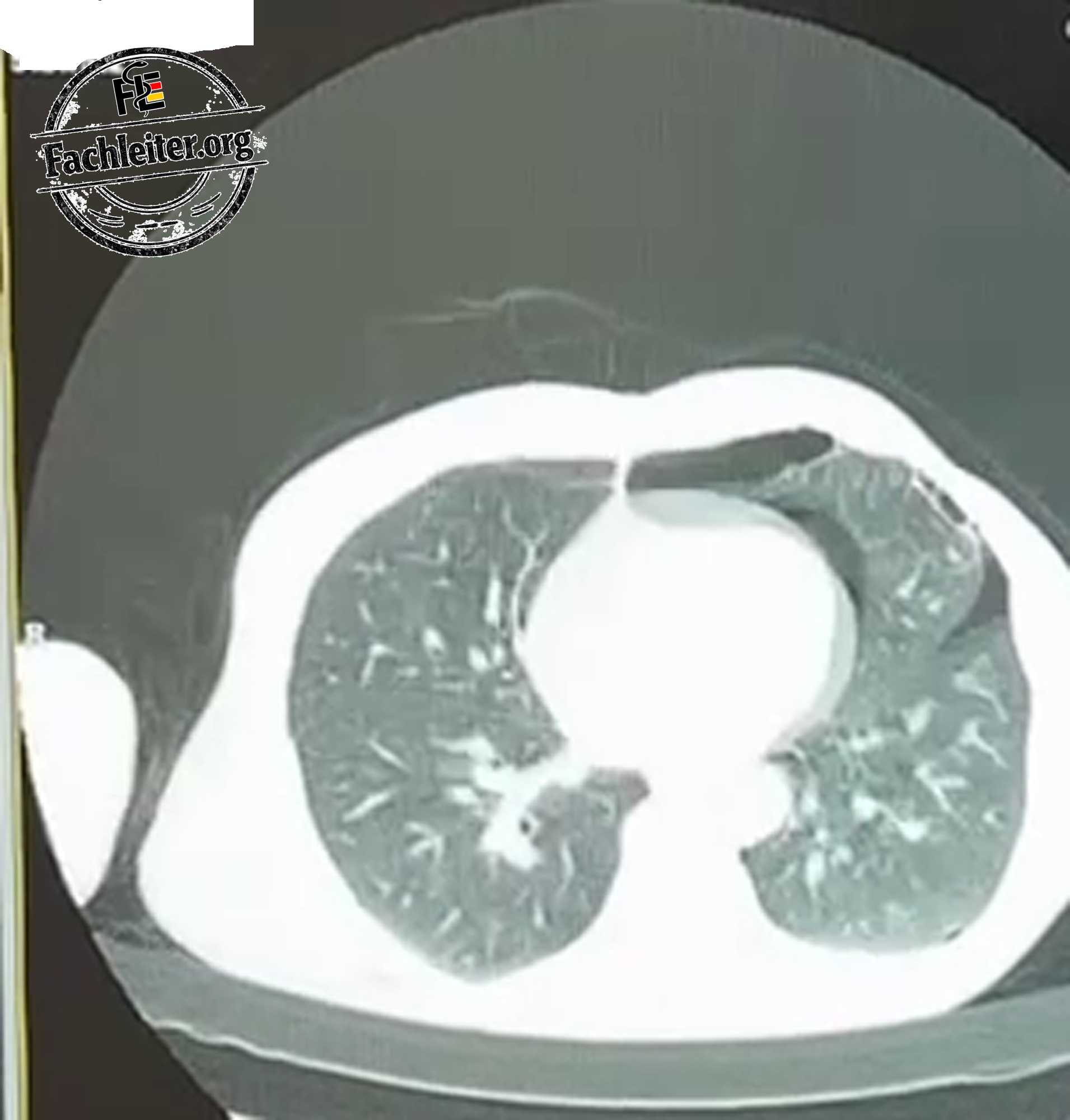
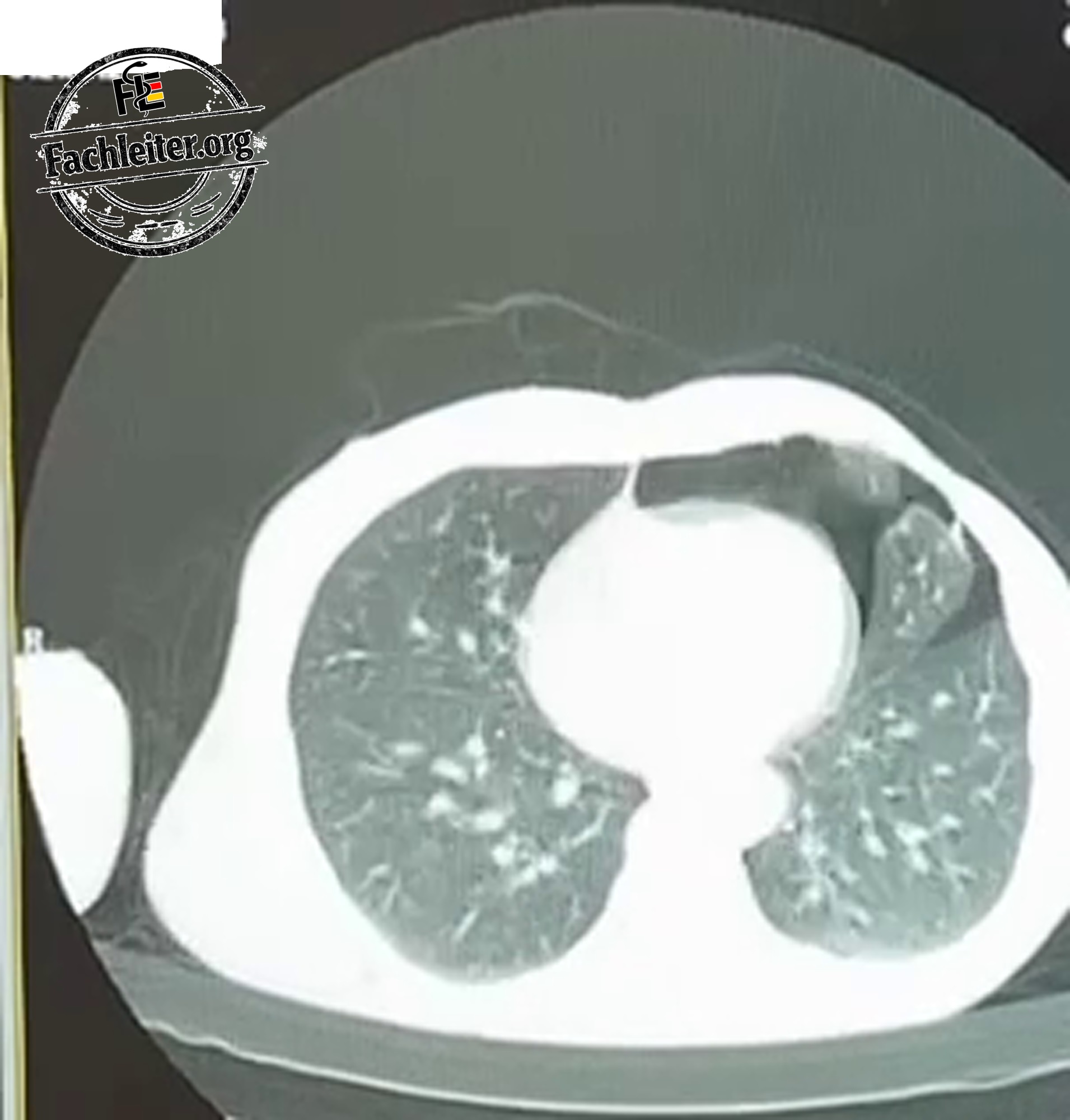
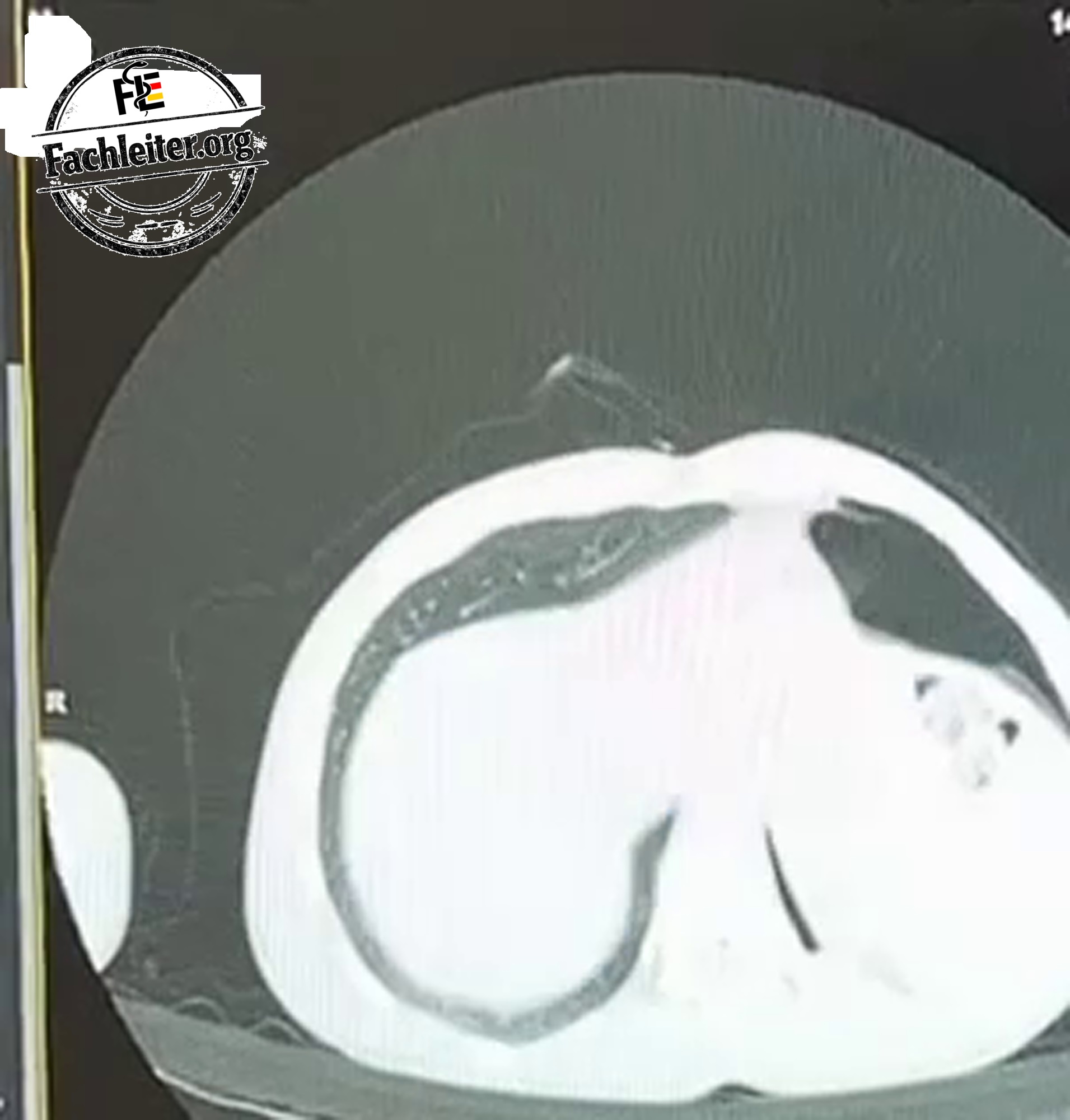
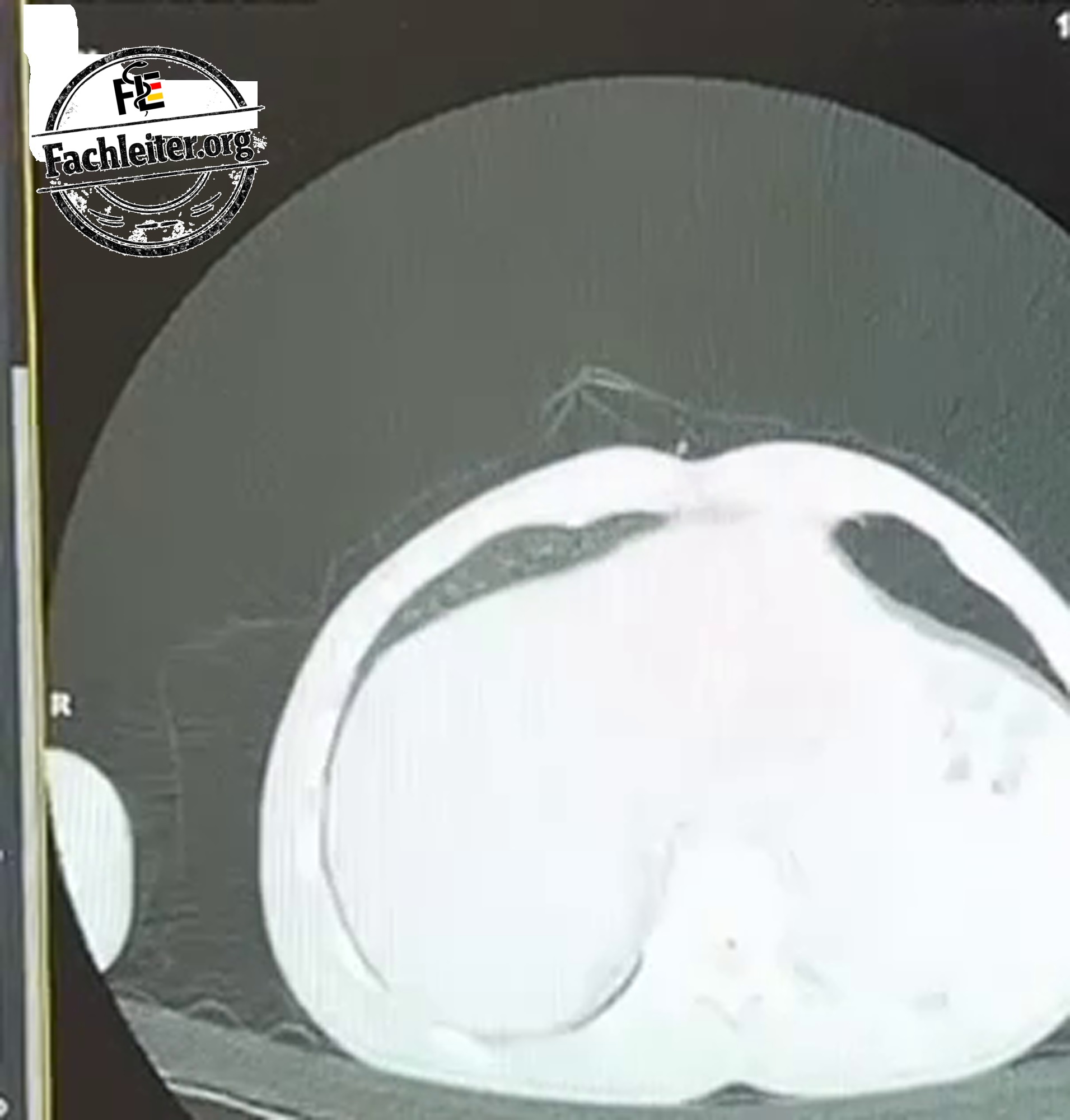
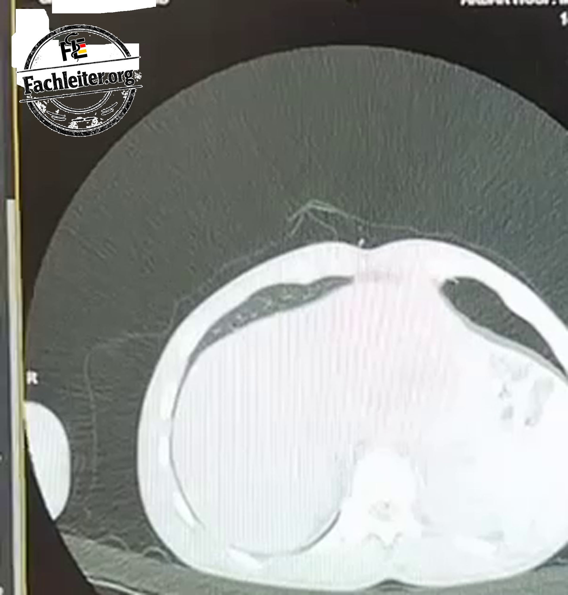
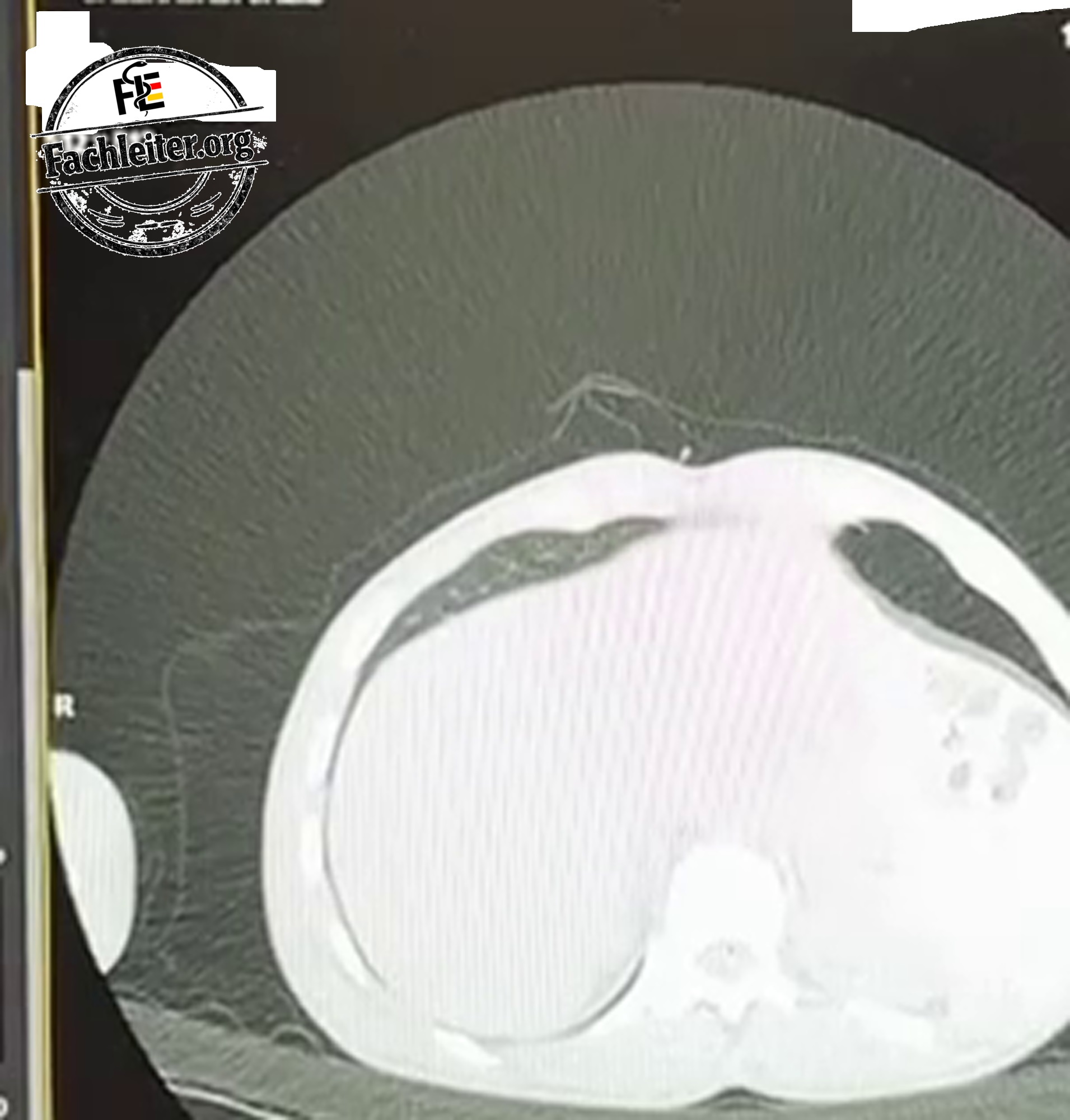
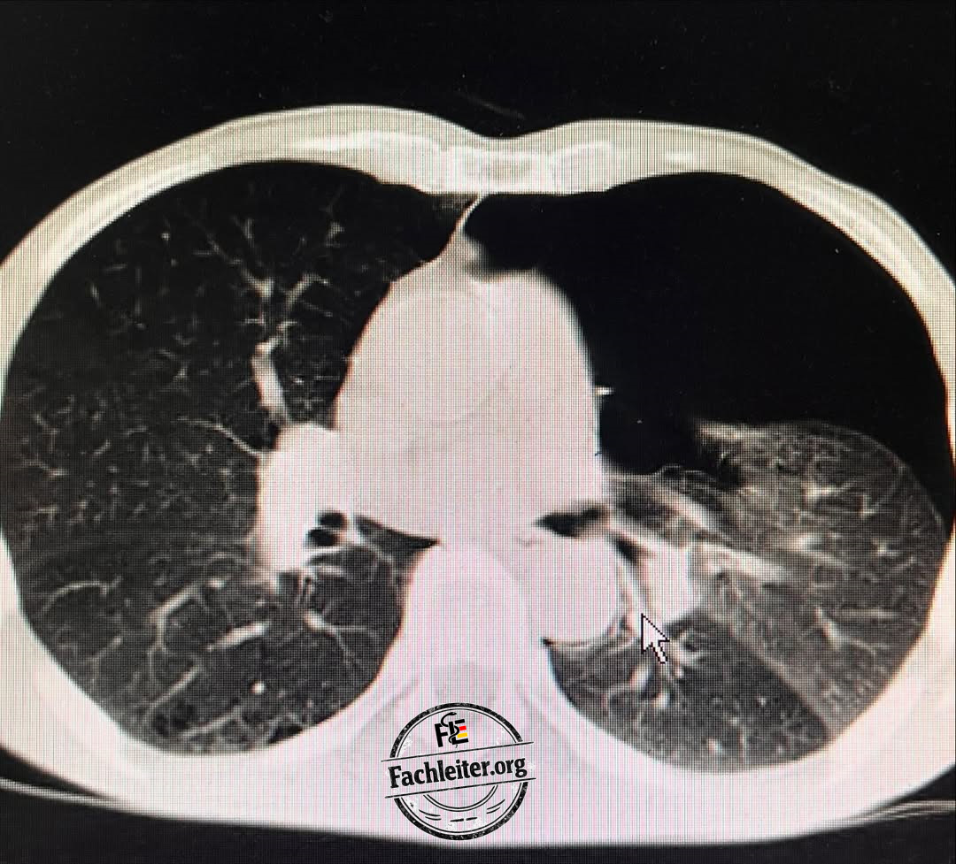
When viewing an image the next question is: is there also a pneumothorax present?
Further imaging
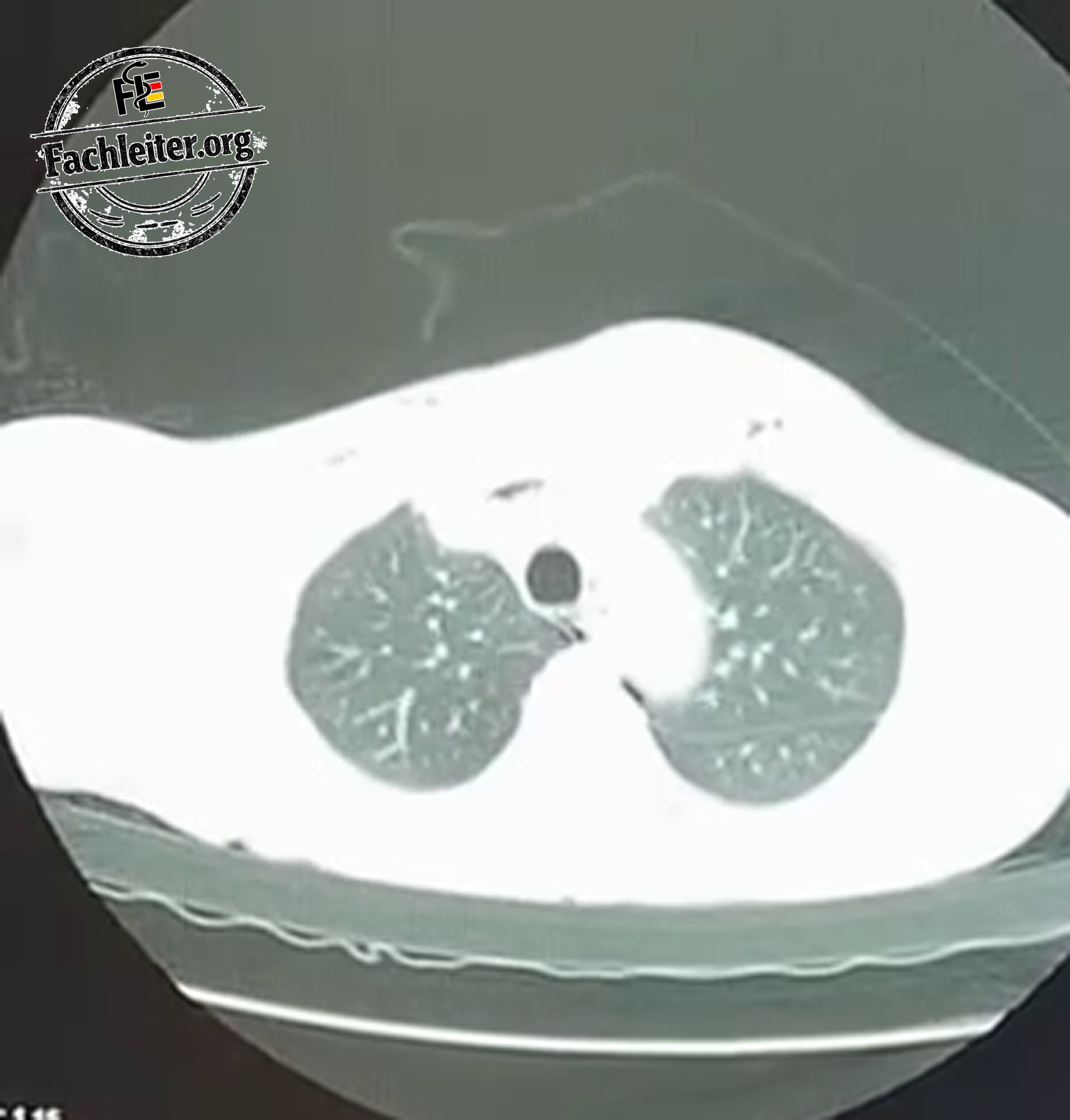
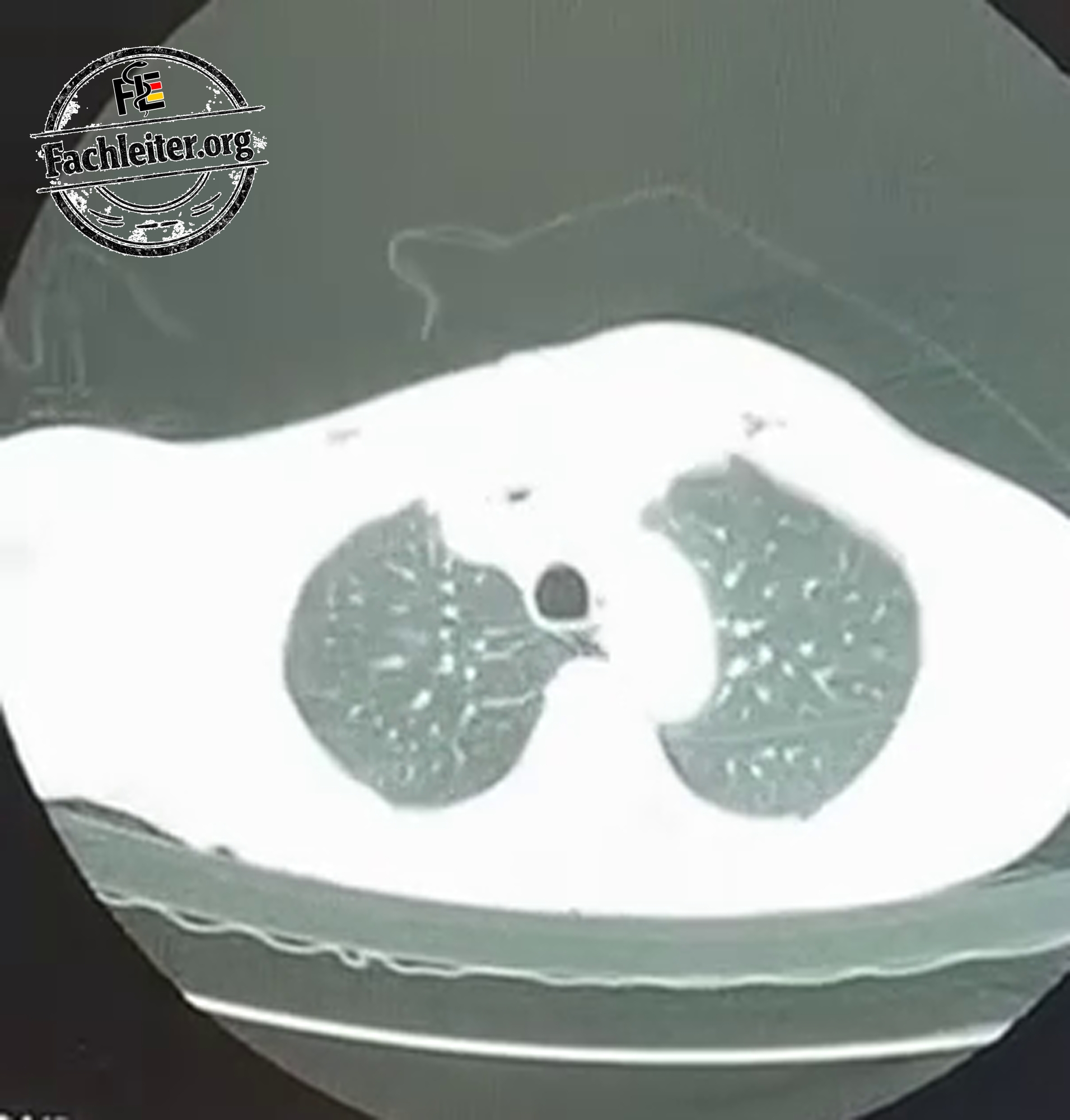
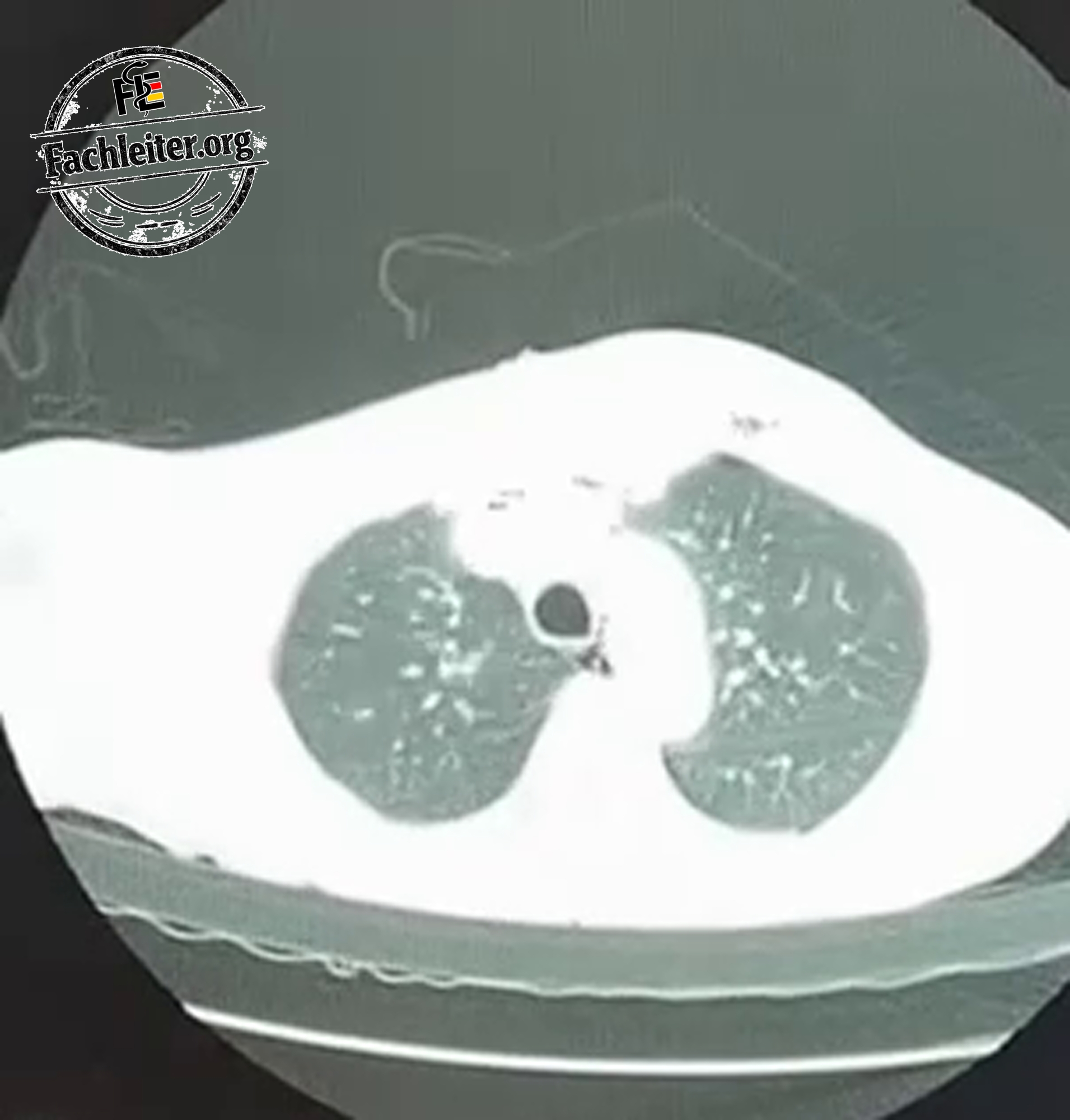
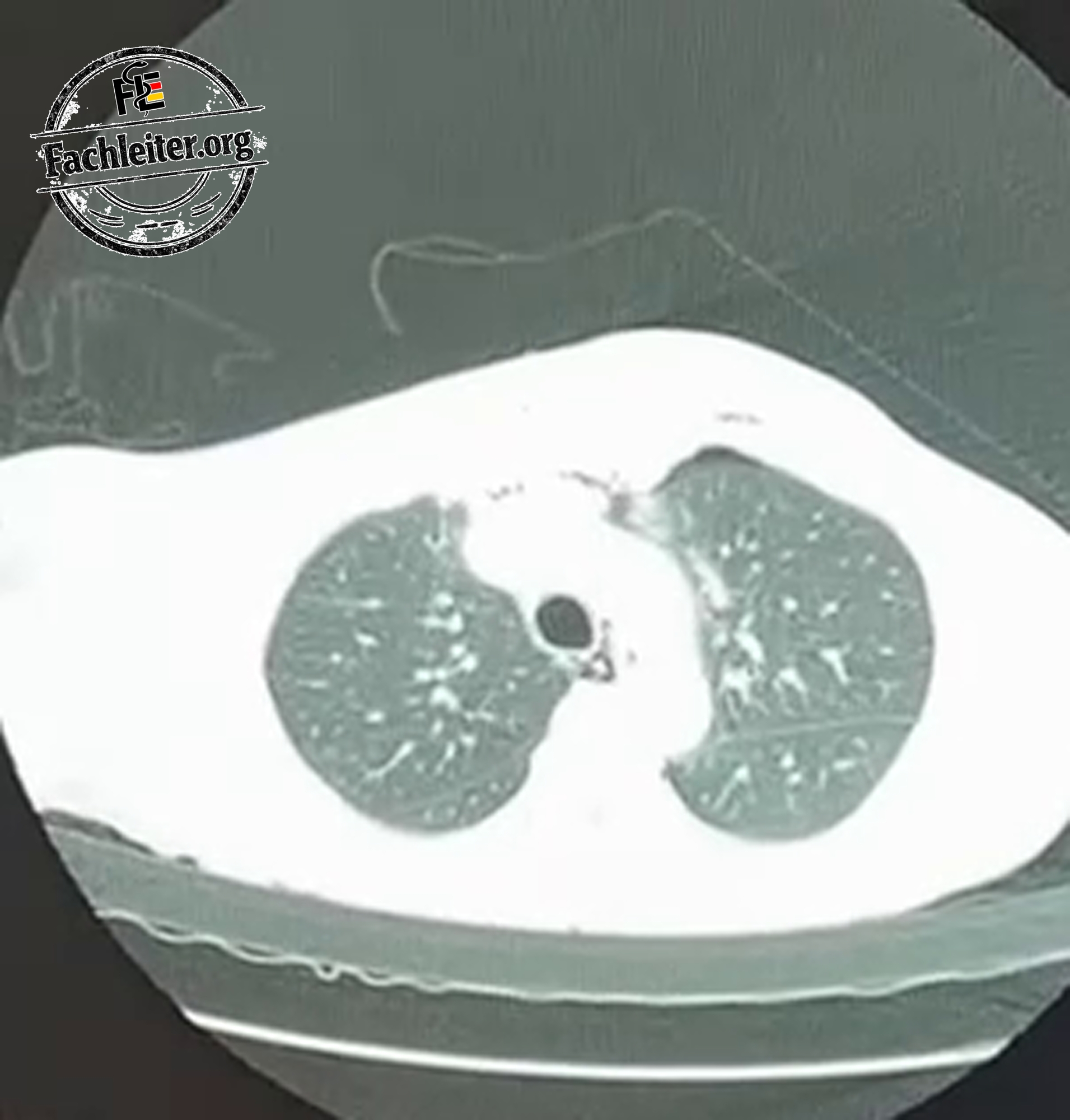
A thoracic computed tomography allows a more precise assessment of the extent of the air and possible accompanying injuries. This is particularly helpful when the cause is unclear after trauma, the air collection is increasing, or there is suspicion of airway injury.
A thoracic ultrasound can help in the search for a pneumothorax. Extensive subcutaneous air, however, can make ultrasound examination more difficult.
Treatment
Treatment is directed at the cause, not just the visible swelling. If a pneumothorax requiring treatment is present, it must be addressed. If a chest drain is already in place and the subcutaneous emphysema increases, its position, patency and function are checked.
A limited subcutaneous emphysema can resolve once no new air is entering. In cases of large air accumulation with increasing symptoms, a procedure to decompress the subcutaneous tissue may be necessary. The treatment team decides based on the overall findings.
Possible complications
If the air source persists, the emphysema can continue to spread. Increasing swelling of the neck and face can make further examination and treatment more difficult; in severe cases breathing can also be impaired. The most important risk, however, is overlooking an accompanying or progressive injury.
After treatment the subcutaneous emphysema may initially remain visible even though the air source has already been sealed. If it increases again, one must look for a persistent air leak or a problem with the drain.
X-ray
Pneumothorax
Definition
A pneumothorax is the presence of air in the pleural space between the lung and the chest wall. Normally the two pleural layers lie close together there. If air enters between them, the lung on the affected side can partially or completely collapse.
A tension pneumothorax is a life-threatening special form: air enters the chest but cannot adequately escape. The rising pressure impairs breathing and eventually the circulation.
How does a pneumothorax occur?
A pneumothorax can occur without external injury, for example from the rupture of a superficial pulmonary bleb. It can also develop in association with an existing lung disease.
Other causes are chest injuries, such as after a fall with rib fracture, and complications of medical procedures. Therefore pneumothorax and subcutaneous emphysema may occur together in trauma.
Symptoms and physical examination
Typical are sudden onset unilateral chest pain and shortness of breath. The pain can worsen with inspiration. A small pneumothorax sometimes causes only minor symptoms; with a larger pneumothorax respiratory rate and pulse can increase and oxygen saturation can fall.
During the physical exam vital signs and respiration are assessed first. The affected side of the chest may move less, and on auscultation breath sounds may be diminished.
Immediate action is necessary if dyspnea and circulatory status rapidly worsen and a tension pneumothorax is suspected. In this situation decompression is not postponed for imaging.
What is seen on the X-ray?
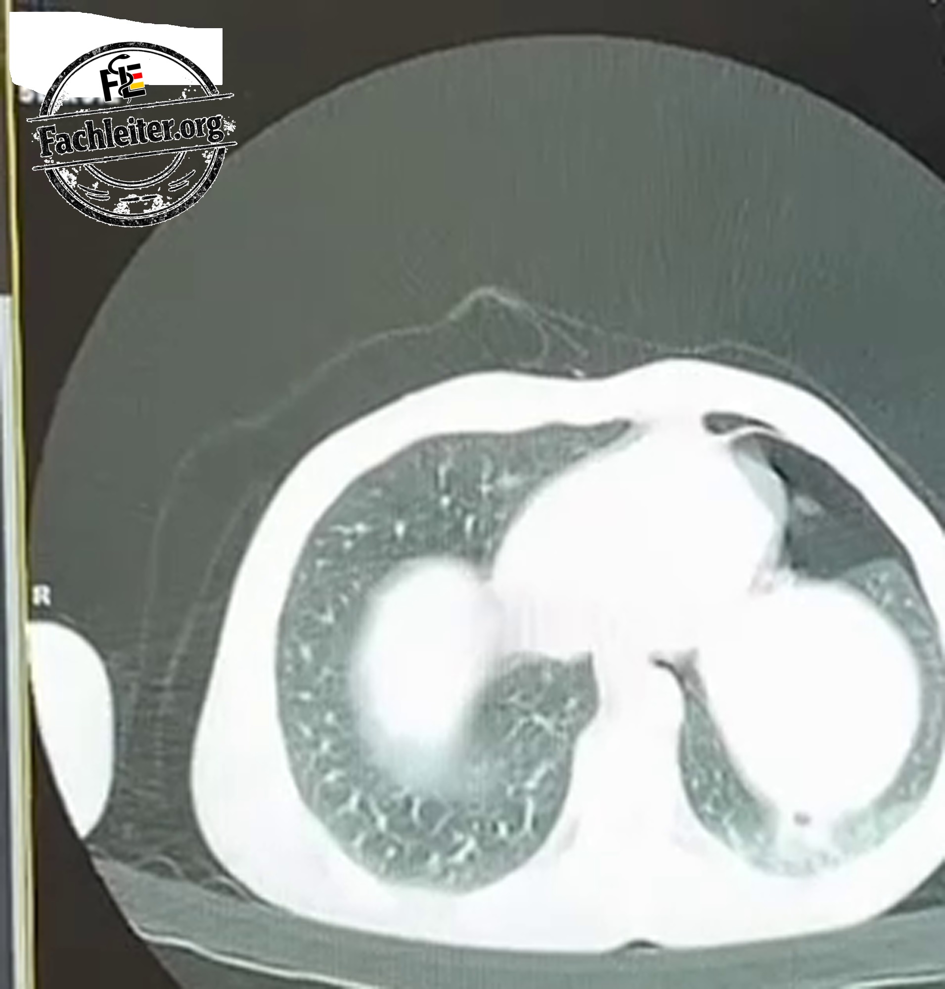
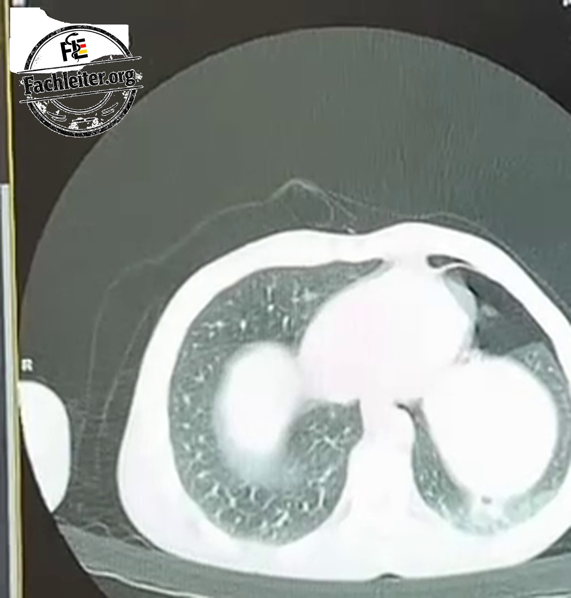
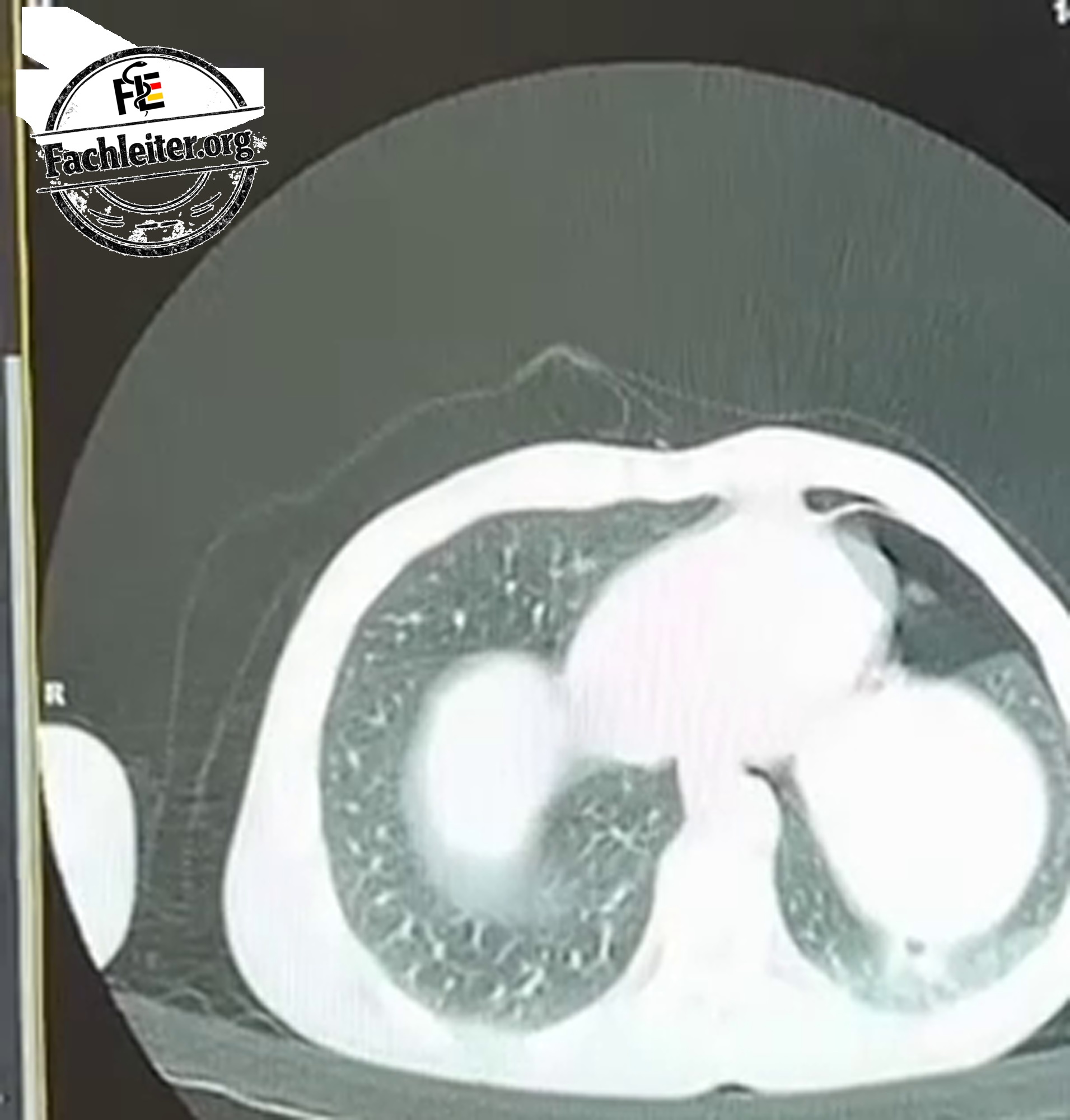
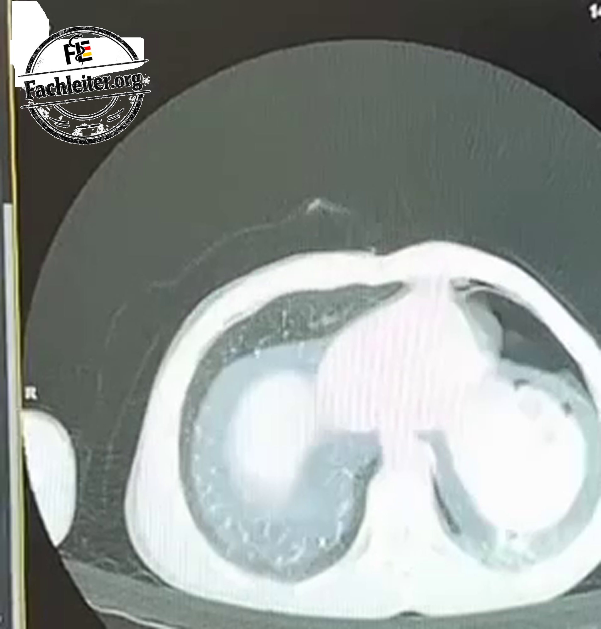
An important sign is the pleural line: it borders the partially collapsed lung. Outside this line no normal lung markings are seen because air is present in the pleural space.
Interpretation also depends on whether the image was taken upright or supine. A small pneumothorax can be more difficult to detect on a supine film.
X-ray
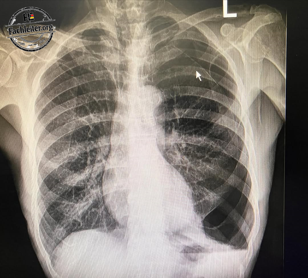
No mediastinal shift to the opposite side as an indication of tension pneumothorax is visible on the X-ray.
Further imaging
A thoracic ultrasound can provide bedside clues to a pneumothorax. Computed tomography is more sensitive for small air collections and also shows possible accompanying injuries after trauma. It is considered when the finding is unclear or further injuries are suspected. If a tension pneumothorax is suspected, immediate treatment takes priority.
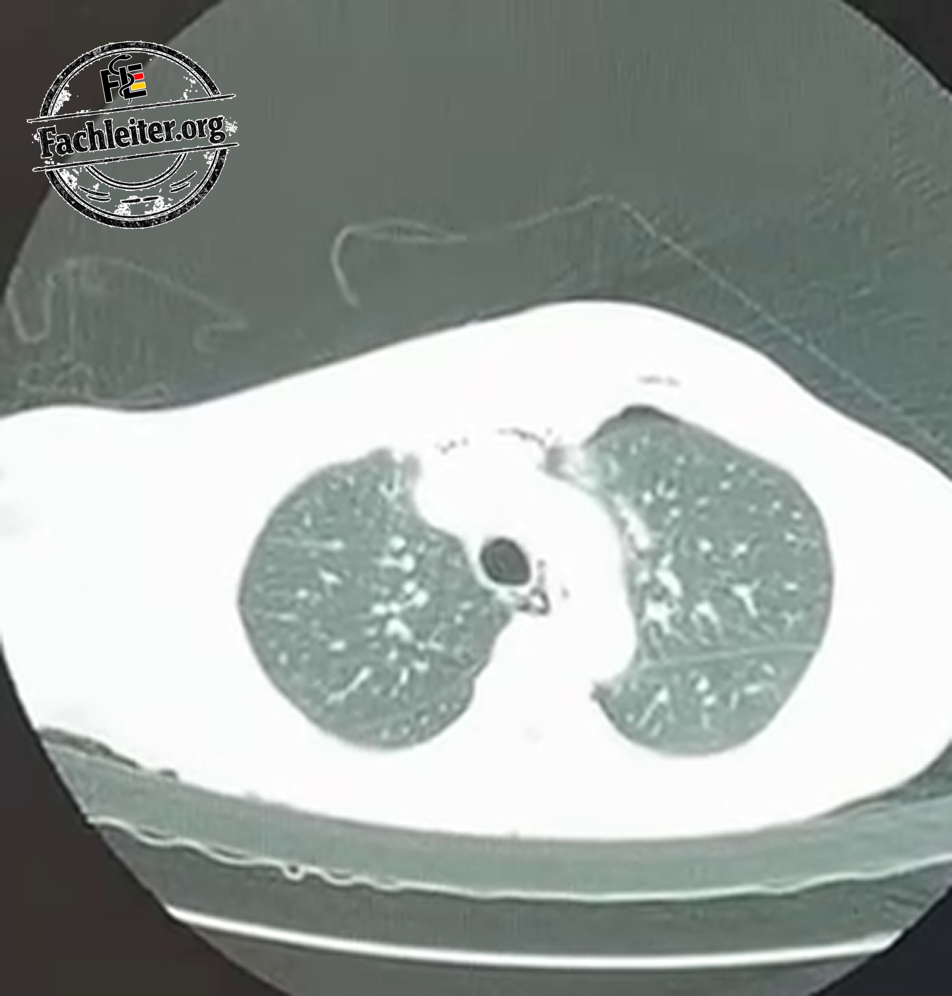
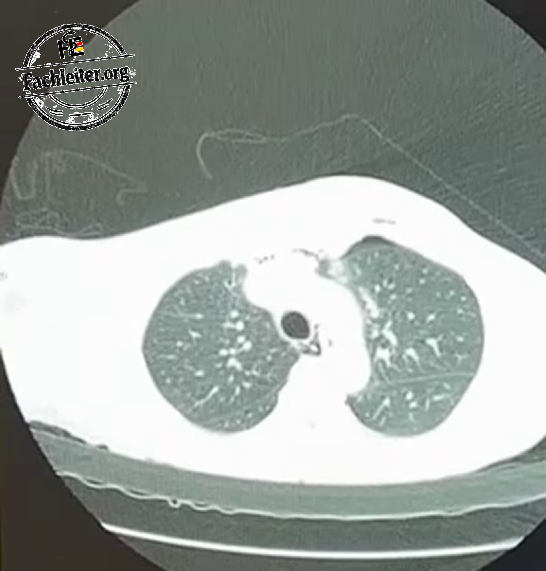
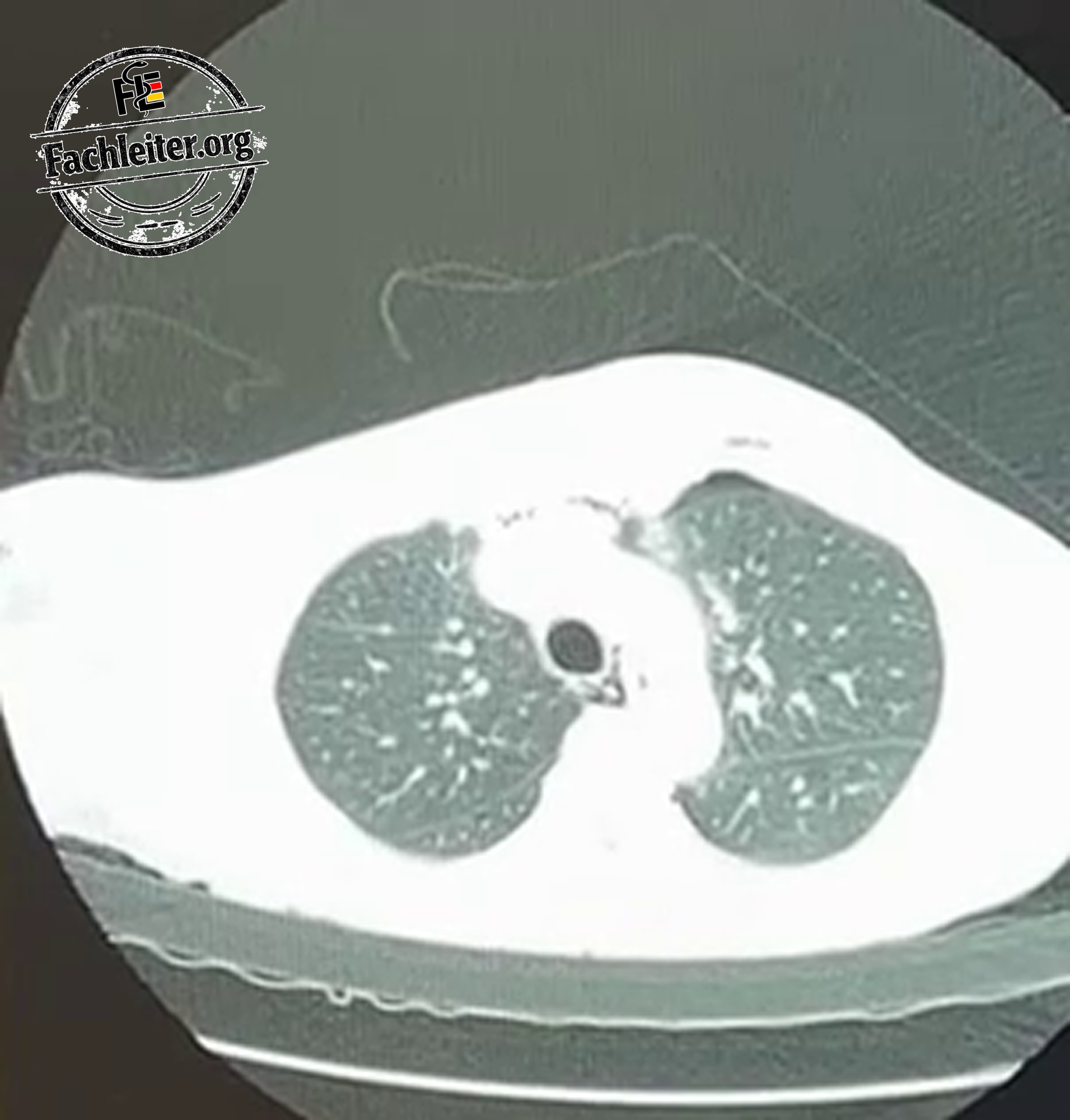
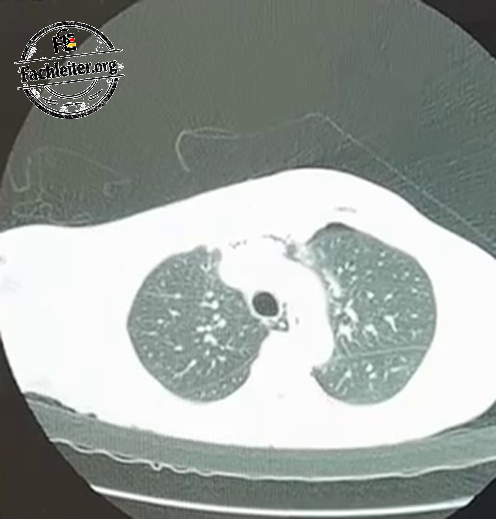
Treatment
Treatment depends on the size of the pneumothorax, the symptoms and circulatory stability.
Immediate measures
- Monitoring of blood pressure, pulse, respiratory rate, ECG and oxygen saturation
- venous access
- adequate pain management
- oxygen administration for low oxygen saturation
- regular clinical and imaging follow-up
A small pneumothorax in a stable patient can initially be managed conservatively under close monitoring. For a larger or symptomatic traumatic pneumothorax a chest drain is required.
Required materials
- sterile gloves, gown and drapes
- skin disinfectant
- local anesthetic
- syringes and needles
- scalpel
- curved clamp
- chest drain, in thoracic trauma usually 24–32 Ch
- suture material
- sterile dressing
- drainage system with water seal
- suction system if required
- monitoring and emergency equipment
Insertion of a chest drain
-
The patient is positioned supine with the upper body slightly elevated. The arm on the affected side is placed above the head.
-
The insertion site is usually in the fourth or fifth intercostal space within the so‑called triangle of safety between the pectoralis major, the latissimus dorsi and the axilla.
-
After skin disinfection and sterile draping the skin, subcutaneous tissue, muscles and pleura are locally anesthetized.
-
The skin is incised parallel to the rib.
-
The tissue is bluntly dissected with a clamp down to the pleura. The access is made immediately above a rib to avoid injuring the vessels and nerves running beneath it.
-
After opening the pleural space, correct placement is checked with the finger.
-
The drain is inserted into the pleural space and oriented anteriorly and superiorly in the case of a pneumothorax.
-
All lateral openings of the drain must lie within the thoracic cavity.
-
The drain is connected to an underwater seal, secured with a suture and dressed sterilely.
-
Subsequently, the function of the drain and respiration are checked. The position of the drain and re-expansion of the lung are verified by ultrasound or radiography.
Tension pneumothorax
In suspected tension pneumothorax, imaging must not be awaited. The chest must be immediately decompressed.
For this, a sufficiently long, large-bore cannula is usually introduced in the fourth or fifth intercostal space between the anterior and midaxillary lines immediately above a rib. Escape of air and clinical improvement are monitored.
The decompression puncture is only a temporary emergency measure. A chest drain must be placed afterward. In severe hemodynamic instability or cardiac arrest, an immediate open decompression of the pleural space may be performed by appropriately trained personnel.
Possible complications
- Bleeding
- Injury to the lung, diaphragm, or abdominal organs
- Malposition, kinking, or obstruction of the drain
- Infection
- persistent air leak
- Pain
- rarely a re-expansion pulmonary edema