
Forearm fractures are among the most common injuries in the emergency department. A systematic assessment of the X‑rays is crucial to reliably detect fractures, malalignment and associated injuries.
In this fracture series you will find six real X‑ray cases. Analyze each image yourself first before checking the diagnosis.
Pay particular attention to:
- Which bone is affected?
- Where is the fracture located?
- Is the fracture displaced?
- Is there joint involvement?
- Are there signs of associated injuries?
- Which therapy would you recommend?
Fachleiter
Case 1
Patient (19 years old) after a fall onto the right forearm with pain, swelling and limited mobility.
Your task
- Which bone is fractured?
- In which segment is the fracture located?
- Is there displacement?
- Conservative or operative treatment?
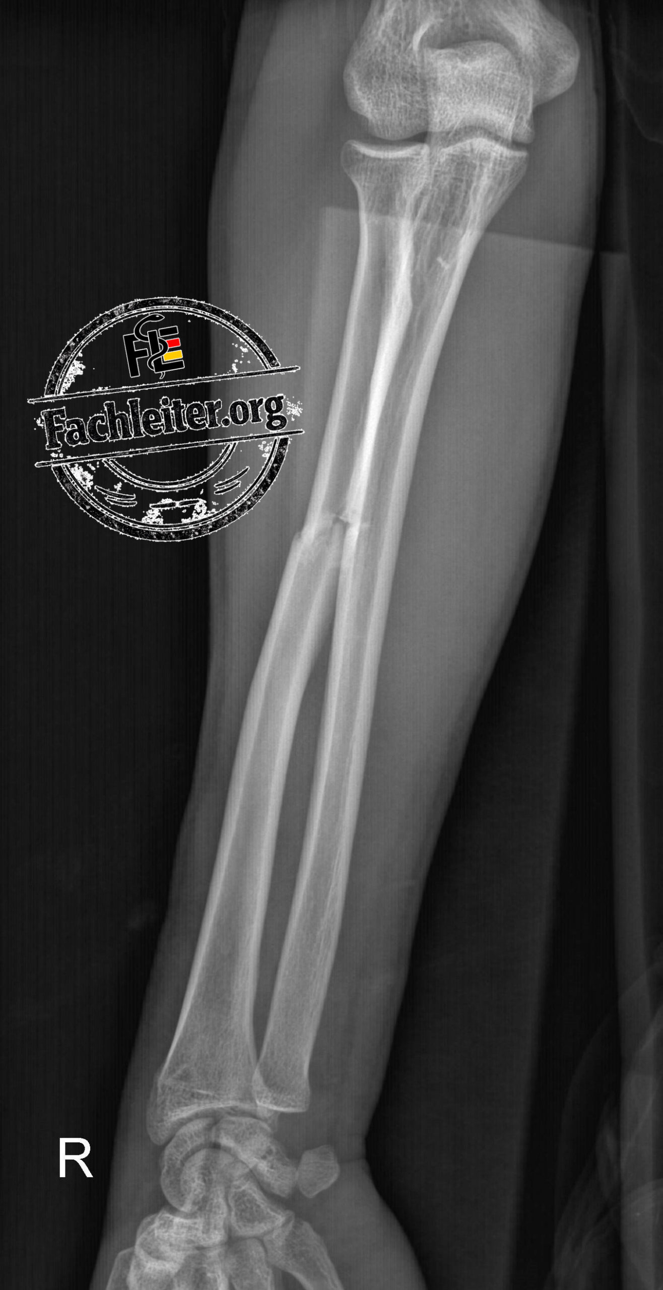
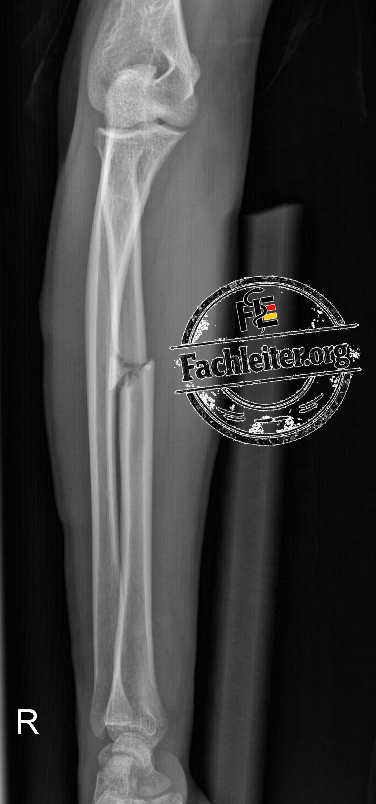
Radiological description
- Localization: Middle third of the radial shaft – the fracture line is located approximately halfway between the elbow and the wrist.
- Fracture type: Oblique fracture with wedge fragment (butterfly fragment) – the fracture line runs obliquely to the bone axis; an additional triangular intermediate fragment is visible.
- Completeness: Complete fracture – the fracture line completely traverses both cortices.
- Displacement: Present (translation and angulation) – the fragments are shifted relative to each other and show an axis deviation.
- Shortening: Slight – the fragments overlap slightly, causing the radius to appear somewhat shortened.
- Associated finding: Soft tissue swelling – increased soft tissue density and contour widening in the area of the fracture as an indication of a posttraumatic hematoma or edema.
Question
- Is there a fracture of the radial or ulnar styloid process?
- Why was the second image (lateral) taken with the elbow extended and not in 90° flexion?
Fachleiter
Case 2
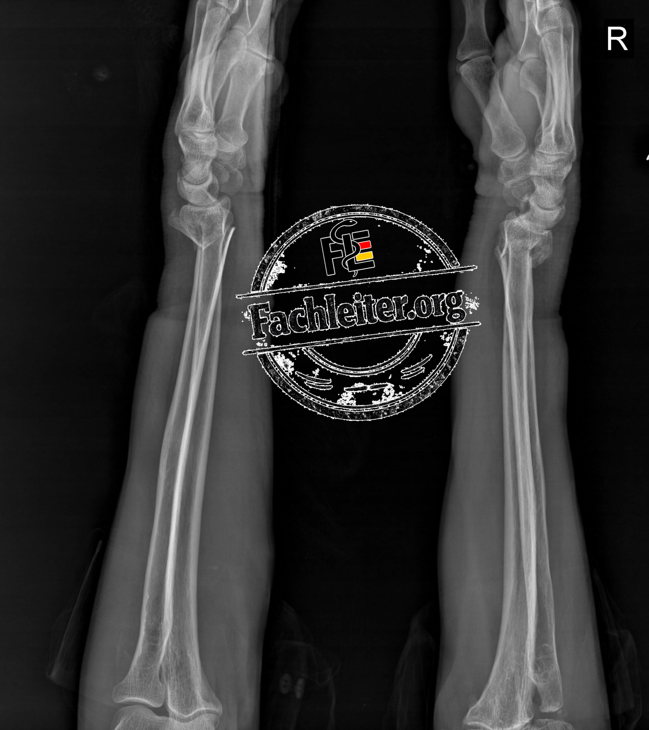
Patient (24 years) after high-energy trauma with pain and deformity of the forearm.
Your task
- Which bones are affected?
- In which segment are the fractures located?
- Is surgical treatment likely?
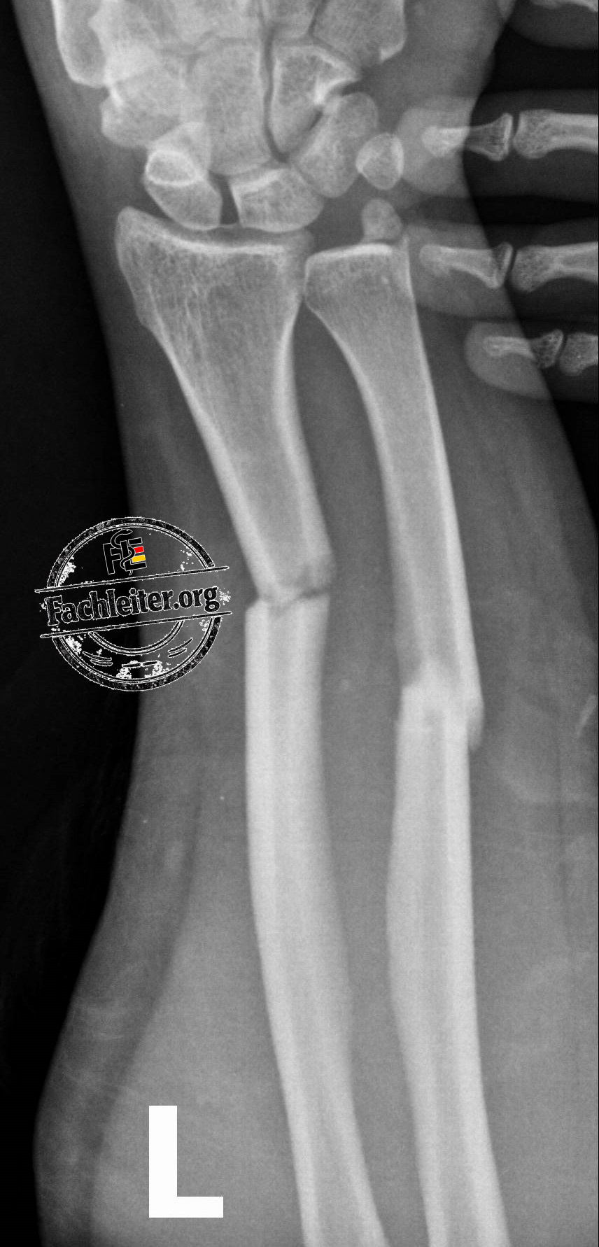
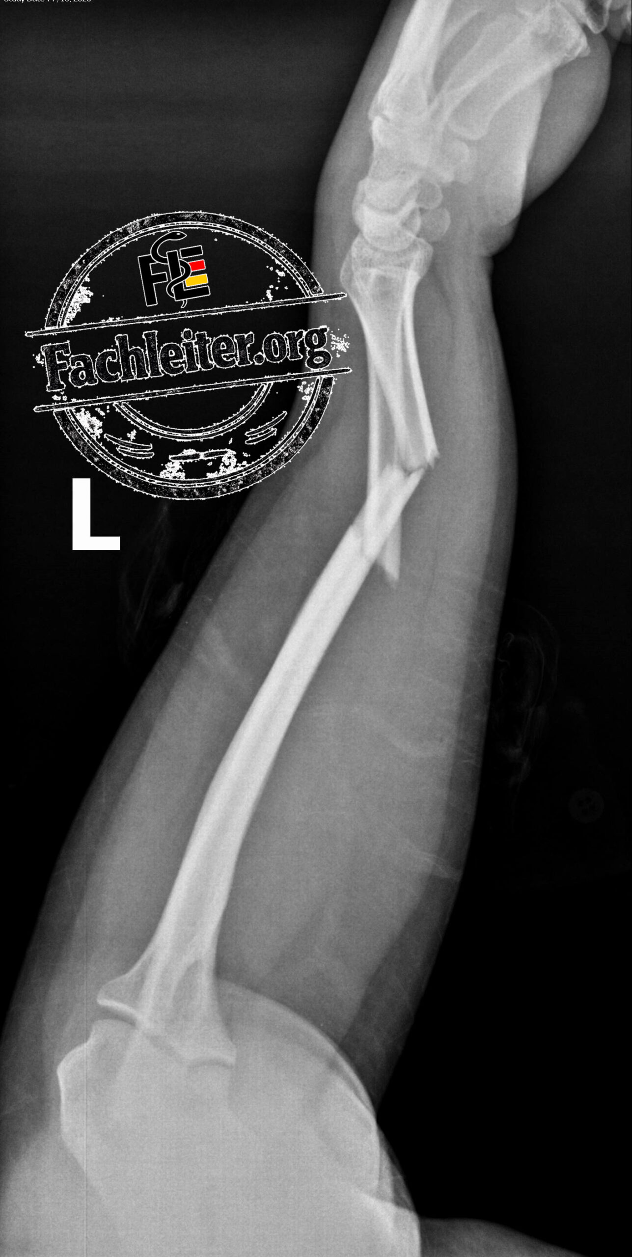
Radiological description
- Location: distal third of the radial and ulnar shafts
- Fracture type radius: transverse fracture – fracture line almost perpendicular to the bone axis.
- Fracture type ulna: short oblique fracture – fracture line slightly oblique to the bone axis.
- Completeness: complete fractures of both bones – both cortices are fully disrupted.
- Displacement: present (translation and slight angulation) – the fragments are displaced relative to each other and slightly tilted.
- Shortening: mild to moderate – due to overlap of the fragments.
- Associated finding: soft tissue swelling.
- Carpal bones: normal alignment, no definite fracture detected.
- Radiocarpal joint and distal radioulnar joint: normal joint alignment, no dislocation visible.
Fachleiter
Case 3
Acute wrist trauma after a fall.
Your task
- Is it the same fracture pattern?
- Which additional structures should be assessed?
- Is the distal radioulnar joint normal?
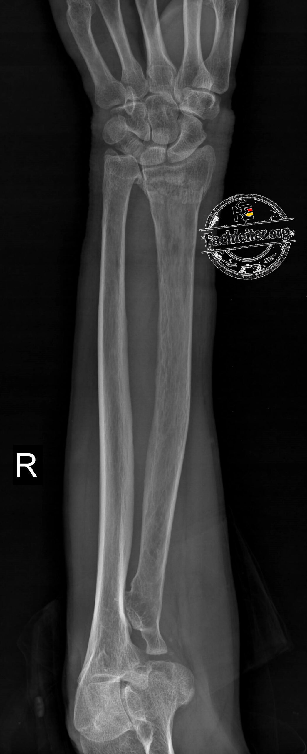
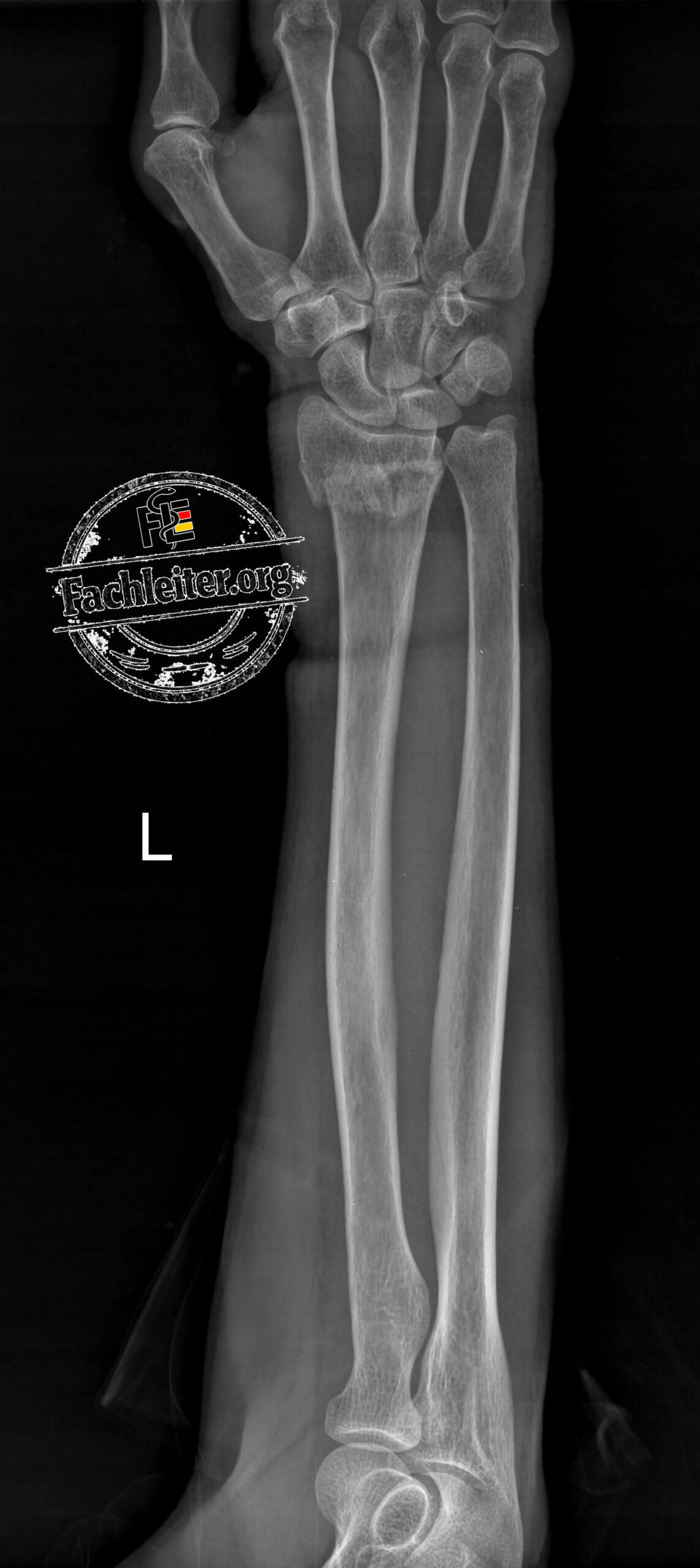
- Location: distal radial metaphysis – the fracture is located immediately proximal to the joint surface.
- Fracture type: transverse fracture (metaphyseal) – the fracture line runs almost transverse through the distal metaphysis.
- Completeness: complete fracture – the fracture line fully transects both cortices.
- Displacement: present (translation and marked dorsal angulation) – the distal fragment is displaced dorsally and tilted.
- Shortening: slight – due to impaction and fragment overlap there is a minor loss of radial length.
- Joint involvement: none – the fracture does not extend into the radiocarpal joint surface.
- Associated finding: soft tissue swelling in the area of the distal forearm.
Which bones are most commonly superimposed on an a.-p. view of the wrist?
- Os trapezium ↔ Os trapezoideum
- Os capitatum ↔ Os hamatum (partially)
- Os pisiforme ↔ Os triquetrum (especially in the AP view)
Fachleiter
Case 4
👧 8-year-old girl
- Fall while playing
- Fell onto an outstretched hand
- Pain in the right wrist
- Swelling
- No open injury
Your task
- Which bone is fractured?
- In which segment is the fracture located?
- Is there displacement?
- Conservative or operative treatment?
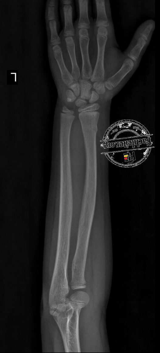
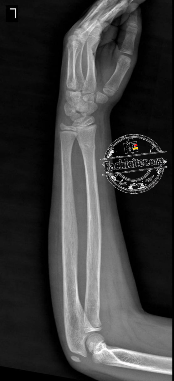
Radiological description
Localization: Distal radial metaphysis – the fracture lies immediately proximal to the distal growth plate.
Fracture type: Buckle fracture (torus fracture) – local cortical bulging or compression of the cortex without a complete fracture line.
Completeness: Incomplete fracture – the cortex is only compressed and not fully disrupted.
Dislocation: None – the bone axis is preserved, there is no relevant fragment displacement.
Shortening: None – the length of the radius is preserved.
Associated finding: Mild soft tissue swelling in the area of the distal wrist. No evidence of concomitant ulnar injury or joint involvement.