
Case description
A 20-year-old patient presents to the emergency department with 3 days of pain in the right hemithorax.
The pain began suddenly, is sharp, pleuritic, and worsens when lying down. The patient reports slight improvement when sitting.
History
- Right-sided chest pain for 3 days
- Pain character: sharp
- Worse with deep inspiration
- Worse when lying down
- Slight improvement when sitting
- Pain intensity: 6/10
Associated symptoms
- ❌ No shortness of breath
- ❌ No fever
- ❌ No cough
- ❌ No recent cold in the past weeks
- ❌ No trauma
- ❌ No syncope
- ❌ No palpitations
Autonomic history
- Unremarkable
Past medical history
- None known
Previous surgeries
- None
Medications
- No regular medications
Allergies
- None known
Substance use
- Non-smoker
- No alcohol or drug use
Family history
- Unremarkable
Vital signs
- Blood pressure: 124/76 mmHg
- Heart rate: 75/min
- Respiratory rate: 15/min
- Oxygen saturation: 99% (room air)
- Temperature: 36.8 °C
Physical examination
- General condition good
- Alert and oriented
- Heart: regular rhythm, no pathological murmurs
- Lungs: bilateral vesicular breath sounds, no crackles or wheeze
- No chest wall tenderness
- No leg edema
- No signs of deep vein thrombosis
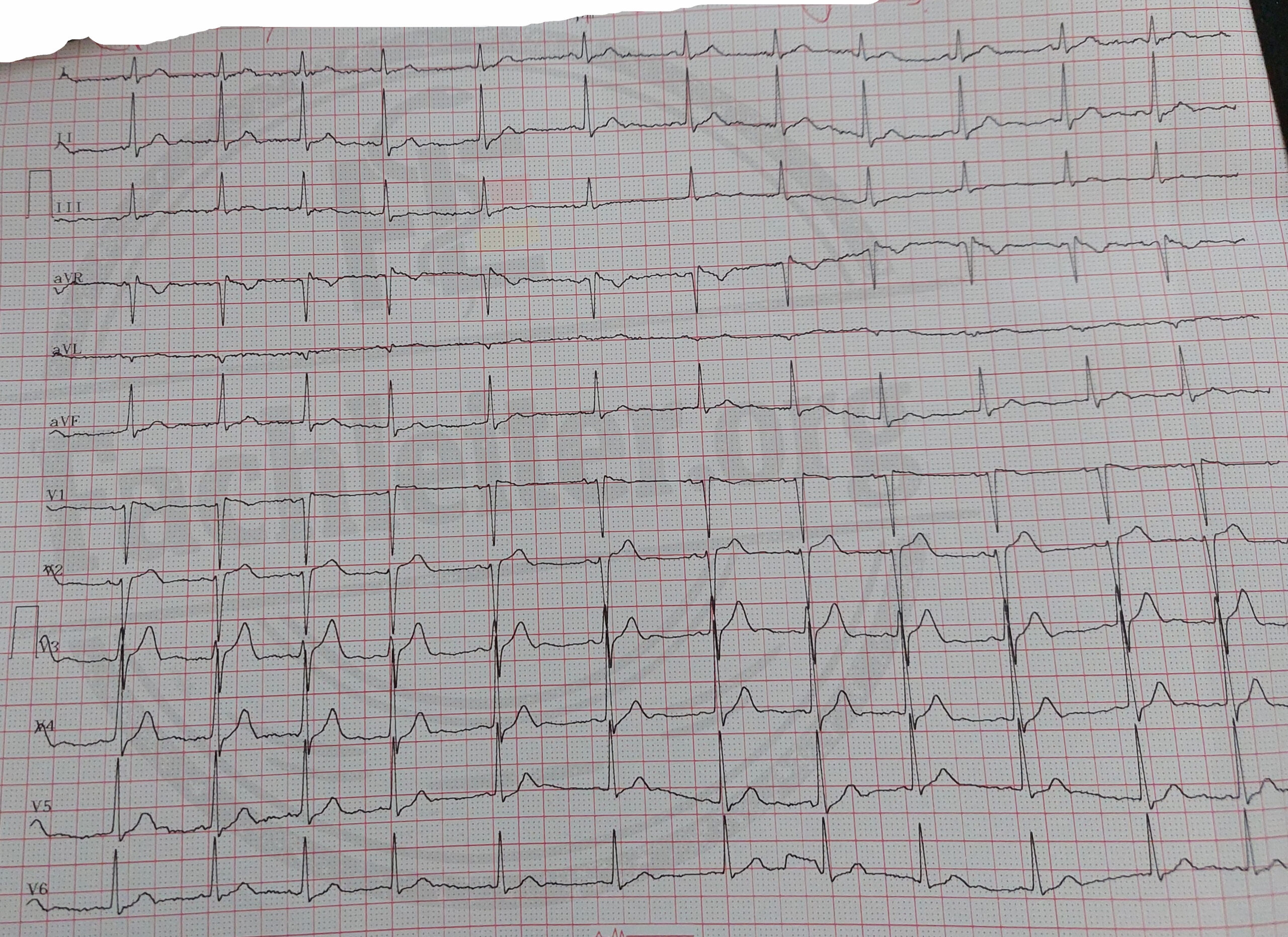
Quiz
1. Which suspected diagnosis is most likely based on the history and the ECG?
A) Acute coronary syndrome
B) Acute pericarditis
C) Pleuritis
D) Spontaneous pneumothorax
E) Pulmonary embolism
2. You work in a general practice and initially have only an ECG available. How would you proceed?
A) Reassure and send home
B) Symptomatic therapy and re-presentation if needed
C) Immediate referral to the emergency department for further diagnostics
D) Prescribe antibiotics and treat as outpatient
E) Arrange only a follow-up ECG in one week
3. Which further investigations would be most useful in the emergency department?
A) Chest X-ray
B) Echocardiography
C) Labs (troponin, CRP, D-dimer depending on suspicion)
D) CT angiography if indicated
E) All of the above investigations may be useful depending on the suspected diagnosis.
4. Which diagnosis cannot be confidently excluded with a normal ECG alone?
5. Which findings in the history argue against an acute coronary syndrome?
6. Which findings point more towards pericarditis or pleuritis?
7. Which warning signs would require immediate hospital referral?