
Children are not small adults – this also applies to the ECG. Heart rate, intervals, electrical heart axis and the morphology of individual leads change with age.
Do you see a difference from a typical adult ECG in these two ECGs?Case 1 – 14-year-old female patient
A 14-year-old female patient presents with headaches and flu-like symptoms.
Clinical information:
- Temperature: 37,9 °C axillary
- Tonsils slightly reddened
- Rhinorrhea
- otherwise no notable physical examination findings
EKG 1
Fachleiter.org
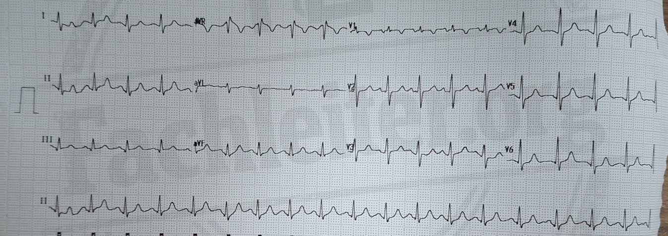
- Rhythm: R-R intervals regular.
- Heart rate: Distance between two R waves approx. 2,5 large boxes → 300 / 2,5 ≈ 120/min → tachycardia.
- QRS complex: narrow → supraventricular origin.
- P waves: positive in lead II and negative in aVR → sinus rhythm.
- Q waves: no pathological Q waves.
- T waves: no abnormal T waves.
- PR interval: within normal range.
- QT interval: with tachycardia only limited assessment based on absolute QT interval; consider QTc.
- ST segment: assessment hindered by higher heart rate; no clear pathological ST changes.
- R progression: normal, transition zone around V3.
Fachleiter
Case 2 – 11-year-old female patient
An 11-year-old female patient presents with mild shortness of breath and nausea.
No known cases of gastroenteritis or influenza in the environment. On physical examination she appears tense and stressed. No other relevant abnormalities are noted.EKG 2
Fachleiter.org
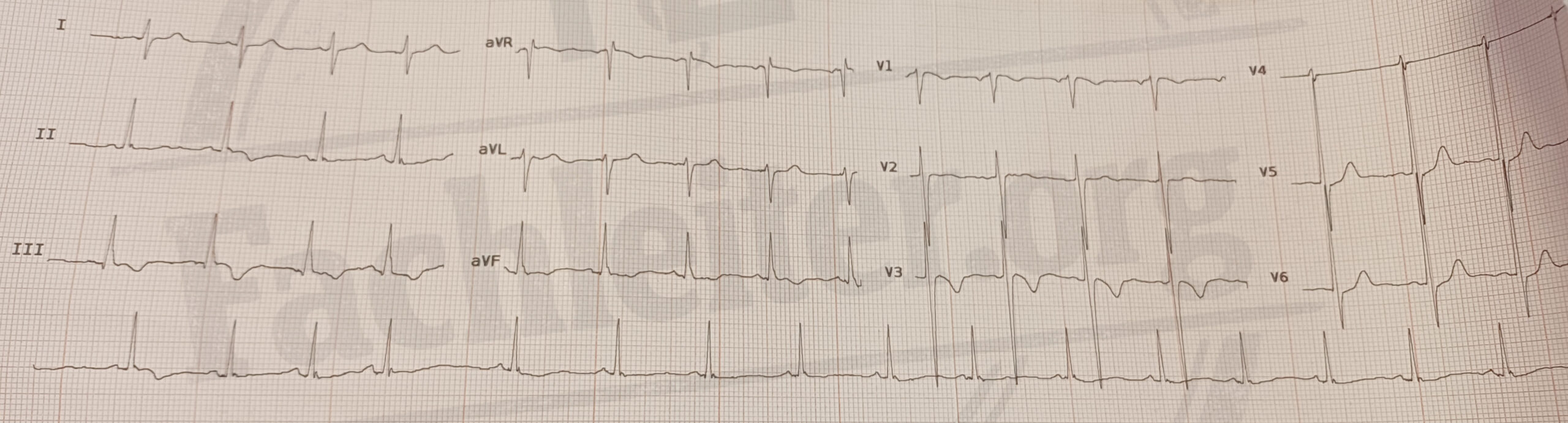
- hythmus: The R-R intervals appear irregular at first glance. The fluctuations are however respiratory-dependent and correspond to a respiratory sinus arrhythmia with shortening of the R-R intervals on inspiration and lengthening on expiration.
- Heart rate: 16 QRS complexes within 57,5 large boxes (= 11,5 seconds at 25 mm/s) → heart rate approx. 83/min.
- P waves: positive in lead II and negative in aVR → sinus rhythm.
- PR interval: within normal range.
- QRS complex: narrow, no pathological Q waves.
- R progression: normal.
- ST segment: no pathological ST-segment changes.
- T waves: no pathological T-wave changes.
- QT/QTc: within normal range.
Fachleiter
Children’s ECG vs. Adult ECG
The younger a child is, the more their normal ECG findings can differ from the adult ECG. With increasing age the ECG gradually approaches the typical adult pattern.
In 11- and 14-year-olds many ECG parameters are already relatively similar to adults. Nevertheless age-dependent reference values must be considered during interpretation.
Note: A finding that appears abnormal in adults can be physiological in children and adolescents – and vice versa.Fachleiter
Do you see an abnormal ECG finding? Ask yourself first:
- Are the electrodes placed correctly?
- Could the finding be caused by artifacts or electrical interference?
- Is the abnormality visible in at least two neighboring leads?