Approach
In ventricular tachycardia, pulse, hemodynamic stability and rhythm morphology determine acute management. This overview shows treatment step by step — with drug dosages, shock energies and repetitions based on the . You can switch between full and short versions depending on learning needs.
Ventricular tachycardia — acute management
First distinguish: Pulse present or pulseless? Hemodynamically stable or unstable? Monomorphic or polymorphic VT?
The recommendations apply to adults under continuous monitoring and with immediate resuscitation readiness. Basis: ERC Guidelines 2025; observe local hospital treatment standards, product information and device-specific energy recommendations.
Immediate actions
1. Assess the patient
Call for assistance, assess consciousness and breathing; if cardiac arrest is suspected, check the pulse for no more than ten seconds.
If unconscious, not breathing normally and pulse absent or not reliably palpable, begin CPR immediately.
2. Monitor and prepare
Attach defibrillator pads; monitor ECG, blood pressure and SpO₂. Establish intravenous access and treat reversible causes.
Obtain a 12-lead ECG if possible, without delaying necessary electrical therapy. Give oxygen for hypoxaemia.
Particularly look for ischaemia, hypoxia and electrolyte disturbances.
Monomorphic VT with pulse — unstable
1. Immediately perform synchronized cardioversion
Initial 120–150 J biphasic. If unsuccessful, increase energy; initially up to three attempts.
If circulation is definitely present, do not perform routine chest compressions.
Signs of instability: Hypotension with hypoperfusion, impaired consciousness or syncope, myocardial ischaemia or acute severe heart failure.
For conscious patients, provide sedation or short general anaesthesia by a qualified team without delaying urgent cardioversion.
Before each shock check SYNC and R-wave markers. If unsuccessful, incrementally increase energy up to the device maximum.
If synchronization cannot be achieved rapidly in critical instability, deliver an unsynchronized shock.
2. Give an antiarrhythmic
Amiodarone 300 mg IV over 10–20 minutes.
OR Procainamide 10–15 mg/kg IV over 20 minutes, maximum 1 g.
Choice guided by contraindications, local availability and SOP. The drugs are alternatives, not routinely combined.
Avoid procainamide in severe heart failure, acute myocardial infarction or end-stage renal failure.
3. Cardioversion again, synchronized
Repeat shock with appropriately increased energy; check SYNC.
After amiodarone consider 900 mg over 24 hours.
Continuously reassess circulation and rhythm. If pulse is lost, immediately enter the resuscitation algorithm.
When circulation is present there is no fixed two-minute CPR interval between cardioversion attempts.
Monomorphic VT with pulse — stable
1. Choose electrical or medical treatment
With structural heart disease or unclear cardiac substrate: synchronized cardioversion, initial 120–150 J biphasic.
Consider medical treatment especially if there is a high risk from sedation or anaesthesia.
For conscious patients, sedation or short general anaesthesia is required for cardioversion. If the shock fails, incrementally increase energy.
2. Antiarrhythmic under monitoring
Procainamide 10–15 mg/kg IV over 20 minutes, maximum 1 g.
If unsuitable or unavailable: Amiodarone 300 mg IV over 10–60 minutes, then if necessary 900 mg over 24 hours.
Avoid procainamide in severe heart failure, acute myocardial infarction or end-stage renal failure.
Do not give routine rapid amiodarone boluses according to the resuscitation regimen when pulse is present.
3. Synchronized cardioversion
Treat instability immediately with electricity; if pulse is lost, perform CPR and unsynchronized defibrillation.
Do not attempt further drug trials if they would delay necessary electrical therapy.
Sustained polymorphic VT
1. Immediately defibrillate unsynchronized
High defibrillation energy per manufacturer recommendation. If pulseless, start CPR immediately as well.
Reliable synchronization is usually not possible in polymorphic VT. Do not wait for synchronization.
2. Assess the cause and the QT interval
Treat ischaemia and electrolyte disturbances. If QT interval is prolonged, apply the torsade de pointes protocol.
QT interval is assessed in rhythm outside the VT. Polymorphic VT is not automatically torsade de pointes.
Torsade de pointes — prolonged QT interval
1. Electrical therapy for ongoing episode
Sustained polymorphic VT — defibrillate immediately unsynchronized; if pulseless, CPR.
The following magnesium administration must not delay necessary defibrillation.
2. Give magnesium and treat triggers
Magnesium sulfate 2 g ≈ 8 mmol magnesium IV over ten minutes. Repeat once if needed.
Correct potassium deficit and stop QT-prolonging drugs. Avoid amiodarone.
Magnesium can be effective even with normal serum magnesium. Amiodarone may further prolong the QT interval.
3. Increase heart rate under specialist care
Consider temporary overdrive pacing.
Further measures under cardiology or intensive care leadership and depending on the cause of QT prolongation.
Pulseless VT / ventricular fibrillation
1. Start CPR and deliver the first shock
Unsynchronized defibrillation: Energy per manufacturer recommendation; with common biphasic devices initially at least 150 J.
If energy recommendation is unknown use the device maximum energy.
With pulsed-biphasic devices initially 130–150 J. If further shocks fail, increase energy if possible.
Continue CPR while preparing and charging; minimize interruption for shock delivery.
2. Two minutes of CPR
Then a brief rhythm check. If VF or pulseless VT persists: second shock.
Immediately after a shock do not routinely check the pulse. If there are clear signs of restored circulation, assess accordingly.
3. Another two minutes of CPR
Then check the rhythm. If ventricular fibrillation or pulseless VT persists: third shock.
Continue vascular access, airway management and treatment of reversible causes during CPR.
4. Give amiodarone and adrenaline
Amiodarone 300 mg i.v./i.o.
Adrenaline 1 mg i.v./i.o., then every three to five minutes repeat.
After two minutes, check the rhythm; if ventricular fibrillation or pulseless VT persists fourth shock.
Give drugs during chest compressions. Administration must not delay CPR or necessary shocks.
5. Two minutes of CPR and rhythm check
If ventricular fibrillation or pulseless VT persists: fifth shock.
Continue giving adrenaline at three- to five-minute intervals and treat reversible causes.
6. Give a second dose of amiodarone
Amiodarone 150 mg i.v./i.o. additional.
Continue treatment in two-minute cycles; only defibrillate again if the rhythm remains shockable.
If an organized rhythm is present, assess for signs of circulation. If PEA or asystole, switch to the non-shockable algorithm; if ROSC, begin post-resuscitation care.
Alternative to amiodarone for ventricular fibrillation or pulseless VT: Lidocaine 100 mg i.v./i.o. after the third shock, further 50 mg after the fifth shock. Use as an alternative, not routinely in addition to amiodarone.
If pulse is lost: Any VT with a pulse can deteriorate into pulseless VT. Then immediately perform CPR and unsynchronised defibrillation.
Special peri-arrest situation: In closely monitored patients with a persistent systolic blood pressure below 50 mmHg despite treatment, chest compressions may already be considered.
Exception in the sequence of shocks: Up to three consecutive immediate shocks are only appropriate for witnessed, monitored cardiac arrest with an immediately available defibrillator. For drug administration, this initial series counts as the first shock of the ALS algorithm.
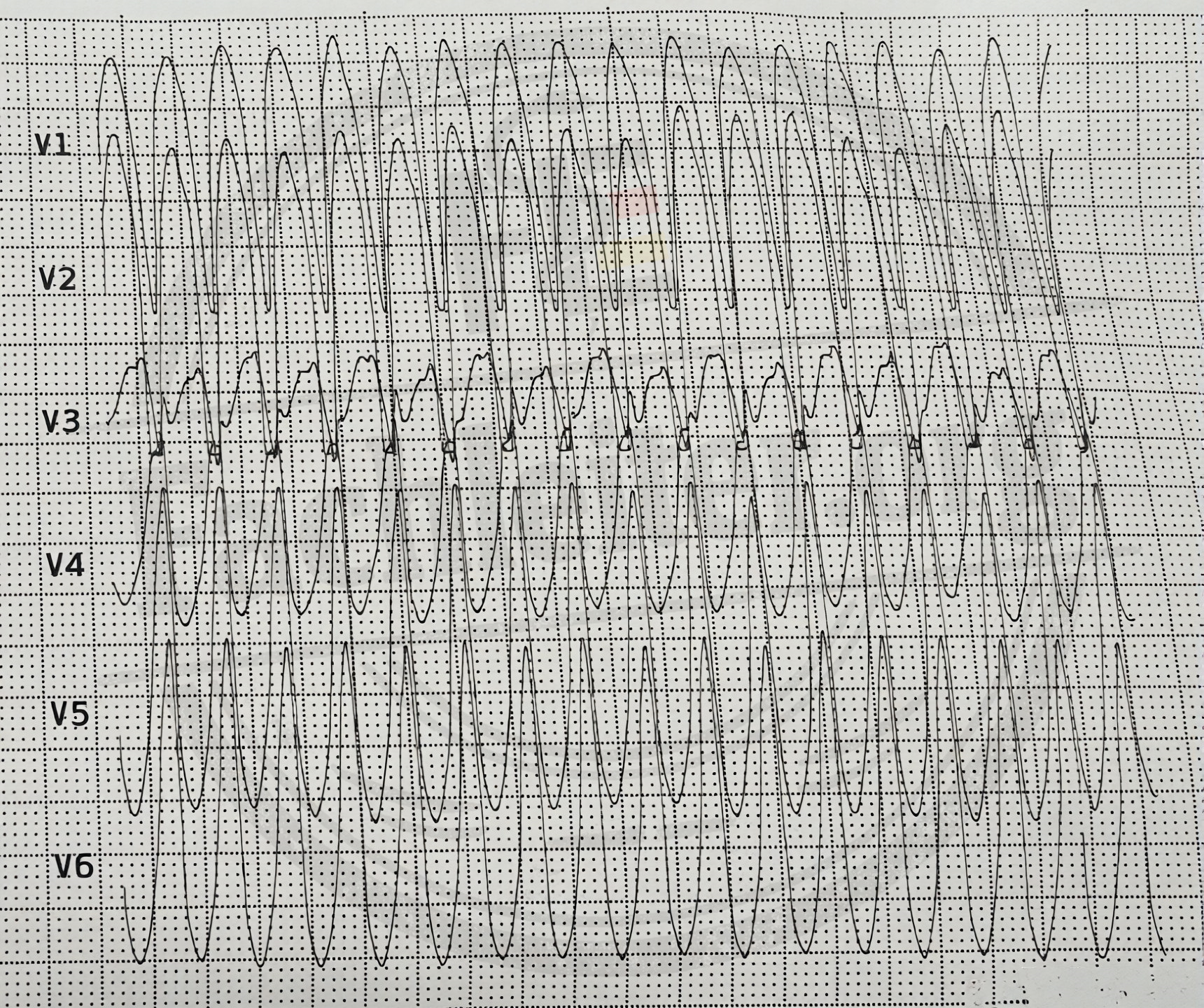
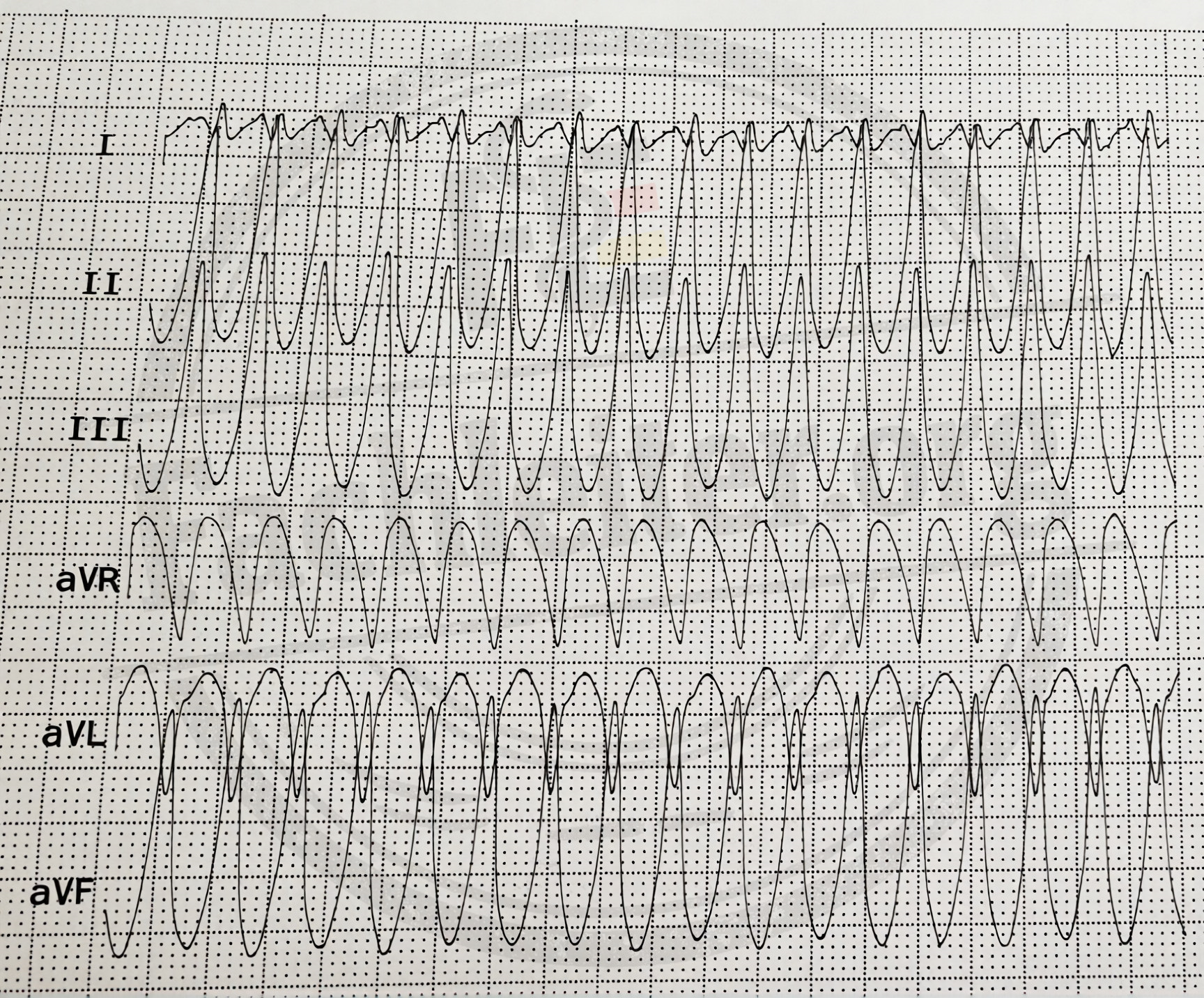
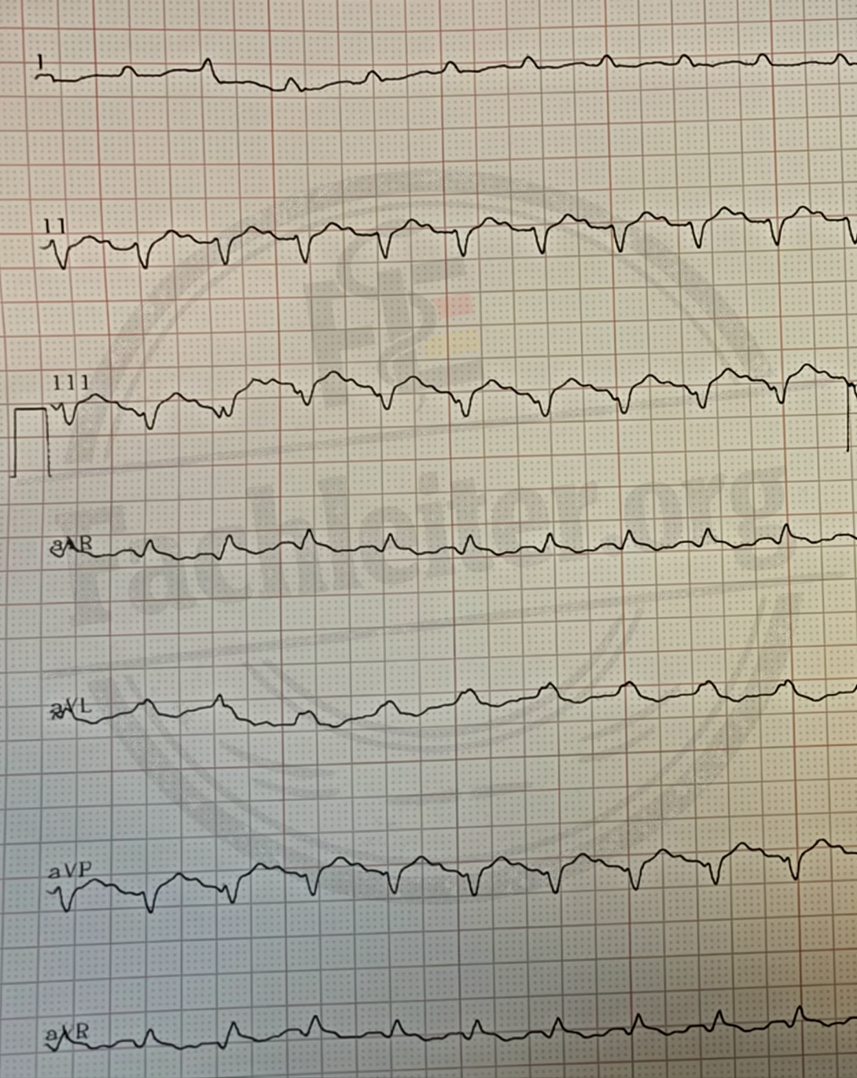
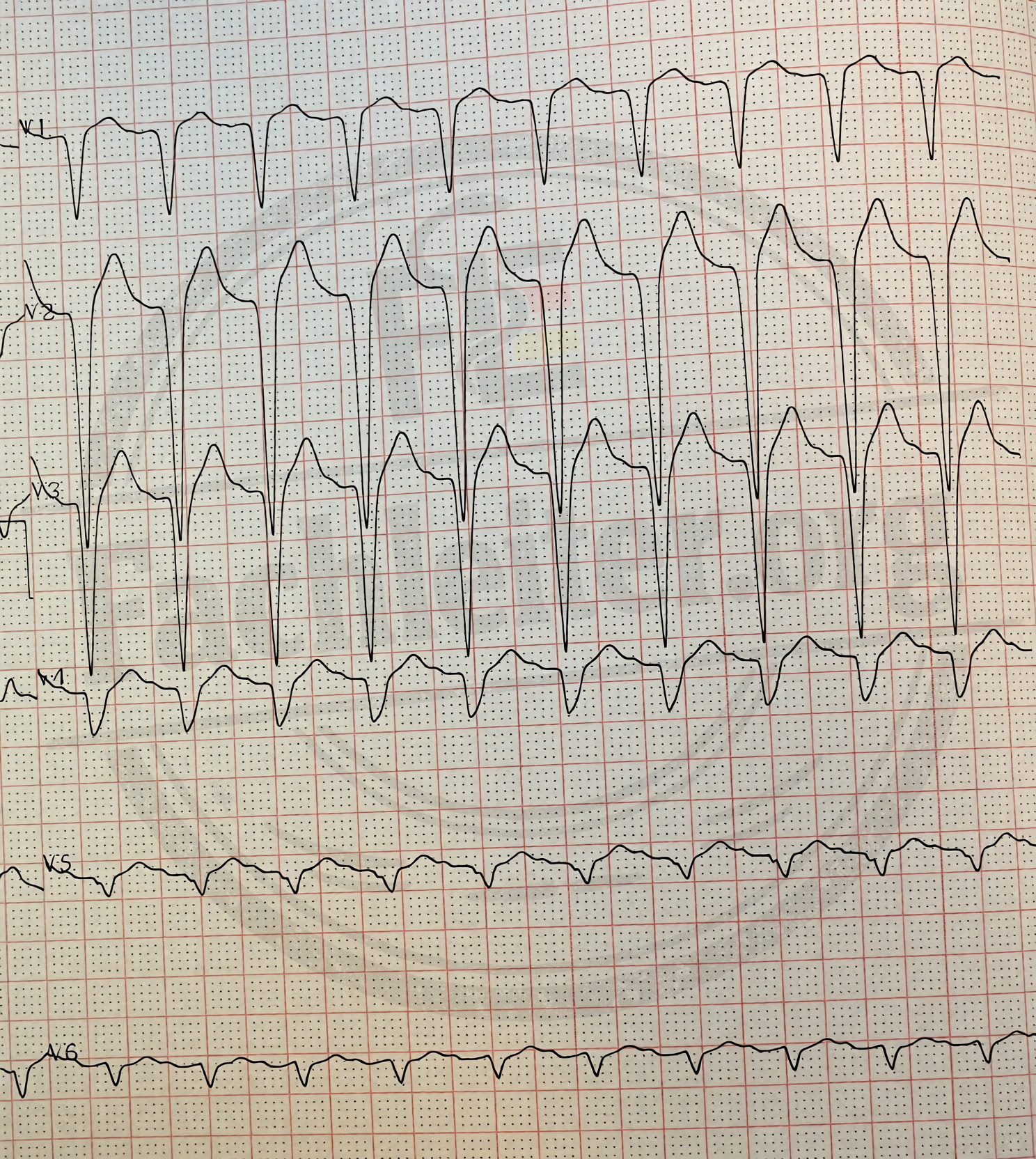
ECGs
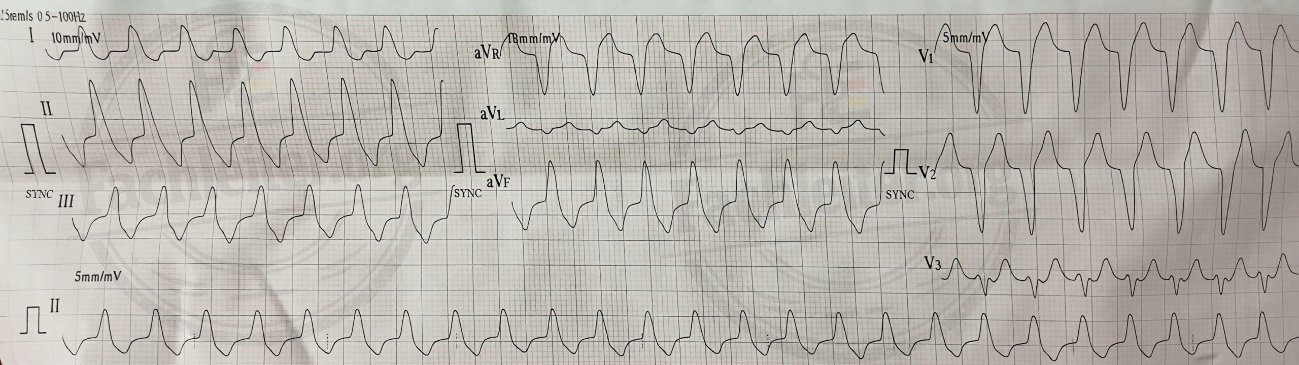
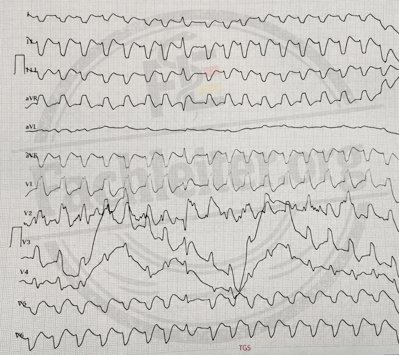
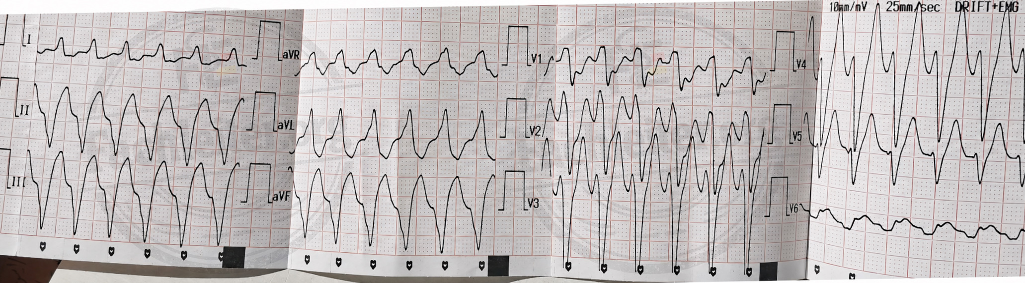
Below you will see several ECGs demonstrating a ventricular tachycardia pattern. Interpret the ECGs, paying particular attention to heart rate, rhythm regularity, QRS width and morphology, and signs of AV dissociation, capture beats or fusion beats. Also distinguish between monomorphic and polymorphic VT.
Also decide how you should proceed best.