
Greeting
Good day, dear Professor Doctor …,
I would now like to present the patient Thomas Münter to you.
He is 45 years old, one meter seventy-four tall and weighs about 68 kg. The body mass index (BMI) is approximately 22.4 kg/m² and is therefore within the normal range.
Current history
The patient presented because of an abscess in the left antecubital region, secondary to intravenous drug use.
The abscess has been present for about one week and has progressively worsened; it is approximately the size of a walnut.
Accompanying constitutional symptoms have now developed, including fatigue, nonspecific cephalgias, myalgias, fever as well as night sweats.
The fever is intermittent.
In addition there is a dry cough, which occurs mainly when lying down and during physical activity.
Cardiac symptoms include exertional palpitations and dyspnea .
The patient reported having to stop after climbing about one and a half flights of stairs and needing one to two minutes to recover until his breathing calmed down.
Gastrointestinally there is loss of appetite and nausea.
On the nails of both hands there were splinter hemorrhages.
The following symptoms were denied:
Osler nodes, Janeway lesions, loss of consciousness, chest pain or tightness, edema, abdominal pain, vomiting, low back pain, hematuria, lymphadenopathy as well as joint involvement.
There are no signs of splenomegaly.
. pharyngitis.
. intravenously heroin for about three years. Administration is usually once or twice daily with a total amount of about 1 g per day. Sterile syringes were not used; he typically uses boiled water and vitamin C powder to dissolve it.
Autonomic history
In the autonomic history there is loss of appetite, constipation and insomnia manifesting as sleep maintenance difficulties due to night sweats.
If needed, the patient also takes valerian tablets for stress.
Pre-existing conditions
His known pre-existing conditions are the following:
About one year ago during an inpatient stay a hepatitis B was diagnosed. Specific antiviral therapy has not been administered so far.
About eight years ago the patient suffered a fracture of the left foot after a car accident, which was treated conservatively with a cast immobilization for about six weeks. The patient was hospitalized for a total of about five days.
Concomitantly there was a concussion with transient dizziness and loss of consciousness.
Overall the injury healed without complications; however, occasional pain still occurs once or twice monthly.
Previous surgeries
About one year ago the patient was hospitalized for about ten days due to a fracture of the right arm and a fracture of the fifth rib on the right.
The right arm was surgically treated with plate and screw osteosynthesis.
About six months later the hardware was removed. Since then the patient has been symptom-free.
Note: When a statement can be objectively confirmed by a medical examination — for example by visible scars — the indicative is appropriate. (No physical examination takes place during the specialist language exam.)
Gynecology
————–
Medications
His prescription lists the following:
Voltaren 50 mg
Valerian tablets
————
The patient reported having received one vaccination against COVID-19.
Note: When a statement can be objectively confirmed by a physician’s observation — for example based on a prescription — the indicative is appropriate.
Allergies
The patient has a known allergy to amoxicillin, manifesting as urticaria and dyspnea. It was prescribed for a pneumonia; he subsequently received azithromycin.
Noxae
The patient has been a smoker for about 15 years and smokes about one pack of cigarettes daily, corresponding to approx. 15 pack-years.
. joint.
Social history
The patient has been divorced for about five years, has no children and has been unemployed for around three years. Previously he worked as a construction worker Additionally, there is no complete health insurance.
Family history
The patient’s mother died at the age of 52 from a breast carcinoma.
There is no information about diseases of the father because there is no contact.
He has a dog.
Suspected and differential diagnosis
In my opinion, the anamnesis data most strongly suggest an endocarditis. The intravenous heroin use as well as the clinical course with multisystem symptoms fit this clinical picture.
With a pericarditis one would typically expect acute, respiration-dependent chest pain that improves when sitting forward.
With a myocarditis we often see exertional dyspnea, palpitations, chest discomfort and rhythm disturbances.
Rheumatic fever typically occurs after an untreated infection with β-hemolytic group A streptococci, usually 1–3 weeks after tonsillopharyngitis. One would expect migratory polyarthritis, carditis, chorea minor or skin manifestations.
A tuberculosis is less likely in this case because no chronic cough, no hemoptysis, no relevant weight loss and no pulmonary focal signs have been described so far. In addition, the overall clinical context with intravenous drug use and a subacute course fits an infectious endocarditis better than tuberculosis.
Diagnostic workup
With suspected infectious endocarditis, hospital admission is required because prompt diagnostic confirmation by multiple blood cultures and echocardiographic examinations is necessary and potentially life-threatening complications such as septic emboli, valvular destruction, heart failure or arrhythmias must be recognized and treated early. In addition, intravenous antibiotic therapy with close clinical and laboratory monitoring is required.
physical examination
Following the medical history, a thorough physical examination should be performed. This begins with recording the vital signs, including heart rate, blood pressure, respiratory rate, oxygen saturation and temperature. Thereafter we perform a focused clinical examination.
Skin and nails should be carefully examined for nonspecific embolic signs such as petechiae, Osler’s nodes, Janeway lesions and splinter hemorrhages. Petechiae are often found on the conjunctivae, palate and extremities; Osler’s nodes are painful on the finger and toe pads, Janeway lesions are painless on the palms and soles, and splinter hemorrhages are linear hemorrhages in the nail bed.
Next, percussion is performed, particularly to exclude or detect splenomegaly.
Finally, auscultation is performed with careful cardiac examination to detect systolic or diastolic, particularly newly arisen murmurs as well as signs of heart failure; additionally the lungs are auscultated.
A thorough neurological examination should be performed to detect focal neurological deficits and at the same time to document a baseline finding for the further in-hospital course; this includes assessment of level of consciousness, cranial nerves, motor function, sensation, coordination, reflexes and gait. In addition, a fundoscopic examination should be performed to detect Roth spots.
Laboratory investigations
Laboratory tests for suspected infectious endocarditis include a complete blood count, inflammatory markers (CRP, ESR), renal retention parameters (creatinine, urea), liver enzymes, electrolytes and a urinalysis to detect proteinuria or hematuria.
To clarify viral infections, serological tests are ordered: for hepatitis B the determination of HBs antigen, anti-HBs and anti-HBc (if necessary HBV-DNA), for hepatitis C the anti-HCV antibody with possible subsequent HCV-RNA determination and for HIV a combined HIV antigen/antibody screening test (if necessary with confirmatory test).
The gold standard for diagnosing infectious endocarditis is pathogen detection from a heart valve, which is usually not possible in everyday practice, so the diagnosis is made clinically.
Blood culture
At least three sets of blood cultures should be taken from different puncture sites, ideally (although this is not always feasible in critically ill patients) at intervals of about one hour.
In most patients with infectious endocarditis who have not previously received antibiotic therapy, all blood cultures are positive because bacteremia in endocarditis is continuous.
A blood culture is a laboratory test used to detect bacteria or fungi in the blood in order to identify the cause of a bloodstream infection. Under strict sterile conditions, blood is drawn from a vein and filled into special aerobic and anaerobic culture bottles. Usually several blood cultures are taken from different puncture sites, preferably before the start of antibiotic therapy. For this one needs sterile blood culture bottles (aerobic and anaerobic), sterile needles or a blood collection system, disinfectant for skin and bottle membranes, sterile gloves as well as appropriate labeling and transport materials.
Electrocardiogram
The electrocardiogram (ECG) should be carefully examined for signs of atrioventricular conduction disturbances, particularly a prolonged PR interval, as this may indicate an aortic ring abscess or a myocardial infarction.
An electrocardiogram (ECG) is a non-invasive test to record the electrical activity of the heart. It is performed by placing multiple electrodes on the chest, arms and legs of the patient; these measure the electrical signals during cardiac action and display them as tracings on a monitor or printout.
Anatomical diagnosis
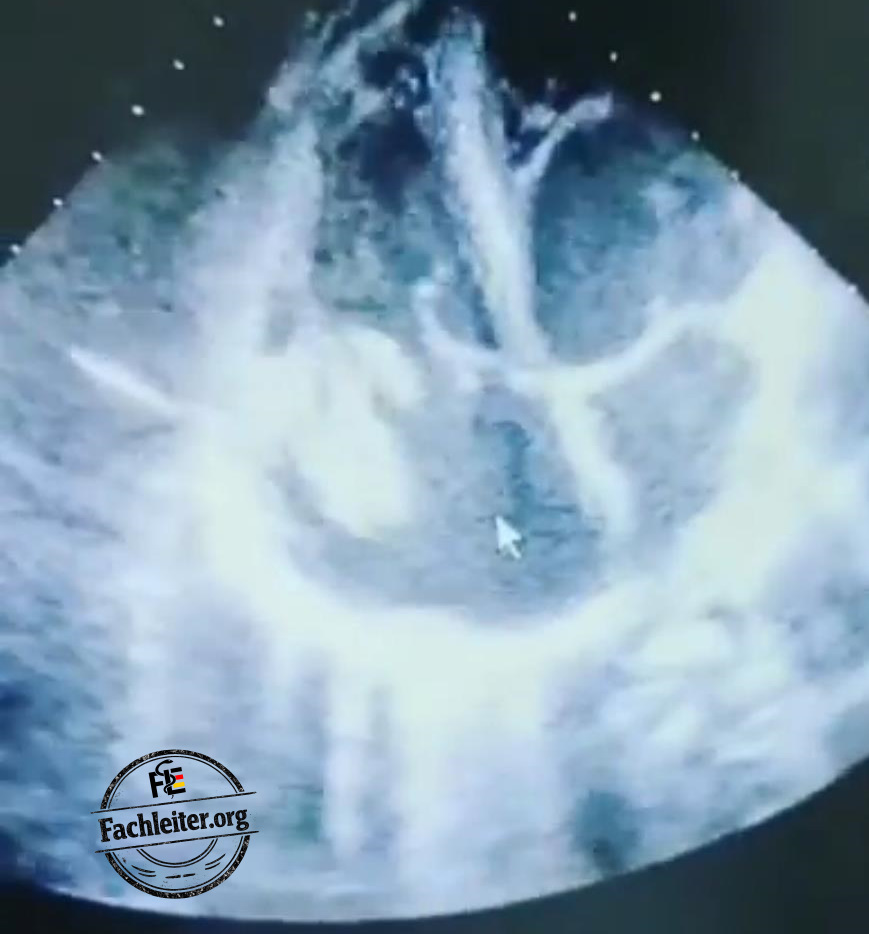
Echocardiography
Echocardiography is an imaging technique for assessing cardiac structure and function, wall motion and movements of the heart valves using ultrasound. For transthoracic echocardiography (TTE) no special preparation is required. The patient usually lies in the left lateral position while the transducer is placed on the chest wall to display various cardiac planes. Among other things, the ejection fraction (EF) can be measured, an important parameter for assessing left ventricular pump function.
An echocardiogram, also called cardiac ultrasound, will show that the patient either has a reduced ejection fraction (less than 40%), a moderately reduced ejection fraction (between 40 and 50%) or a preserved ejection fraction.
(For transesophageal echocardiography (TEE) the patient must be fasting for about six hours. Before the examination, information is provided and sedation is given if necessary. During TEE an ultrasound probe is introduced via the esophagus to obtain detailed images of the heart, particularly the heart valves and atria.)
X-ray
A chest X-ray is an imaging test that uses X-rays to depict the heart, lungs, ribs and mediastinum.
The examination is usually performed standing in two planes (PA and lateral); the patient holds their breath briefly while the image is taken. It is quick, painless and lasts only a few seconds.
Possible findings:
• Septic pulmonary emboli, especially in right-sided endocarditis (i.v. drug use): multiple peripheral infiltrates, possibly cavitations
• Signs of heart failure: pulmonary congestion, interstitial or alveolar edema
• Pleural effusions
Abdominal ultrasound
Abdominal ultrasound is an ultrasound examination of the abdomen that depicts organs such as the liver, spleen, kidneys, gallbladder and free fluid.
The examination is performed in the supine position; a transducer is applied to the abdominal skin with contact gel and moved across the abdomen. It is painless, radiation-free and usually takes a few minutes.
Possible sonographic findings:
• Splenomegaly, especially in subacute course
• Splenic infarcts or splenic abscesses due to septic emboli
• Liver enlargement in right heart failure
Treatment
Targeted antibiotic treatment of infectious endocarditis is based on susceptibility testing of the identified pathogen.
Until the definitive culture results are available, empirical therapy is not required for all patients with suspected endocarditis, but mainly for high-risk patients, for example in prosthetic heart valve carriers.
broad-spectrum empirical regimens
• Nafcillin (or Oxacillin) + Penicillin + Gentamicin
• Vancomycin + Gentamicin
→ in case of immediate-type penicillin allergy or MRSA risk
Consultations
• Cardiology consultation
• Infectious diseases consultation
• Cardiac surgery consultation
Duke criteria
|
Duke criteria for infectious endocarditis |
|
|
Category |
Criteria |
|
Major criteria |
Positive blood cultures with typical pathogens (Staphylococcus aureus, streptococci, enterococci) from ≥2 separate cultures or persistent bacteremia Endocardial involvement on echo (vegetations, abscess, new partial prosthetic dehiscence) or newly arisen valvular insufficiency |
|
Minor criteria |
Predisposition (e.g., IV drug use, valvular defect, prosthetic valve) Fever ≥ 38 °C Vascular phenomena (arterial emboli, Janeway lesions, splinter hemorrhages) Immunologic phenomena (Osler nodes, Roth spots, glomerulonephritis) Microbiological finding not meeting a major criterion |
|
Diagnosis |
Definitive Endocarditis: 2 major or 1 major + 3 minor or 5 minor Possible endocarditis: 1 major + 1 minor or 3 minor |
Complications:
Complications of infective endocarditis include direct valvular damage with consequent valvular insufficiency or stenosis as well as locally invasive spread such as paravalvular abscesses, fistulas or conduction disturbances. In addition, embolic complications can occur, for example cerebral or peripheral emboli and septic pulmonary emboli. Metastatic infections resulting from bacteremia include, among others, spondylodiscitis, osteomyelitis, splenic abscesses or septic arthritis. Immunologically mediated phenomena such as glomerulonephritis, Osler nodes or Roth spots may also occur.
Epidemiology
The majority of community-acquired infective endocarditis on native valves is caused by staphylococci, streptococci or enterococci, which belong to the normal flora of the skin, oropharynx and the urogenital and gastrointestinal tracts and regularly enter the bloodstream.
In patients without intravenous drug use or prior healthcare contact, streptococci are the most common pathogens, whereas when these risk factors are present, predominantly Staphylococcus aureus is detected.
Among streptococci, in particular viridans streptococci from the oropharynx are the leading causes of native valve endocarditis.
Group B streptococci, as β-hemolytic members of the oro- and urogenital flora, occur more frequently in patients with liver cirrhosis, diabetes mellitus and in intravenous drug users.
Fungal endocarditis is rare, diagnostically and therapeutically challenging, and occurs mainly with IV drug use, after valve surgery or with long-term intravascular catheters; the most common pathogens are Candida and Aspergillus.
Risk factors for infective endocarditis
• Intravenous drug use
• Preexisting valvular heart disease (e.g., valvular defects, bicuspid aortic valve)
• Valve prostheses or foreign material on the heart
• Previous endocarditis
• Intracardiac devices (e.g., pacemaker, ICD)
• Long-term intravascular catheters
• Frequent medical procedures or recent hospitalization
• Immunosuppression (e.g., HIV, steroid therapy, chemotherapy)
• Chronic diseases (e.g., diabetes mellitus, liver cirrhosis)
• Poor oral hygiene / dental infections
Extra
Names:
Gudrun Munte,
Details:
Oxy/ Morphine
Homeless
Pain in the left inguinal area (abscess/ phlebitis)
Intake of paracetamol: no effect
Past medical history:
Hepatitis C for 10 years, untreated
Multiple abscess drainages on both arms due to injections; most recently about two years ago.
Osteosynthesis for left radius fracture about three years ago.
Daily methadone intake as part of a substitution program for …………. months.
Liver biopsy is planned.
Questions
1. What do I have and what is it?
You most likely have an inflammation of the inner lining of the heart, called endocarditis. Bacteria have attached to a heart valve or the inner heart wall and are causing an infection there.
2. Why did I get this?
The disease occurs when bacteria enter the bloodstream and settle on the heart. This can happen with infections, via injections or in the presence of existing risk factors. In your case, intravenous drug use and previous infections favor the occurrence of this disease.
3. Should I stay in the hospital?
Yes, inpatient treatment is necessary because we must closely monitor the infection, treat it specifically and detect possible complications early. Therapy is generally with several weeks of intravenous antibiotics.
4. What is sepsis? What symptoms occur with sepsis?
Sepsis is a life-threatening body response to infection, resulting in organ dysfunction.
Typical symptoms are fever or low body temperature, chills, tachycardia, rapid breathing, low blood pressure, altered consciousness, decreased urine output and a general feeling of illness.
5. How is sepsis treated?
Treatment is immediate and inpatient, usually in the intensive care unit, and includes rapid administration of intravenous broad-spectrum antibiotics, fluid therapy, if necessary vasopressors, oxygen therapy and control of the infection source.
6. When must a patient with endocarditis be admitted to the ICU or CCU?
Admission to the ICU/CCU is required for hemodynamic instability or septic shock, acute heart failure due to severe valvular insufficiency, relevant arrhythmias or high-grade AV block, neurological complications such as stroke or altered consciousness, extensive paravalvular abscesses or fistulas, respiratory failure, multiorgan failure and for perioperative monitoring before or after cardiac surgery.
7. What complications does hypoalbuminemia have?
Hypoalbuminemia can lead to peripheral edema and ascites, hypotension, reduced drug binding, increased infection risk, impaired wound healing and worsening renal function.
8. What is liver cirrhosis?
Liver cirrhosis is the end stage of chronic liver diseases in which healthy liver tissue is replaced by connective tissue (fibrosis) and nodular restructuring, leading to impaired liver function and portal hypertension.
Causes:
• Chronic alcohol consumption
• Chronic viral hepatitis (HBV, HCV)
• Non-alcoholic fatty liver disease (NASH)
• Autoimmune diseases (autoimmune hepatitis, PBC, PSC)
• Toxic-drug induced
• Metabolic diseases (e.g., hemochromatosis, Wilson’s disease)
Complications:
• Portal hypertension with ascites, esophageal varices and splenomegaly
• Hepatic encephalopathy
• Hepatorenal syndrome
• Coagulation disorders
• Increased susceptibility to infections (e.g., spontaneous bacterial peritonitis)
Notes
BMI
Definition:
Body mass index (BMI) is a relative measure of body weight in relation to height. It is calculated by dividing body weight in kilograms by the square of height in meters.
Meaning and limitations:
BMI serves as a rough screening tool to classify underweight, normal weight and overweight in the adult population. It does not distinguish between fat and muscle mass and is less informative for children, pregnant women and very muscular individuals.
Abscess
Definition:
An abscess is a localized collection of pus in a tissue space, usually as a result of a bacterial infection.
Clinical features and causes:
Typical findings are a well‑defined swelling, pressure and throbbing pain, as well as redness and warmth over the focus; systemic accompanying symptoms can also occur. Common triggers are bacterial skin infections, small injuries, foreign bodies or inoculation through injections; intravenous drug use increases the risk of soft tissue abscesses.
Possible complications:
If untreated, the infection can spread into deeper tissue layers, lead to fistula formation or cause systemic infection up to sepsis.
Fatigue
Definition:
Fatigue denotes persistent, overwhelming exhaustion and decline in performance that go beyond normal tiredness and are not fully relieved by rest.
Cephalalgia
Definition:
Cephalalgia is the medical term for headache and includes various manifestations of pain in the head and facial area.
Classification and notes:
Headaches are broadly divided into primary forms (e.g. migraine, tension-type headache) and secondary causes (e.g. infection, vascular event, inflammatory processes). Acute, very severe or newly occurring headaches, as well as those with neurological deficits, are alarm symptoms and require further evaluation.
Myalgias
Definition:
Myalgias are pains in the muscles, which can be localized or diffuse.
Cough
Definition:
Cough is a reflex protective mechanism that clears mucus, foreign bodies or irritants from the airways. It can be dry (nonproductive) or with sputum (productive).
Palpitations
Palpitations
Subjective perception of increased, rapid or irregular heartbeats, often described as palpitations or pounding. The manifestation can range from harmless extrasystoles or stress‑induced tachycardia to organic causes such as arrhythmias or systemic diseases. Differentiation by duration, triggers and accompanying symptoms is important for further evaluation.
Dyspnea
Dyspnea
Subjective sensation of shortness of breath or difficulty breathing, which can occur on exertion or at rest. Causes can include cardiac diseases (e.g. heart failure), pulmonary disorders (e.g. obstructive or restrictive lung diseases), anemia or psychogenic factors; course and exertion dependence help with clinical classification.
Nausea
Nausea
Subjective feeling of sickness, often with the sensation of imminent vomiting, which does not necessarily lead to vomiting. Common triggers are diseases of the gastrointestinal tract, vestibular disorders, medications, metabolic disturbances or pregnancy.
Splinter hemorrhages
Splinter hemorrhages
Longitudinal, linear bleeding under the nail plate that corresponds with the direction of nail growth. They arise from small vessel injuries, microemboli or vasculitic processes and can occur in infectious endocarditis, but are not specific to that disease.
Osler nodes
Osler nodes
Small, painful, reddish‑violet nodules on the fingertips or toe tips that can be tender to pressure and are usually transient. Pathophysiologically they are associated with immunologically mediated vasculitis; classically they are described in infectious endocarditis but are not exclusively specific to it.
Janeway lesions
Janeway lesions
Painless, flat erythematous‑hemorrhagic skin lesions that preferentially occur on the palms and soles. Pathophysiologically they are explained by septic microemboli or organism‑associated microinfarcts of the skin. Clinically they are frequently associated with infectious endocarditis; in contrast to Osler nodes, Janeway lesions are not tender to pressure.
Edema
Edema
An edema is a pathological accumulation of fluid in the interstitium or in body cavities. Common mechanisms include increased hydrostatic pressure (e.g., in heart failure), decreased colloid osmotic pressure (e.g., hypoalbuminemia), lymphatic drainage disturbance, or increased vascular permeability. Clinically, peripheral edemas often show a pressure dent (depending on location and cause) and especially occur in the lower extremities with venous congestion. Edemas are a nonspecific sign and require investigation of the underlying cause.
Emesis
Emesis
Emesis denotes vomiting, i.e., the forced emptying of stomach contents through the mouth. It can be triggered by gastrointestinal diseases, intoxications, vestibular disorders, metabolic or central causes. Clinically relevant are possible consequences such as dehydration, electrolyte disturbances and the risk of aspiration, which is why the underlying cause should be investigated.
Low back pain
Low back pain
Low back pain denotes pain in the lumbar spine area or lower back. Causes range from mechanical and degenerative changes through disc pathologies to rarer infectious, inflammatory or malignant diseases. Important distinctions concern acute versus chronic course and the presence of red flags (e.g., neurological deficits, fever, weight loss) that require further diagnostics.
Hematuria
Hematuria
Hematuria denotes the presence of blood in the urine; it can be macroscopically visible (reddish‑brown or rust‑colored urine) or detectable only microscopically. Possible causes include urological infections, urinary stones, tumors, glomerular diseases and coagulopathies. The symptom is nonspecific but clinically significant and usually requires diagnostic clarification to determine the cause.
Splenomegaly
Splenomegaly
Splenomegaly denotes an enlarged spleen. It can arise from various causes, including infectious diseases, hematological disorders, portal hypertension or metabolic and storage diseases. Clinically, an enlarged spleen can cause a feeling of pressure in the left upper abdomen, a palpable finding below the rib cage and occasionally cytopenias in the blood due to increased sequestration. The cause and severity determine further evaluation and treatment.
Pharyngitis
Pharyngitis
Definition
Inflammation of the pharynx (throat), characterized by sore throat and difficulty swallowing.
Clinical features
Typical signs are sore throat, reddened throat, difficulty swallowing and possibly fever or swollen cervical lymph nodes.
Causes and significance
Common causes are viral infections; bacterial infection is less common. Allergies, reflux or irritants can occasionally underlie it. With certain bacterial pathogens, further complications can occur, so medical evaluation is indicated for severe or persistent symptoms.
intravenous
Intravenous
Definition
Intravenous denotes the administration or introduction of a substance directly into a vein, whereby the substance immediately enters the bloodstream.
Characteristics and risks
Intravenous administration leads to a very rapid onset of effect and high bioavailability. Non‑sterile applications carry an increased risk of local inflammation and thrombosis, systemic infections, bloodstream infections and transmission of bloodborne diseases. Other possible complications include emboli and permanent vascular damage. Especially with intravenous use of illegal substances, the risk of severe infectious and toxic consequences is markedly increased.
Heroin
Heroin
Group
Semi‑synthetic opioid (diacetylmorphine), chemically closely related to morphine.
Mechanism of action
Acts primarily as an agonist at opioid receptors (mainly μ‑receptors), thereby influencing pain perception, consciousness and respiratory regulation.
Clinical effects
Short‑term effects can include intense euphoria, sedation and analgesia; the effect begins rapidly and wanes again.
Important risks
High likelihood of physical and psychological dependence. Acute dangers include respiratory depression up to fatal respiratory arrest in overdose. With parenteral use, severe infections (e.g., endocarditis, septic infections), transmission of blood diseases and local tissue damage also occur. Adulterants or contaminations can cause additional toxic reactions.
Constipation
Constipation
Definition
Decreased frequency of bowel movements or difficult, hard stools, commonly described as constipation.
Causes and significance
Causes are varied and range from diet and activity habits through medication side effects (including opioids) to metabolic or neurological diseases. Chronic constipation can lead to discomfort, strain and in rare cases to complication‑requiring conditions such as fecal impaction.
When to seek medical evaluation
Sudden changes, blood in the stool, unexplained weight loss or severe pain should be medically evaluated.
Insomnia
Insomnia
Definition
A sleep disorder characterized by difficulty falling asleep, staying asleep or nonrestorative sleep resulting in daytime impairment.
Possible consequences
Persistent sleep disturbances can lead to daytime sleepiness, concentration problems and reduced quality of life; medical advice should be sought if concerns arise.
Hepatitis
Hepatitis — liver inflammation
Definition and causes: Hepatitis denotes inflammation of liver tissue. Common causes are viral infections (e.g., hepatitis viruses), but also toxic influences such as alcohol, certain medications or other poisons as well as autoimmune processes.
Clinical features: Possible complaints are persistent fatigue, nausea, upper abdominal pain and, with bilirubin‑related involvement, jaundice with dark urine and pale stool. Courses can be acute or chronic; some infections are oligosymptomatic or asymptomatic.
Diagnostics: Assessment is based on liver function laboratory parameters, specific serological or molecular tests to differentiate pathogens, imaging procedures such as ultrasound and, if necessary, a liver biopsy for further clarification.
Voltaren
Voltaren — diclofenac (nonsteroidal anti‑inflammatory drug)
Drug class and mechanism: Voltaren contains the active ingredient diclofenac, a nonsteroidal anti‑inflammatory drug that exerts anti‑inflammatory and analgesic effects mainly by inhibiting cyclooxygenase enzymes and thereby reducing proinflammatory mediators.
Indications: It is used for short‑term relief of pain and inflammation in musculoskeletal complaints, rheumatic diseases and postoperative pain.
Important risks and interactions: Relevant side effects include gastrointestinal complaints up to bleeding and ulcer formation, impaired renal function and an increased cardiovascular risk with long‑term use. Interactions with anticoagulant medications and other NSAIDs should be considered.
Note: Specific dosage information is not provided here.
Amoxicillin
Amoxicillin
Group: Aminopenicillin, β‑lactam antibiotic.
Action: Inhibits bacterial cell wall synthesis by binding to penicillin‑binding proteins and thereby acts bactericidally against many gram‑positive and some gram‑negative pathogens.
Indications: Commonly used for respiratory tract infections, urinary tract infections and skin and soft tissue infections and can be part of combination regimens (e.g., for Helicobacter pylori eradication).
Important risks: Common adverse effects are gastrointestinal complaints and skin rashes. Severe allergic reactions (urticaria, bronchospasm, anaphylaxis) are possible complications; the drug is contraindicated in known penicillin allergy. Additionally, disturbance of the gut flora with increased risk of Clostridioides difficile infections is possible.
Pack years
Pack years
Definition: Pack years are a measure of cumulative tobacco exposure.
Formula: (Number of cigarettes smoked per day ÷ 20) × number of smoking years.
Significance and limitations: This value quantifies mainly cigarette smoking (1 pack = 20 cigarettes). It is useful for estimating the risk of diseases such as COPD, lung cancer and cardiovascular disease, but does not necessarily account for cigar, pipe or pure cannabis smoke consumption or mixing cannabis with tobacco.
Joint
Joint
Term: A joint refers to a hand‑rolled smoking product that usually contains cannabis (marijuana or hashish) and is often mixed with tobacco.
Effects and risks: Cannabis produces psychoactive effects through the active ingredient Δ9‑THC (intoxication, perceptual and concentration disturbances). Smoking can cause acute effects such as tachycardia and coordination disorders; with longer use dependence, impairments of cognitive performance and possible psychotic symptoms in predisposed individuals have been described. Inhaled contaminants can also burden the airways.
Construction worker
Construction worker
Job description: A construction worker performs manual tasks on construction sites, such as masonry, concrete and assembly work as well as transport and demolition activities.
Strains and work environment: The activity is generally physically demanding and can involve exposure to noise, dust, vibrations and heavy loads; typical occupational medicine risks are musculoskeletal strain, accident hazards and occupation-related respiratory burdens.
Mammary carcinoma
Mammary carcinoma
Definition: Mammary carcinoma is a malignant tumor of the breast gland.
Risk factors: Increased prevalence with advancing age; family history (e.g., BRCA mutations) as well as hormonal influences and lifestyle factors can increase risk.
Clinical signs: Abnormalities can include a palpable lump, changes in skin texture (retractions, peau d’orange), nipple retraction or discharge, and regionally enlarged axillary lymph nodes.
Diagnostics: Imaging procedures (mammography, sonographic examination, possibly MRI) are used for visualization; definitive confirmation is achieved by tissue sampling and histological examination.
Endocarditis
Endocarditis
Definition: Endocarditis is an inflammation of the inner lining of the heart, often involving the heart valves, and is predominantly caused by microbial pathogens.
Pathogenesis and causes: It is usually preceded by bacteremia; in addition to bacteria, fungi can also be considered as pathogens. Certain situations such as preexisting valvular disease, prosthetic heart valves or intravenous drug use increase the risk.
Consequences: Possible complications include valve destruction with heart failure, septic emboli to various organ systems and systemic infections.
Pericarditis
Definition:
Pericarditis is an inflammation of the double-layered tissue surrounding the heart (pericardium).
Causes:
Viral infections are common; bacterial pathogens, autoimmune processes or irritation after a myocardial infarction or medical procedures can also be responsible.
Clinical presentation:
Typical features are acute sharp chest pain that increases with inhalation or coughing and whose intensity often decreases when leaning forward or sitting. On examination a friction rub (pericardial friction) may be audible.
Diagnostics:
Workup includes ECG examinations, echocardiography to detect pericardial effusions, measurements of nonspecific inflammatory markers and, if indicated, further imaging or microbiological/serological tests depending on the suspected cause.
Myocarditis
Definition:
Myocarditis refers to an inflammation of the heart muscle (myocardium) that can impair the heart’s pumping function and electrical excitability.
Causes:
Common triggers are viral infections; less commonly bacterial, toxic or autoimmune mechanisms may be involved.
Clinical presentation:
The spectrum ranges from nonspecific fatigue and chest discomfort to dyspnea, heart rhythm disturbances and signs of myocardial weakness. The course can be acute or chronic and vary in severity.
Diagnostics:
For evaluation, blood tests (e.g., troponin to detect myocardial injury), ECG, echocardiography, cardiac MRI for tissue characterization and, in selected cases, an endomyocardial biopsy are used.
Tuberculosis
Pathogen and clinical picture:
Tuberculosis is a bacterial disease caused by Mycobacterium tuberculosis that predominantly affects the lungs but can also involve other organ systems.
Clinical presentation:
Typical indicators are a prolonged cough, night sweats, unintended weight loss, fever and in some cases bloody sputum. The clinical picture can vary depending on the site of infection.
Diagnostics:
Diagnosis is based on imaging procedures (e.g., chest X-ray), microbiological detection of the pathogen in sputum or other samples (microscopy, PCR, culture) and immunological tests such as IGRA or the tuberculin skin test.
Respiratory rate
Definition:
The respiratory rate is the number of breaths per minute and is one of the vital parameters.
Normal ranges and deviations:
In adults the normal respiratory rate typically ranges around 12–20 breaths per minute. Increased values are called tachypnea, decreased values are called bradypnea.
Clinical significance:
Deviations in respiratory rate can indicate pulmonary, metabolic or neurological disorders and are an important parameter for assessing the severity of an acute clinical condition.
Oxygen saturation
Term:
Oxygen saturation (SpO2) indicates the percentage of hemoglobin bound with oxygen in arterial blood and is often measured noninvasively by pulse oximetry.
Measurement and normal values:
Measurement is done using a finger sensor and provides an estimate of arterial oxygen content; for most healthy adults values in the high 90 percent range are considered normal.
Limitations:
Measurements can be distorted by poor peripheral perfusion, movement, nail polish, certain hemoglobin variants or carbon monoxide poisoning. If measurement is questionable, an arterial blood gas analysis is the more informative method.
Temperature
Body temperature
Body temperature is a vital sign and provides information about the body’s thermal state. Measurements can be taken at different sites: axillary measurements typically lie below core temperature and may underrepresent it; oral temperatures are usually closer to core temperature but can be affected by breathing and food intake; rectal measurements most reliably reflect core body temperature and are often considered the reference. Differences between measurement sites are variable and depend on the measurement method and patient circumstances.
Hepatitis C
Hepatitis C
Hepatitis C is an inflammation of the liver caused by the hepatitis C virus, which in many affected individuals becomes chronic. The main transmission routes are blood contact, for example by sharing needles for injectable drugs or through unsafe medical procedures. Clinically, symptoms are often nonspecific or absent; possible signs include persistent fatigue and pressure or pain in the right upper abdomen. Diagnostics include serological tests for antibodies against HCV and direct detection of viral RNA to confirm active infection. Modern direct-acting antiviral therapies usually allow viral cure in most patients.
Heart valves
Heart valves
Heart valves are anatomical structures that ensure unidirectional blood flow through the heart chambers and atria. Their function is to coordinate the opening and closing of the valves at the correct time in the cardiac cycle and thus prevent backflow. In inflammatory processes such as bacterial endocarditis, adhesions or focal lesions can develop on the valves; echocardiography can detect such changes as well as resulting valve insufficiencies, tissue defects (e.g., perforations), periannular abscesses or problems with valve prostheses.
PatientSalutation: Mr/Ms Senior Physician ……….., I would like to present the patient Thomas Münter to you. |
Date15/12/2025 |
| Age45 years | Weight68 kg | Height174 cm |
Allergies (keywords)
Amoxicillin allergy (urticaria, dyspnoea); pneumonia treated with azithromycin.
Substance use / drugs (keywords)
Smoker for about 15 years (≈15 PY)
occasional alcohol consumption
daily cannabis use.
Intravenous heroin use for about three years (1–2× daily, approx. 1 g/day) using non-sterile syringes with boiled water and vitamin C powder.
Social history (keywords)
divorced for about 5 years
childless
unemployed for about 3 years; previously a construction worker
no comprehensive health insurance.
Family history (keywords)
Mother † at 52 y. (breast carcinoma)
Father: no information available
Pet: dog
Medications (keywords)
Voltaren 50 mg
valerian tablets
vaccinated against COVID-19 once
History (full sentences)
The patient presented due to an abscess in the left antecubital region following intravenous drug use.
The abscess has been present for about a week and has progressively worsened; it is approximately walnut-sized.
He now has constitutional symptoms including fatigue, nonspecific headaches, myalgias, fever and night sweats.
The fever is intermittent.
Additionally, there is a dry cough, particularly when lying down and with physical activity.
Cardiac symptoms include exertional palpitations and dyspnoea.
The patient reported having to stop after climbing about one and a half flights of stairs and needing one to two minutes of recovery time before his breathing settled.
Gastrointestinally there is anorexia and nausea.
Splinter haemorrhages were observed on the nails of both hands.
The following symptoms were denied:
Osler nodes, Janeway lesions, loss of consciousness, chest pain or tightness, abdominal pain, emesis, low back pain, haematuria, lymphadenopathy and joint involvement.
There are no signs of splenomegaly.
He had neither recent travel abroad nor pharyngitis.
In the review of systems there was anorexia, constipation and insomnia in the form of sleep maintenance problems due to night sweats.
His known past medical/surgical history includes the following:
Hepatitis B known for about 1 year, untreated so far
Car accident about 8 years ago with fracture of the left foot (cast 6 wk.) and concussion with brief loss of consciousness; inpatient approx. 5 days, overall uncomplicated, occasional pain 1–2×/month.
Fracture of the right arm and fracture of the 5th right rib about 1 year ago; surgical fixation with plate/screw osteosynthesis, hardware removal after approx. 6 months, symptom-free since; inpatient approx. 10 days.
Working diagnosis – differential diagnosis (full sentences)
In my opinion the history is most suggestive of endocarditis. The intravenous heroin use and the clinical course with multisystem symptoms fit this condition.
In pericarditis one would expect acutely onset, pleuritic chest pain that improves when sitting forward.
Rheumatic fever typically occurs 1–3 weeks after an untreated tonsillopharyngitis.
Further management – therapy (full sentences)
With suspected infectious endocarditis, fever and tachycardia are common; skin manifestations such as petechiae, Osler nodes, splinter haemorrhages and Janeway lesions can be seen, and with a subacute course splenomegaly may occur. Diagnostics include multiple blood cultures, CBC, inflammatory markers (ESR, CRP), urinalysis and, if indicated, serologies or PCR; EKG and echocardiography are performed. After obtaining blood cultures, an empiric intravenous antibiotic therapy targeting the most likely pathogen is started.
Signature
First Name Last Name
Kind regards
1. Typical pathogens of endocarditis by risk factors
1) Endocarditis in i.v. drug use
→ primarily Staphylococcus aureus
2) Nosocomial / healthcare-associated endocarditis
→ after i.v. catheters, TPN, pacemakers;
primarily Staphylococcus aureus, coagulase-negative staphylococci, enterococci
1. Typical pathogens of endocarditis by risk factors
2. Age
3) Prosthetic valve endocarditis (PVE)
• early (≤ 60 days): Staphylococcus aureus, coagulase-negative staphylococci
• late (> 60 days): viridans streptococci, enterococci (pathogen spectrum similar to native valves)
2. Age: usually over 30 years
(depending on risk factors)
3. Chief complaint
1) Abscess at the injection site (antecubital fossa or groin)
2) Fatigue, nonspecific headaches, myalgias, fever and night sweats
4. Associated symptoms
Cardiac symptoms: chest pain or tightness, cough (esp. when lying down), palpitations, dyspnoea, oedema
Gastrointestinal: anorexia, nausea, abdominal pain, emesis
CNS: concentration problems, confusion, focal neurological deficits
Others: low back pain, petechiae
5. Drug use
What? Since when? How much? How often? Route of administration (syringe)? Shared injection equipment/solvent? Withdrawal symptoms? Intoxications?
6. Physical examination
fachleiterinfo@gmail.com
instagram.com/fachleiterinfo
Vital signs
Splenomegaly
Auscultation
neurological examination
Level of consciousness, cranial nerves, motor, sensation, coordination, reflexes
Fundoscopy
6. Physical examination
1) Osler nodes
2) Janeway lesions
3) Splinter haemorrhages
1) painful, tender, reddish nodules on finger or toe pads; immunologically mediated.
2) painless, erythematous macules on palms or soles; expression of septic microemboli.
3) elongated, splinter-like bleeding under the fingernails; consequence of microemboli.
7. Differential diagnoses
1. Pericarditis
2. Myocarditis
3. Rheumatic fever
1. acute onset, pleuritic chest pain, improvement when sitting forward
2. exertional dyspnea, palpitations, chest complaints, arrhythmias
3. 1–3 weeks after untreated streptococcal A tonsillopharyngitis; migratory polyarthritis, carditis, chorea minor, skin manifestations
8. Duke criteria
Major criteria are typical positive blood cultures and echocardiographic signs (vegetations, abscess, new valvular regurgitation).
Minor criteria include predisposing factors (e.g., valvular heart disease, IV drug use), fever ≥ 38 °C, vascular phenomena (emboli, Janeway lesions), immunologic phenomena (Osler nodes, glomerulonephritis) and nonspecific microbiological findings.
The diagnosis is considered definite with 2 major, 1 major + 3 minor, or 5 minor criteria.
9. Laboratory
1) Complete blood count, inflammatory markers (CRP, ESR), renal retention values (creatinine, urea), liver enzymes, electrolytes and a urinalysis
2) Blood culture: from different puncture sites > bacteremia
HBs antigen, anti-HBs and anti-HBc, anti-HCV antibodies, HIV antigen/antibody
10. Further management
11. Complications
Electrocardiogram
Echocardiography
10. Valvular damage (valvular regurgitation), paravalvular abscesses, fistulae, conduction disturbances, cerebral or peripheral emboli, spondylodiscitis, osteomyelitis, splenic abscesses
12. Treatment
fachleiterinfo@gmail.com
instagram.com/fachleiterinfo
12.
inpatient admission
intravenous antibiotic therapy
supportive therapy
addiction medicine consult for drug use
13. How would you typically present a patient with endocarditis?
The patient presented due to an abscess in the area of the left antecubital fossa following intravenous drug use.
Constitutional symptoms are now present, such as fatigue, nonspecific headaches and myalgias, as well as intermittent fever and night sweats.
13. How would you typically present a patient with endocarditis?
Cardiac symptoms include exertion-related palpitations and dyspnea.
Additionally, there is a dry cough that occurs especially when lying down and during physical activity.
Gastrointestinally there is loss of appetite and nausea. Splinter hemorrhages were seen on the nails of both hands.
1. Inflammation of the inner lining of the heart
2. valve in the heart that controls blood flow
3. bacterial deposit on the heart valve
4. bacteria in the blood
5. severe blood poisoning (sepsis)
6. ultrasound examination of the heart
7. heart failure
1. the endocarditis
2. the heart valve
3. the vegetation
4. the bacteremia
5. the sepsis
6. the echocardiography
7. the heart failure
8. irregular heartbeat
9. death of splenic tissue due to impaired blood supply
10. pus collection in the spleen
11. splenomegaly
12. inflammation of the kidney filters
13. bone inflammation
8. the arrhythmia
9. the splenic infarction
10. the splenic abscess
11. the splenomegaly
12. the glomerulonephritis
13. the osteomyelitis
To run an AI simulation, download the PDF with the prompt, then upload it to an AI application such as ChatGPT.
Page 1 · 1–20 / 66
DOCTOR (female): Good day,
My name is Doctor Schulze.
I am working today as the junior doctor on the ward.
I would like to take a medical history with you.
If you don’t understand something or have questions, please let me know.
First I need some personal information, then we can continue.
What is your name?
PATIENT: My name is Thomas Münter.
DOCTOR (female): Do you spell your first name with “u” or with an umlaut?
PATIENT: With an umlaut.
DOCTOR (female): How old are you, Mr. Münter?
PATIENT: I am forty-five years old.
DOCTOR (female): When exactly were you born?
PATIENT: I had my birthday three days ago, so I was born in 1980.
DOCTOR (female): Then belated happy birthday.
That means on the fourteenth of December?
PATIENT: Yes, that’s correct.
DOCTOR (female): How tall are you?
PATIENT: I am one seventy-four tall.
DOCTOR (female): How much do you weigh?
PATIENT: I weigh about sixty-eight kilograms.
DOCTOR (female): What is the name of your general practitioner?
PATIENT: My GP is Doctor Karla Scholz.
DOCTOR (female): What brings you to us today?
PATIENT: Well, for a few days I have had a painful lump on my arm, it’s red and really hurts.
Also I feel totally exhausted, have hardly any strength, I’m constantly tired, and I’ve also repeatedly had a fever.
DOCTOR (female): Since when have you had the lump, and how did it come about — from an injection, an injury or a sting?
PATIENT: So, it’s been about a week now.
At first there was only a small, hard spot on my arm that hardly hurt.
I thought it would go away on its own.
After two or three days it got bigger, became red and really tender to the touch, and the arm felt warm.
Since yesterday it really hurts, is noticeably swollen, and overall I feel worse, with fever and total weakness.
I usually fix Brown Sugar into my left arm, mostly in the crook of my arm.
DOCTOR (female): Thank you for your openness.
Can you tell me since when you have been injecting, how often you do it and how much each time?
PATIENT: So, I’ve been injecting it for about three years now.
Mostly once or twice a day, depending on how I can get to it.
How much exactly is hard to say, I don’t really measure it.
Usually it’s a small sachet, and each time I take about half of it.
DOCTOR (female): Do you use a sterile syringe, or is it used/ shared with others?
PATIENT: Well, I usually don’t use a truly sterile syringe.
Often I take one that I’ve used before, and sometimes I share the syringe with others.
DOCTOR (female): What do you use as the solvent?
PATIENT: Mostly I use boiled water and vitamin C powder to help it dissolve better.
DOCTOR (female): Can you tell me whether you also experience withdrawal symptoms? How often does that happen?
PATIENT: Not that often. Actually only when I don’t have anything on hand.
DOCTOR (female): One more question, if I may: Do you use other drugs as well?
PATIENT: Yes, well I’ve been smoking a pack of cigarettes a day for about 15 years and also regularly cannabis, actually a joint every day.
DOCTOR (female): How big is the lump?
PATIENT: So about the size of a walnut, maybe a bit bigger.
It is hard, feels tight and hurts when you press on it.
DOCTOR (female): Since when have you had other symptoms?
PATIENT: For about three to four days I’ve also had other symptoms.
DOCTOR (female): Have you measured the fever?
Where and how high?
PATIENT: No, I haven’t measured it.
But I can clearly tell that I have a fever.
At first it was rather mild, now it has become stronger, and it fluctuates – sometimes it gets better, sometimes worse again.
DOCTOR: Do you have headaches, dizziness, or problems with your vision?
PATIENT: Yes, I’ve had headaches for a few days.
They are rather dull and feel like pressure throughout my head.
Sometimes I also feel dizzy, especially when I stand up.
With my eyes I occasionally have the feeling that things go briefly blurry, but then it goes away.
DOCTOR: Do you have symptoms like cough, palpitations, or shortness of breath?
PATIENT: Yes, I also have a cough.
It’s rather dry, without phlegm, and occurs mainly when I exert myself or lie in bed in the evening.
I get palpitations and shortness of breath mainly with exertion, for example when I climb stairs or lift something heavy.
Page 2 · 21–40 / 66
DOCTOR: How long can you climb stairs without stopping?
PATIENT: About one and a half flights, then I have to stop and take a break of one to two minutes until my breathing calms down again.
Also, I feel totally wiped out, as if someone had beaten me, with a pulling sensation in my arms and legs.
DOCTOR: Do you have gastrointestinal complaints, such as nausea, vomiting, or diarrhea?
PATIENT: Yes, I’ve been feeling nauseous off and on for a few days, especially if I haven’t eaten.
I haven’t had to vomit so far.
I don’t have diarrhea either, but my appetite is significantly less than usual.
DOCTOR: Have there been situations recently in which you suddenly lost consciousness or briefly blacked out?
PATIENT: No, I haven’t fainted.
DOCTOR: Have you noticed anything unusual, for example swelling or redness of the joints, legs, or arms?
PATIENT: No, I haven’t really noticed anything unusual.
My legs or joints are not noticeably swollen or red.
I don’t have joint pain either, just this general beaten-down feeling all over my body.
DOCTOR: Have you noticed anything unusual on your nails, for example small reddish streaks or bleeding under the nails?
PATIENT: Yes, on a few fingers I saw fine red lines under the nails.
I noticed them a few days ago and they don’t hurt.
DOCTOR: Have you had small painful nodules or tenderness on the fingertips or toes?
PATIENT: No, I haven’t had such painful nodules.
DOCTOR: Have you noticed anything on the palms of your hands or soles of your feet, such as red spots or dots that don’t hurt?
PATIENT: No, I haven’t noticed anything special there.
DOCTOR: Have you noticed any changes when urinating, for example blood in the urine?
PATIENT: No, I haven’t noticed anything unusual there.
DOCTOR: Have you noticed anything in the left upper abdomen, such as pressure, pain, or a feeling of fullness, even when you eat little?
PATIENT: No, I haven’t noticed anything particular there.
I don’t have pain or pressure on the left side, and my abdomen feels normal there.
DOCTOR: Do you have back pain, for example in the lower or upper back?
PATIENT: No, I don’t have back pain.
DOCTOR: Have you noticed enlarged lumps or nodes, for example on the neck or in the groin?
PATIENT: No, I haven’t noticed anything.
DOCTOR: Have you had a sore throat or pharyngitis recently?
PATIENT: No, I haven’t had that.
DOCTOR: Have you traveled recently, especially abroad?
PATIENT: No.
DOCTOR: Has anyone around you had similar symptoms?
PATIENT: No, nobody.
DOCTOR: Are you accompanied?
PATIENT: No, I came alone.
DOCTOR: How did you get to the hospital?
PATIENT: By taxi.
DOCTOR: Do you have fever, night sweats, or chills?
PATIENT: Yes, especially at night I sweat excessively, the bed sometimes gets really wet.
And occasionally I have such strong shaking, like a real chill.
DOCTOR: How do you eat – vegan, vegetarian, or do you eat everything?
PATIENT: I basically eat everything.
DOCTOR: Have you recently lost or gained weight unintentionally?
PATIENT: Yes, for about a week now I have almost no appetite.
But to be honest, I didn’t really have a good appetite even before that.
Over the last four to five months I’ve lost a few kilos, I don’t know exactly — but my clothes definitely fit more loosely.
DOCTOR: Do you have problems with bowel movements or urination?
PATIENT: Yes, since I baller, I almost always have problems with bowel movements.
I often feel like I don’t properly finish on the toilet and then have to push very hard.
Page 3 · 41–60 / 66
DOCTOR: Please try not to strain strongly, that can be harmful.
It’s important that you consume enough fiber, so eat plenty of fresh fruit and vegetables, and drink about six to eight glasses of water a day.
Do you suffer from chronic diseases such as high blood pressure, diabetes, or elevated blood lipid levels?
PATIENT: No, I don’t have such chronic illnesses.
About a year ago I was in the hospital because I broke my right arm and also a rib.
At that time the doctors took blood before a planned operation, and a liver inflammation was also detected.
DOCTOR: What treatment did you receive for that exactly?
PATIENT: The arm was operated on with screws and a plate.
After about six months the material was removed again, and since then everything has been fine.
DOCTOR: Which rib and which side were affected?
PATIENT: The fifth rib on the right. But that did not require surgery.
DOCTOR: Do you still remember the exact name of the liver inflammation?
PATIENT: I’m not entirely sure, but I think it had something to do with “B”.
DOCTOR: Have you received any treatment for the liver inflammation so far?
PATIENT: Unfortunately no. I don’t have proper health insurance, and the costs were simply too high. That’s why I haven’t pursued further treatment so far.
DOCTOR: I understand, that’s a difficult situation for many people. It’s important that you know you will receive medical care here regardless of insurance issues. We’ll take care of your health first. Afterwards we can look together at what options there are for further treatment and support.
Were any other viral diseases detected at that time?
PATIENT: No, not as far as I know.
DOCTOR: Is there anything else important in your medical history?
PATIENT: Yes, about eight years ago I had a fracture of the left foot after a car accident.
It was treated with a cast for about six weeks at that time.
I also had a concussion then, so I was briefly dizzy and lost consciousness for a while.
DOCTOR: How well has it healed?
PATIENT: Overall it has healed well.
Sometimes I still have pain, maybe once or twice a month.
DOCTOR: What do you do about it?
PATIENT: If necessary, I take Voltaren.
DOCTOR: How long were you in the hospital on each occasion?
PATIENT: About a year ago I was in the hospital for roughly ten days.
And eight years ago it was about five days.
DOCTOR: Do you take any other medications?
PATIENT: Yes, I take Baldrian-Dragees for stress as needed.
DOCTOR: In what dosage do you take Voltaren and Baldrian?
PATIENT: I usually take one tablet of Voltaren with 50 mg, at most once or twice a day.
I usually take one to two Baldrian-Dragees in the evening when I’m restless or have trouble falling asleep.
DOCTOR: Do you have a prescription for your medications?
PATIENT: Yes, I do.
DOCTOR: Have you received all important vaccinations since childhood?
PATIENT: I can’t say for sure, I can’t concentrate very well at the moment.
However, I did receive one vaccination against COVID.
DOCTOR: Do you have known allergies or intolerances, for example to certain foods, medications, animal hair, or other substances?
PATIENT: Yes, I am allergic to Amoxicillin.
DOCTOR: How does your allergy manifest?
I get an itchy rash all over my body and shortness of breath.
DOCTOR: For what reason did you take the medication?
In what dosage?
PATIENT: That was 20 years ago because of pneumonia.
I took a 500-milligram capsule at the time.
DOCTOR: Which antibiotic did you take afterwards?
PATIENT: Afterwards I was given Azithromycin.
PHYSICIAN: Do you drink alcohol?
If so, how often, how much and what type of alcohol do you drink?
PATIENT: Only rarely.
Occasionally I have a glass of beer.
PHYSICIAN: May I ask whether there was any particular trigger for your heroin use?
PATIENT: Yes, it started for me back then with stress and problems.
At first it was curiosity, later it became more, and eventually I just slipped into it.
PHYSICIAN: Thank you for your openness.
As you surely know, drug use brings many health and social problems.
We can talk more about that in detail at the end of the conversation.
There are different options and support services.
PATIENT: Gladly.
Page 4 · 61–66 / 66
PHYSICIAN: Who do you live with?
PATIENT: I live alone, I have been divorced for five years.
PHYSICIAN: Do you have children?
PATIENT: No, I don’t have any children.
PHYSICIAN: What is your occupation?
PATIENT: At the moment I am unemployed.
I used to work as a construction worker.
PHYSICIAN: Are there any relevant illnesses in your family, for example in your parents?
PATIENT: Yes, my mother died of breast cancer at the age of 52.
PHYSICIAN: I’m sorry to hear that.
PATIENT: Unfortunately I don’t know about my father’s illnesses because I don’t know him.
PHYSICIAN: Do you have any pets?
PATIENT: I have a dog.
PHYSICIAN: Mr Münter, we have finished the medical history.
Do you have any more questions?
PATIENT: Yes, what is actually wrong with me?
Do I have to stay in the hospital?
PHYSICIAN: Based on your symptoms and the information so far, there is suspicion of a serious infection that we need to clarify further.
Further investigations are necessary for this, such as blood tests and an echocardiogram.
This should be done in the hospital.
Therefore you will probably have to stay here initially so that we can clarify everything calmly and, if necessary, treat you directly.