
What is the function of the clavicle?
The clavicle (collarbone) connects the sternum to the scapula. It holds the shoulder away from the thorax laterally and thus enables a large range of motion of the arm, in particular lateral raising (abduction), raising forward (anteversion) and raising above the horizontal to above the head (elevation). The clavicle moves with these actions and supports rotation of the scapula.
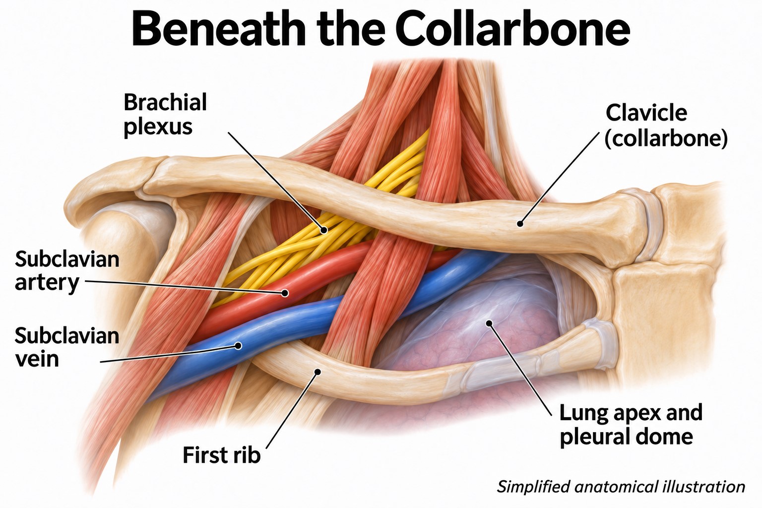
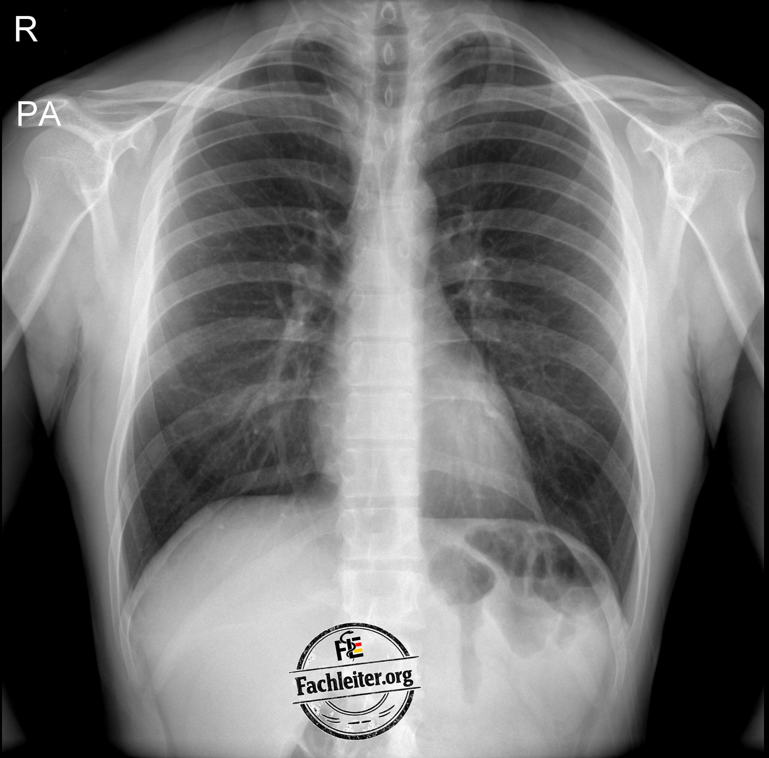
It also transmits forces from the arm to the trunk and protects the underlying structures. These include in particular the subclavian artery and vein, more laterally the axillary vessels, as well as the brachial plexus. Posterior to the medial portion of the clavicle lies the lung apex with the pleural dome.
Mechanism of injury and typical complaints
The most common mechanism of injury is a direct fall onto the lateral shoulder. Less often the fracture is caused by a direct blow to the collarbone or a fall onto the outstretched arm.
Typical complaints and findings are:
- Pain, swelling and hematoma over the collarbone.
- Pressure pain and painfully limited shoulder mobility.
- Visible deformity or palpable step-off.
- Protective posture: the injured arm is often supported with the healthy hand.
Examination includes assessment of the skin and checking circulation, motor function and sensation of the affected arm. With more severe trauma, additional injuries of the shoulder girdle and thorax must be excluded.
Diagnostics
Basic diagnostics are performed by X-ray of the clavicle:
-
Anteroposterior view – a. p.
In medial fractures, unclear joint involvement or suspicion of posterior displacement, computed tomography (CT) is often required. If vascular injury is possible, CT angiography may be considered.
Fracture classification
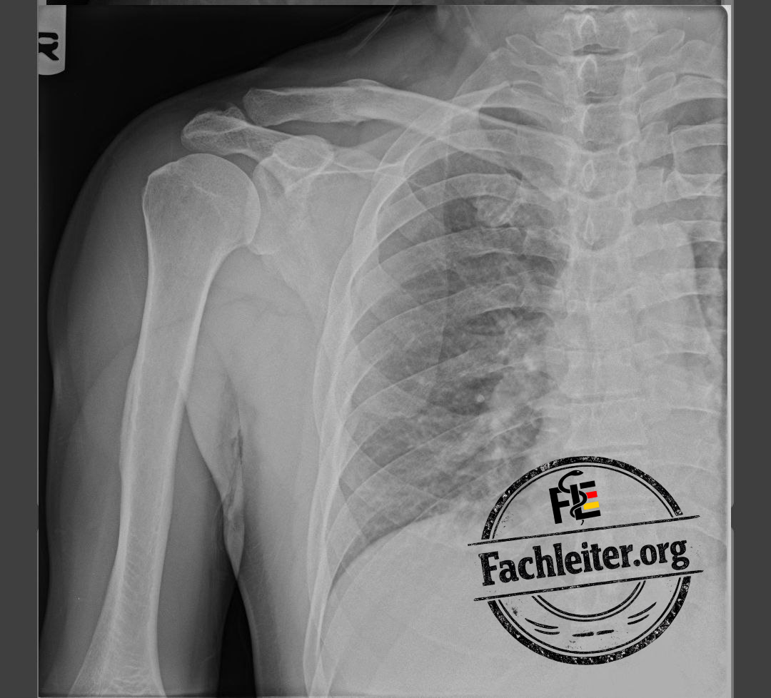
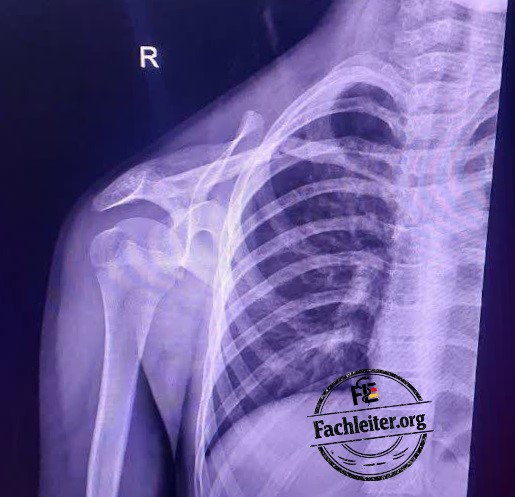
The middle third is most commonly affected.
Conservative treatment
Non-displaced or minimally displaced fractures are usually treated conservatively.
Treatment comprises an arm sling, adequate pain management and gradual mobilization. The arm sling is usually worn for about two to four weeks, depending on pain, fracture stability and the physician’s follow-up plan. In some cases, longer support up to about six weeks is reasonable.
Operative treatment
Urgent surgical management is required or should be considered particularly in the following findings:
-
Open fracture.
-
Impending skin perforation or threatened skin over a fragment.
-
Vascular injury or relevant nerve injury.
-
Unstable concomitant injury of the shoulder girdle.
In adults, completely displaced fractures, pronounced comminution or marked shortening may also argue for surgery. Shortening alone, however, is not an automatic indication for operation. The decision takes into account fracture pattern, symptoms, activity level and the individual advantages and disadvantages.
Stabilization is usually achieved with a plate and screws
For fractures in the lateral third it is crucial whether the coracoclavicular ligaments continue to sufficiently stabilize the shoulder girdle.
To assess the stabilizing function of the coracoclavicular ligaments, X-rays are first taken. The ligaments themselves are not directly visible on these. Clues to an unstable injury are the fracture course, displacement of fragments and an increased distance between the clavicle and the coracoid process. Intact ligaments can also attach to a broken bone fragment and thereby lose their stabilizing effect. In unclear cases, an MRI can be performed additionally to directly assess the ligaments if the result would influence the treatment decision.
Conservative treatment
Stable, non-displaced or minimally displaced fractures are usually treated with an arm sling, pain management and subsequent mobilization. The duration of sling treatment depends on stability and symptoms; it is often about two to four weeks.
Operative treatment
For unstable and markedly displaced fractures, in particular Neer type II and often type V, surgical treatment is considered. These injuries have an increased risk of failure of bony healing.
Possible procedures are an angular-stable lateral plate and, if necessary, additional coracoclavicular stabilization.
Medial fractures are rare. It is particularly important to determine whether a fragment is displaced posteriorly toward the mediastinum.
Conservative treatment
Non-displaced and stable fractures can usually be treated with an arm sling, pain management and cautious mobilization. The duration of immobilization is determined by symptoms and fracture stability.
Operative treatment
With marked displacement, instability, open injury or threat to adjacent structures, operative treatment should be considered.
Posterior displacement can endanger large vessels, the trachea or the esophagus. Shortness of breath, swallowing difficulties, hoarseness or circulatory disturbances require immediate evaluation.
X-ray
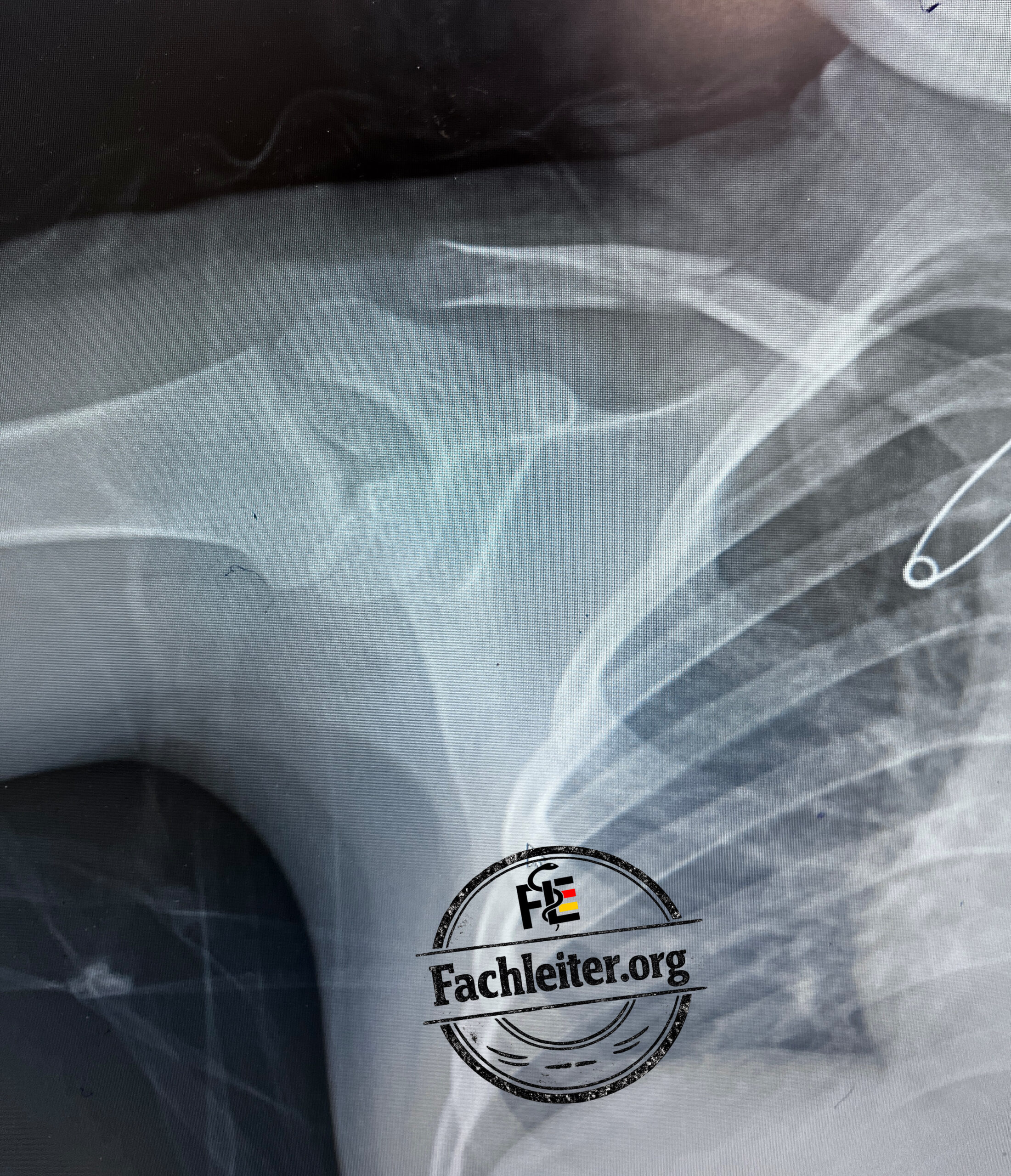
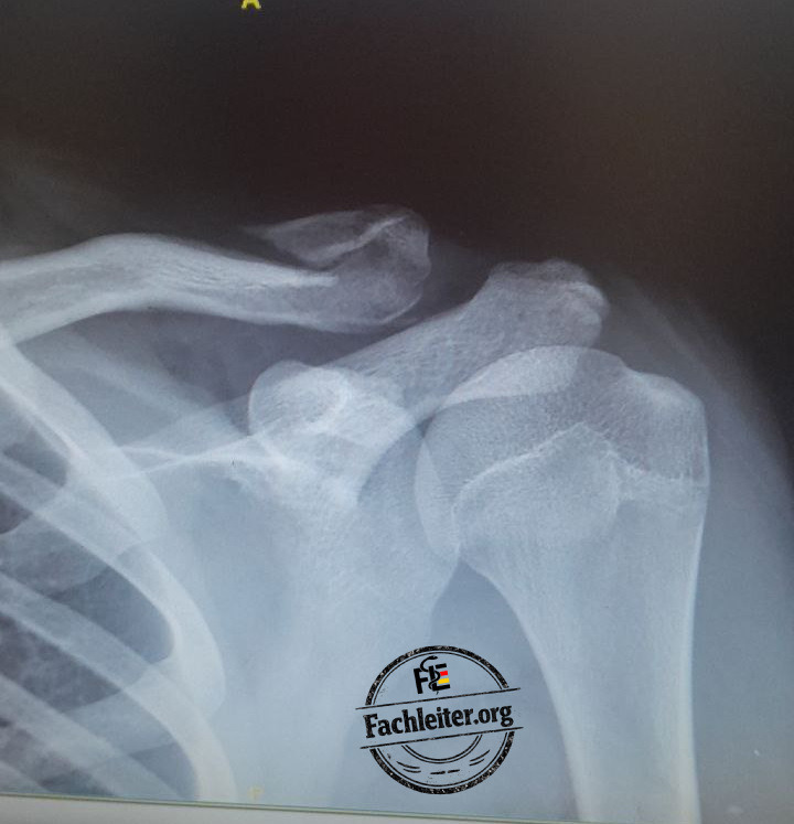
Describe the findings on each image. Pay particular attention to the fracture location, fracture course, number of fragments and the extent and direction of displacement. Also assess any angulation, shortening or overlap of the fragments as well as joint position and recognizable concomitant injuries of adjacent bony structures and soft tissues.
Pain management at home
| Medication, oral | Single dose | Interval as needed | Maximum dose in 24 hours |
|---|---|---|---|
| Ibuprofen | 400 mg | At least 6 hours | 1,200 mg without medical prescription |
| Paracetamol | 500–1,000 mg | At least 6 hours | 4,000 mg |
| Metamizole, prescription only | 500–1,000 mg | 6–8 hours | 4,000 mg |
| Naproxen | 250 mg | 8–12 hours | 750 mg without medical prescription |