Deep vein thrombosis (DVT) is one of the most important differential diagnoses for acute unilateral leg pain. In this article you will learn risk factors, typical symptoms, differential diagnoses, diagnostics, therapy and possible complications such as pulmonary embolism — ideal for FSP, knowledge exam, M3 and clinical practice.
Tiefe Venenthrombose (TVT) – Differenzialdiagnosen, Risikofaktoren und Komplikationen
Main complaint
- Typically, a patient over 40 presents with acute pain in one lower leg.
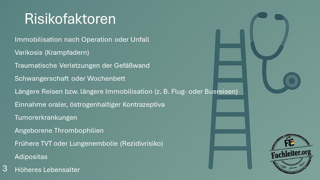
Risk factors
- Immobilization after surgery or trauma
- Varicose veins
- Traumatic injury to the vessel wall
- Pregnancy or puerperium
- Long journeys or prolonged immobilization (e.g., long flights or bus trips)
- Use of oral, estrogen-containing contraceptives
- Malignancy
- Inherited thrombophilias
- Previous deep vein thrombosis or pulmonary embolism (risk of recurrence)
- Obesity
- Advanced age
Differential diagnoses
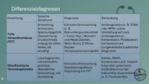
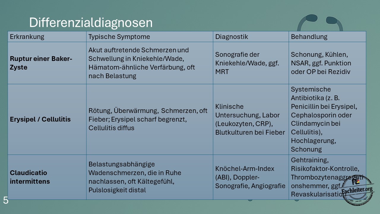
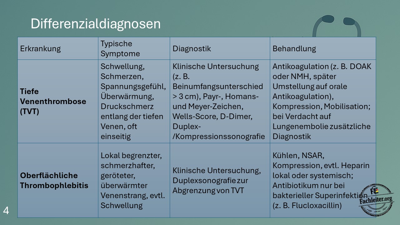
| Condition | Typical symptoms | Diagnostics | Treatment |
|---|---|---|---|
| Deep vein thrombosis (DVT) | Swelling, pain, a feeling of tightness, warmth, tenderness along the deep veins, usually unilateral | Clinical examination (e.g., calf circumference difference > 3 cm), Payr, Homans and Meyer signs, Wells score, D-dimer, duplex/compression ultrasound | Anticoagulation (DOAC or LMWH, followed by oral anticoagulation), compression, mobilization; if pulmonary embolism is suspected, additional diagnostics |
| Superficial thrombophlebitis | Localized painful, reddened and warm venous cord, possibly swelling | Clinical examination, duplex ultrasound to exclude DVT | Cooling, NSAIDs, compression, topical or systemic heparin if indicated; antibiotics only for bacterial superinfection |
| Rupture of a Baker’s cyst | Sudden onset of pain and swelling in the popliteal fossa or calf, bruise-like discoloration, often after exertion | Ultrasound of the popliteal fossa or calf, possibly MRI | Rest, cooling, NSAIDs, aspiration or surgical therapy for recurrent cases |
| Erysipelas / Cellulitis | Redness, warmth, pain, often fever; erysipelas is sharply demarcated, cellulitis is diffuse | Clinical examination, laboratory (CRP, leukocytes), blood cultures if febrile | Systemic antibiotics, elevation and rest |
| Intermittent claudication (PAD) | Exercise-dependent calf pain, improvement at rest, often coldness and absent distal pulses | Ankle-brachial index (ABI), Doppler ultrasound, angiography | Walking exercise, risk factor control, antiplatelet therapy, revascularization if indicated |
Further notes
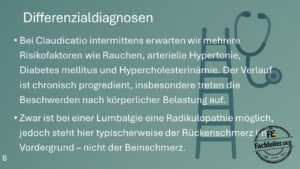
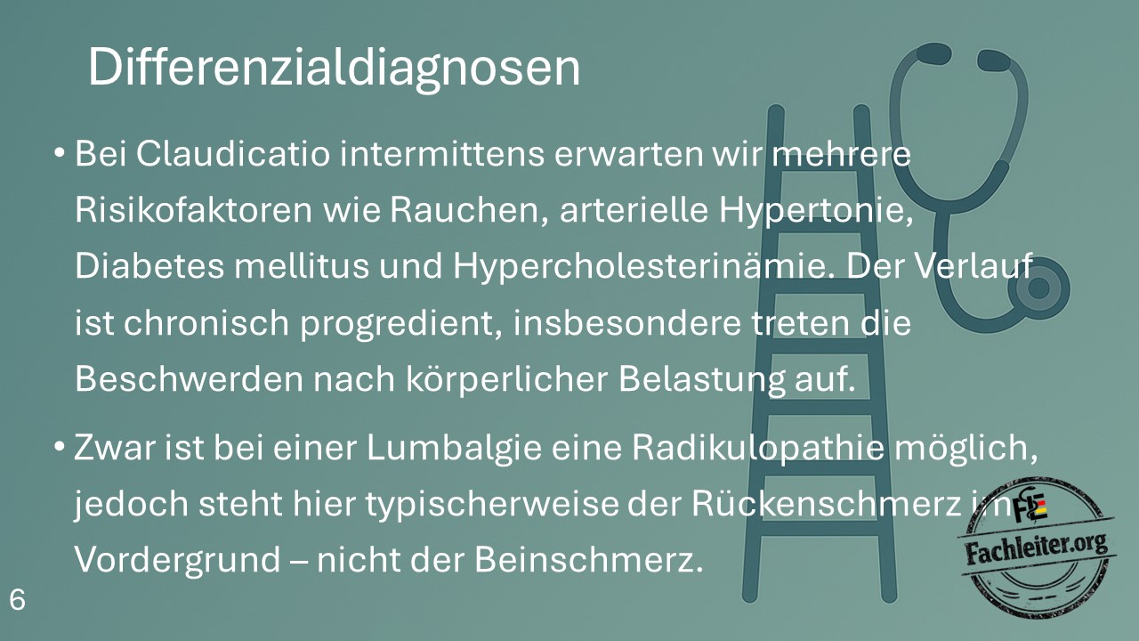
- In intermittent claudication multiple risk factors such as smoking, arterial hypertension, diabetes mellitus and hypercholesterolemia are often present.
- The course is chronically progressive; symptoms typically occur on exertion.
- In lumbosciatic pain a radiculopathy may be present, but back pain usually predominates rather than primary leg pain.
Complications
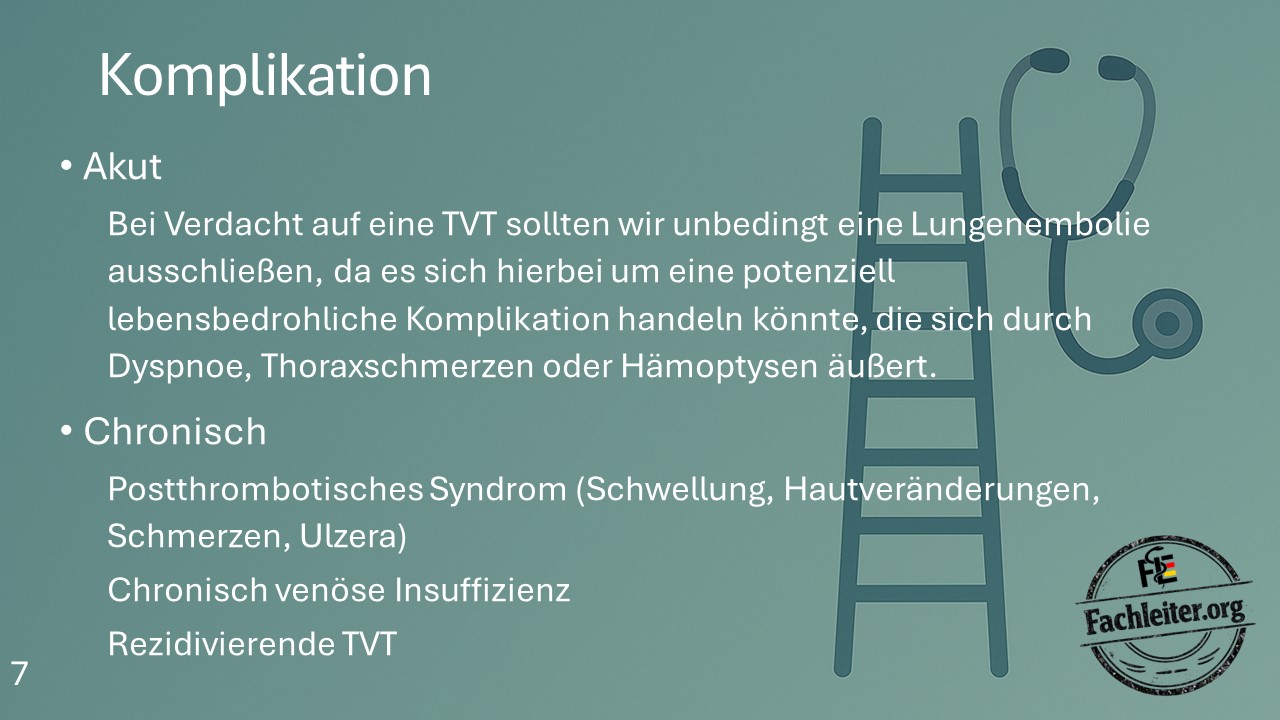
Acute
- If DVT is suspected, pulmonary embolism must always be excluded.
- Typical symptoms
Chronic
- Post-thrombotic syndrome (swelling, skin changes, pain, ulcers)
- Chronic venous insufficiency
- Recurrent deep vein thromboses