
Current medical history
A 68-year-old woman presents to the emergency department because of left-sided chest pain.
The complaints began at rest about 2 hours ago. The patient describes a persistent, stabbing pain in the left hemithorax. There is no clear radiation to the left arm, the back, the lower jaw, or the upper abdomen.
She rates the pain intensity as 5 out of 10 points.
The pain is:
- not related to respiration,
- not related to movement,
- not position-dependent,
- not reproducible by pressure on the thorax.
The patient cannot reliably assess improvement or worsening with physical exertion because the complaints began at rest and have persisted since then.
Associated symptoms
The patient denies:
- shortness of breath,
- nausea or vomiting,
- cold sweats,
- palpitations,
- dizziness or syncope,
- cough,
- fever,
- hemoptysis,
- recent chest trauma.
There are also no signs of deep vein thrombosis such as unilateral leg swelling or calf pain.
Relevant medical history
Known is arterial hypertension, which is treated with ramipril.
No known coronary artery disease is documented. No coronary angiography has been performed in the past. No percutaneous coronary intervention or bypass surgery has been performed either.
The patient denies:
- a previous myocardial infarction,
- diabetes mellitus,
- chronic renal insufficiency,
- known retinopathy,
- peripheral arterial occlusive disease,
- stroke or transient ischemic attack.
Long-term medication
- Ramipril, dosage not recalled with certainty
There is no regular intake of antiplatelet agents or anticoagulants.
Allergies and noxae
No known drug allergies.
Smoking and alcohol consumption are denied.
Vital signs on admission
- Blood pressure: 130/80 mmHg
- Heart rate: 76/min
- Respiratory rate: 14/min
- Oxygen saturation: 97 % on room air
- Body temperature: 36.8 °C
The patient is awake, responsive and hemodynamically stable.
Physical examination
The patient is in a generally good condition for her age. She is oriented to time, place, person and situation.
Cardiovascular system
- Heart sounds clear and regular
- No abnormal heart murmurs
- Peripheral pulses palpable bilaterally
- No jugular venous distension
- No peripheral edema
- Capillary refill time normal
Lungs
- Vesicular breath sounds bilaterally
- No crackles
- No wheeze
- No side-to-side difference
- No signs of acute respiratory failure
Thorax
- No tenderness to pressure
- No visible injuries
- No swelling or skin changes
- Pain not reproducible by palpation and movement
Abdomen
- Soft and non-tender
- No guarding
- No palpable pulsatile mass
- Bowel sounds present
Extremities
- No unilateral leg swelling
- No calf tenderness to pressure
- No clinical signs of deep vein thrombosis
ECG
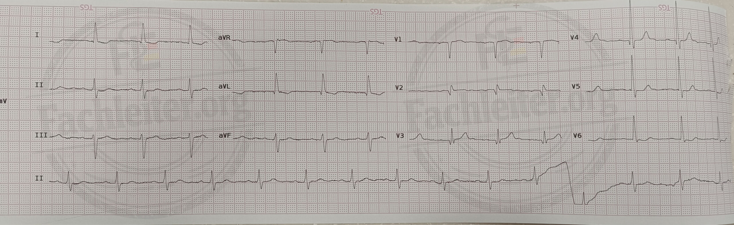
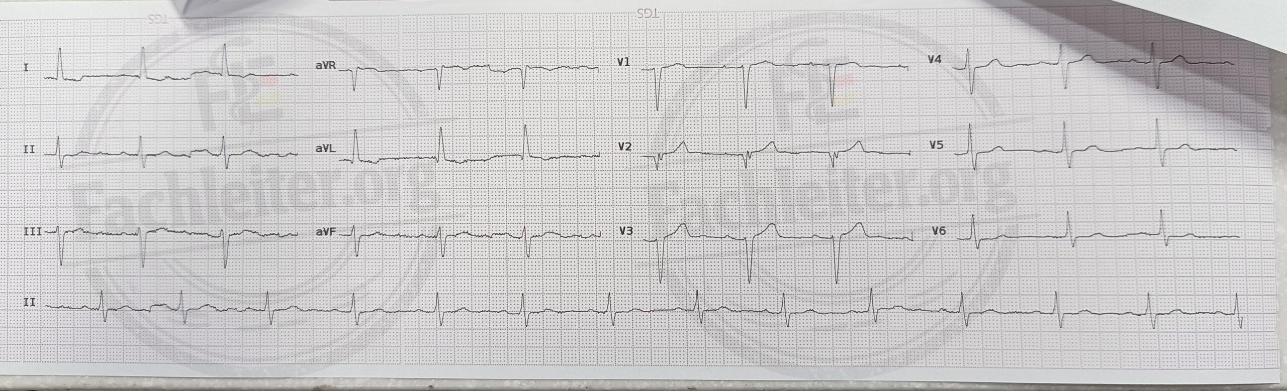
Quiz: How would you proceed?
Question 1
Which suspected diagnosis must initially be excluded despite the non-classical pain description?
Question 2
How do you assess the Q-waves in the inferior leads?
- Definite acute inferior myocardial infarction
- Possible indication of a prior infarction
- Definite normal finding
- Proof of acute pericarditis
Question 3
What causes could explain the lack of R-wave progression?
Question 4
Would you start a full antithrombotic acute therapy immediately based solely on the Q-waves and the PRWP?
Question 5
Which investigations should be performed immediately?
Question 6
With a symptom duration of about 2 hours, is a single normal hs-troponin value sufficient to rule out an acute myocardial infarction?
Question 7
After what symptom duration can, in a suitable clinical constellation and with a validated assay, a single very low hs-troponin value be used to rule out an acute myocardial infarction?