Back pain and lumbar disc prolapse explained practically: This article shows typical complaints, red flags, radicular symptoms in L4, L5 and S1 lesions, important differential diagnoses and therapy principles. Ideal for FSP, knowledge exams, M3 and clinical practice.
Back pain
Main complaint
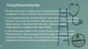
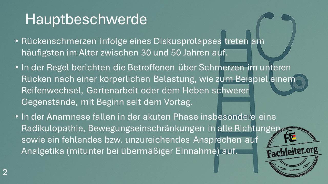
- Back pain due to a disc prolapse occurs most frequently between the ages of 30 and 50 years.
- Typically, affected patients report lower back pain after physical exertion, for example after changing a tire, gardening or lifting heavy objects, with onset since the day before.
- In the history, during the acute phase in particular radiculopathy, movement restrictions in all directions and a lack of or insufficient response to analgesics are notable, sometimes despite excessive intake.
Red Flags
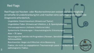
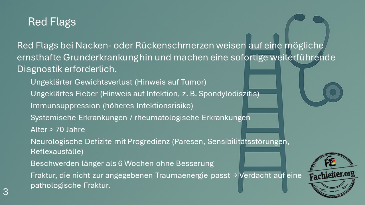
Red Flags in neck or back pain indicate a possible serious underlying condition and require immediate further diagnostics.
- Unexplained weight loss (suggestive of tumor)
- Unexplained fever (suggestive of infection, e.g. spondylodiscitis)
- Immunosuppression
- Systemic or rheumatologic diseases
- Age > 70 years
- Progressive neurological deficits (paresis, sensory disturbances, loss of reflexes)
- Complaints lasting longer than 6 weeks without improvement
- Fracture not consistent with reported trauma energy → suspicion of pathological fracture
Radiculopathy
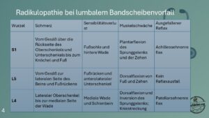
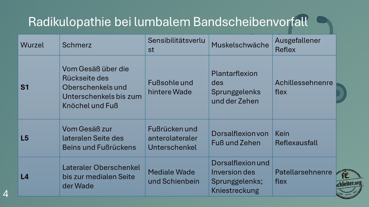
| Root | Pain | Sensory loss | Muscle weakness | Lost reflex |
|---|---|---|---|---|
| S1 | From the buttock along the back of the thigh and calf to the ankle and foot | Plantar surface of foot and posterior calf | Plantarflexion of the ankle and toes | Achilles tendon reflex |
| L5 | From the buttock to the lateral aspect of the leg and dorsum of the foot | Dorsum of the foot and anterolateral lower leg | Dorsiflexion of foot and toes | No typical reflex loss |
| L4 | Lateral thigh to the medial side of the calf | Medial calf and shin | Dorsiflexion and inversion of the ankle; knee extension | Patellar tendon reflex |
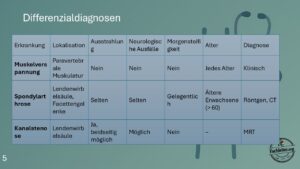
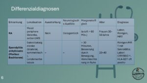
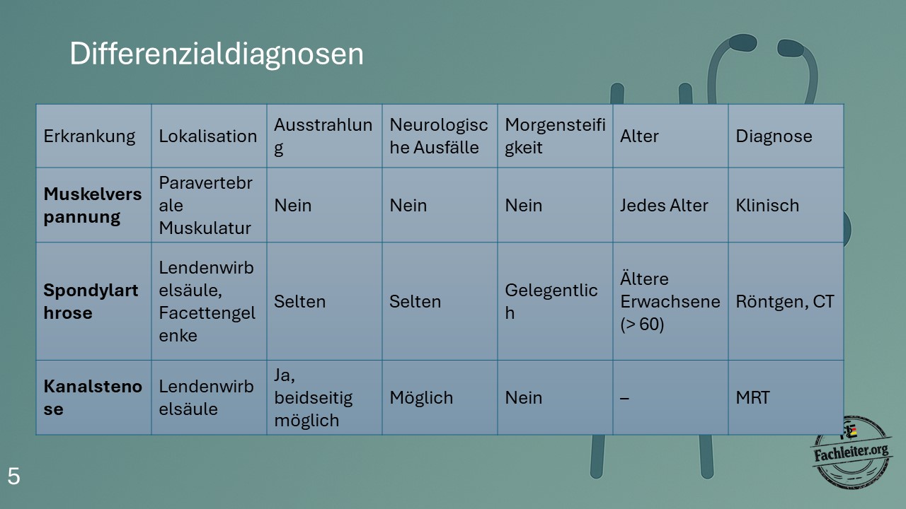
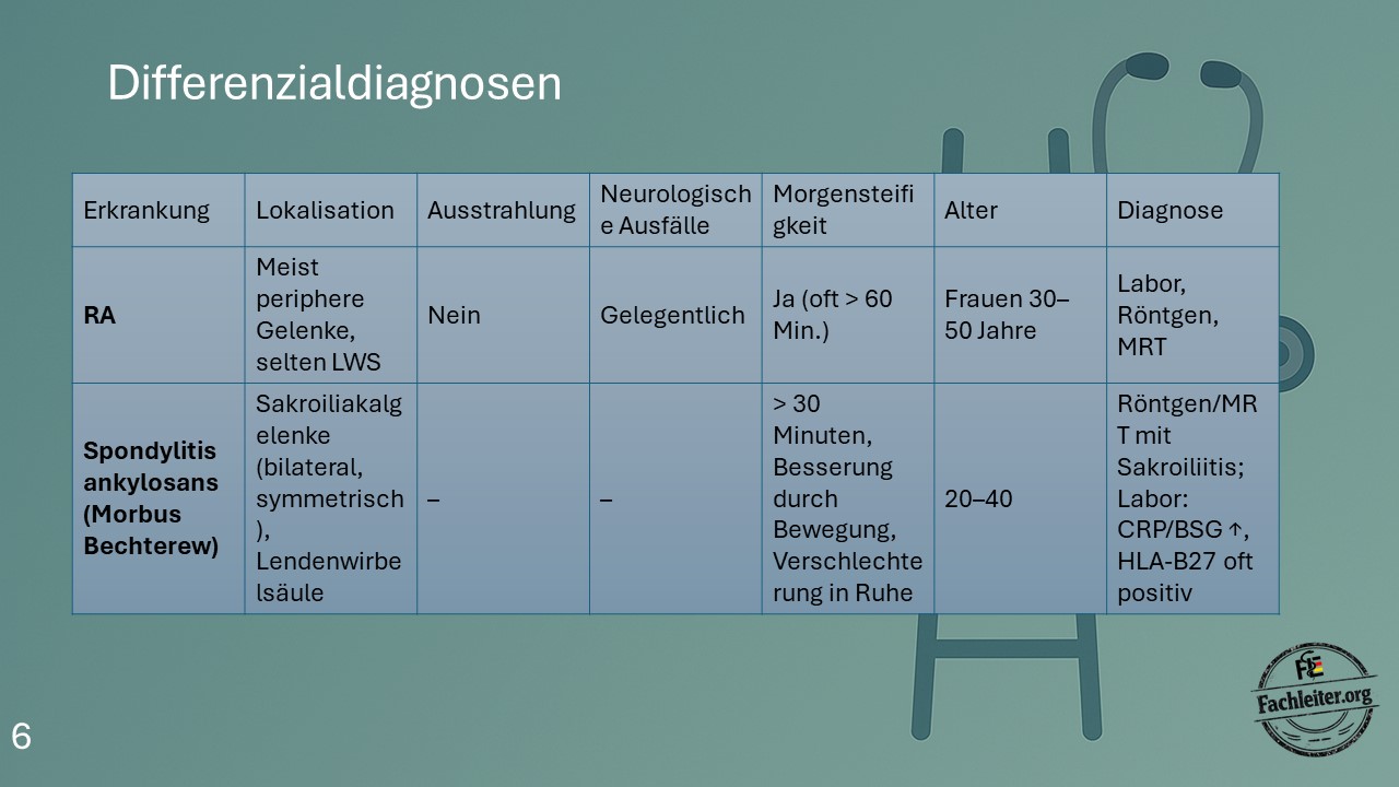
Differential diagnoses
| Condition | Location | Radiation | Neurological deficits | Morning stiffness | Age | Diagnosis |
|---|---|---|---|---|---|---|
| Muscle tension | Paravertebral muscles | No | No | No | Any age | Clinical |
| Spondylarthrosis | Lumbar spine, facet joints | Rarely | Rarely | Occasionally | Older adults (> 60) | X-ray, CT |
| Spinal canal stenosis | Lumbar spine | Yes, bilateral possible | Possible | No | – | MRI |
| Rheumatoid arthritis | Mostly peripheral joints, rarely lumbar spine | No | Occasionally | Yes, often > 60 minutes | Women 30–50 years | Laboratory, X-ray, MRI |
| Ankylosing spondylitis (Morbus Bechterew) |
Sacroiliac joints bilateral/symmetrical, lumbar spine | – | – | > 30 minutes, improvement with movement, worsening at rest | 20–40 years | X-ray/MRI with sacroiliitis; labs: CRP/ESR ↑, HLA-B27 often positive |
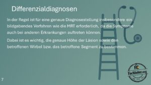
Diagnostic note
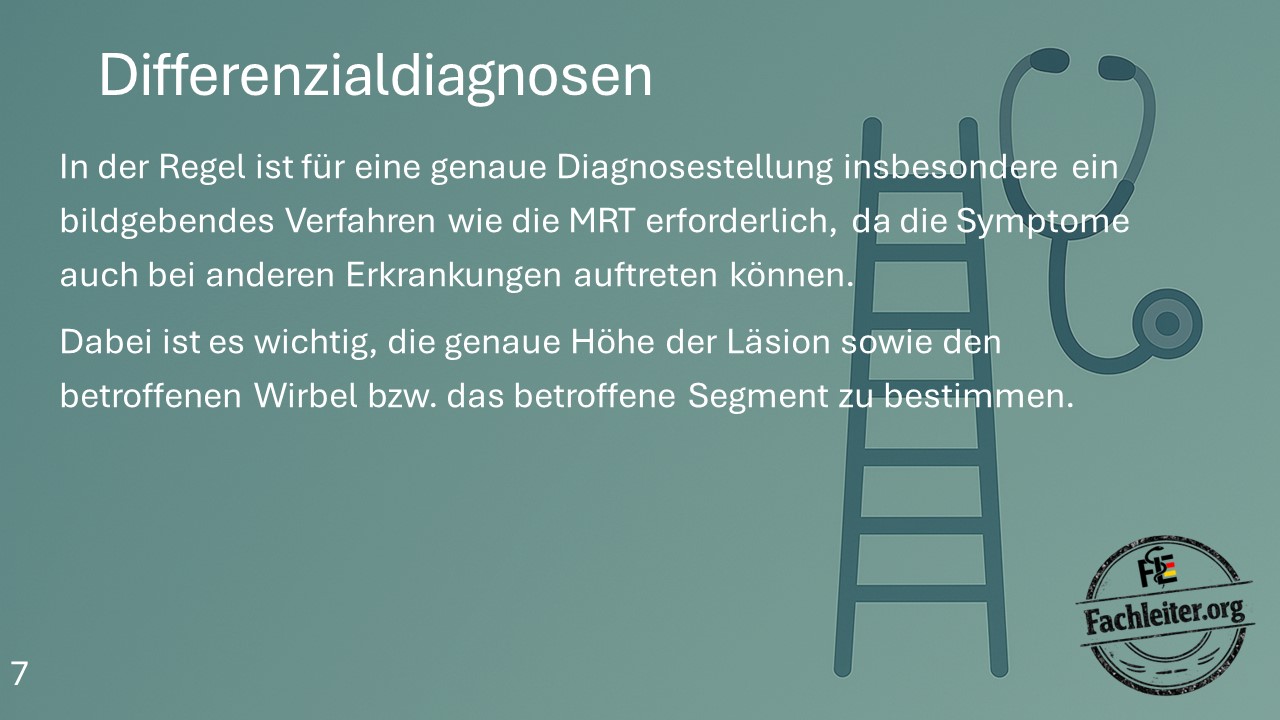
In general, imaging—particularly MRI—is required for an accurate diagnosis, since the symptoms can also occur in other conditions.
It is important to determine the exact level of the lesion and the affected vertebra or segment.
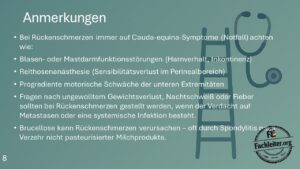
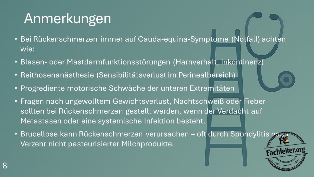
Notes
- Always watch for cauda equina symptoms in patients with back pain.
- Bladder or rectal dysfunctions, e.g. urinary retention or incontinence, are emergency signs.
- Saddle anesthesia means loss of sensation in the perineal area.
- Progressive motor weakness of the lower extremities is also a warning sign.
- Ask about unintended weight loss, night sweats or fever if metastases or a systemic infection are suspected.
- Brucellosis can cause back pain—often due to spondylitis after consumption of unpasteurized dairy products.
Therapy
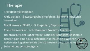
Therapeutic recommendations:
- Stay active – movement is recommended; avoid bed rest if possible.
- Medications: NSAIDs, e.g. ibuprofen or naproxen.
- Muscle relaxants: e.g. diazepam or tizanidine.
- In about 90% of patients with a lumbar disc herniation, an acute sciatica improves within 6 weeks and typically resolves completely within 12 weeks under conservative treatment.