Anemia explained — from symptoms through causes to diagnostics, laboratory values and therapy. In this article you will learn the most important forms of anemia, understand the significance of MCV, MCH and MCHC, find out when a blood transfusion or an intravenous iron therapy is necessary, and receive a practical overview of the diagnostics of iron deficiency anemia. Ideal for preparation for the Fachsprachprüfung (FSP), Kenntnisprüfung (KP), M3 as well as for everyday clinical practice.
Anemia – Symptoms, Causes, Diagnostics, Laboratory and Therapy
An anemia denotes a reduction in the hemoglobin concentration and/or the erythrocyte count. It is not an independent disease, but a symptom with numerous possible causes.
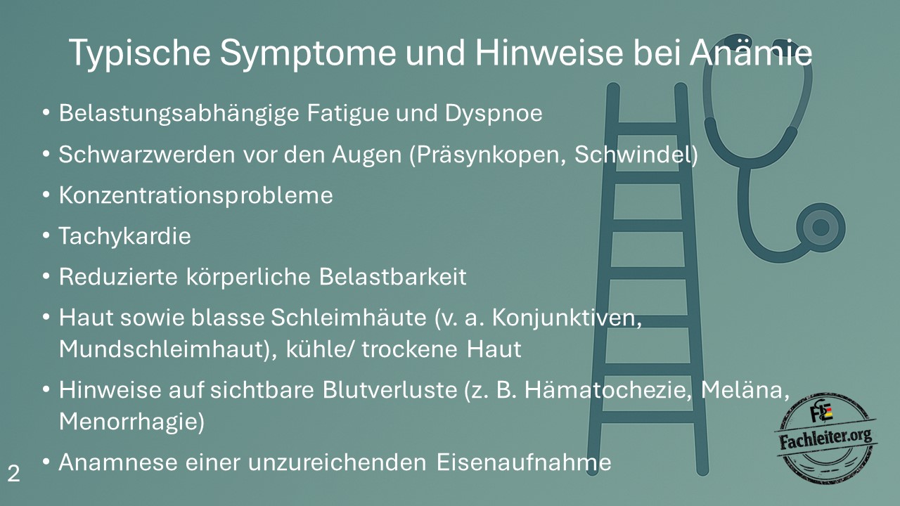
Typical Symptoms and Clues in Anemia
- Exertion-related fatigue and dyspnea
- Blackening of vision, presyncope or dizziness
- Concentration problems
- Tachycardia
- Reduced physical performance
- Pale skin and pale mucous membranes, especially conjunctivae and oral mucosa
- Cool and dry skin
- Signs of visible blood loss, e.g. hematochezia, melena or menorrhagia
- History of insufficient iron intake or malnutrition
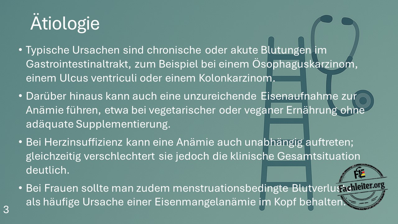
Etiology
Blood loss
The most common cause of iron deficiency anemia is chronic blood loss.
- Gastrointestinal bleeding
- Esophageal carcinoma
- Gastric ulcer
- Colon carcinoma
- Acute hemorrhages
- Chronic occult bleeding
In women, menstrual bleeding should additionally always be considered as a frequent cause.
Reduced iron absorption
- Vegetarian or vegan diet without sufficient iron intake
- Malnutrition
- Malabsorption syndromes, e.g. celiac disease or status post gastric surgery
Chronic diseases
- Heart failure
- Chronic inflammatory diseases
- Rheumatologic diseases
- Malignant diseases
An existing anemia significantly worsens the overall clinical situation, especially in heart failure.
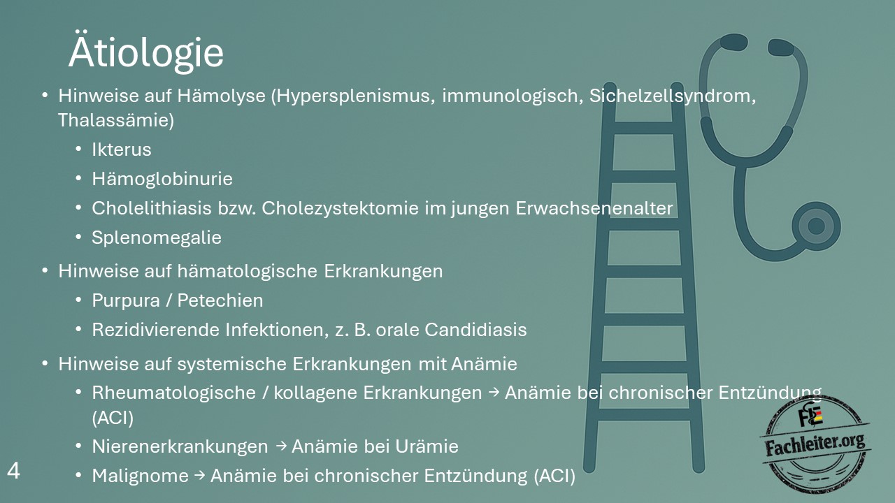
Hemolysis
Hemolytic anemia should be considered in cases of:
- Hypersplenism
- Autoimmune hemolysis
- Sickle cell disease
- Thalassemia
Typical clues are:
- Jaundice
- Hemoglobinuria
- Splenomegaly
- Cholelithiasis or cholecystectomy in young adulthood
Hematological diseases
- Petechiae
- Purpura
- Recurrent infections
- Oral candidiasis
- Leukemias
- Bone marrow disorders
- Aplastic anemia
Systemic diseases
- Rheumatologic diseases → anemia of chronic inflammation
- Chronic renal insufficiency → reduced erythropoietin production
- Malignant diseases → anemia of chronic inflammation
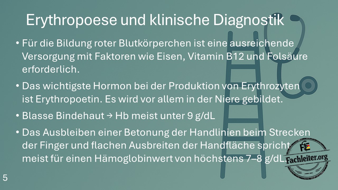
Erythropoiesis and Clinical Diagnostics
Formation of red blood cells requires:
- Iron
- Vitamin B12
- Folic acid
The most important hormone of erythropoiesis is Erythropoietin. It is produced predominantly in the kidney.
Clinical clues
- Pale conjunctivae often indicate an Hb value below 9 g/dL.
- Absence of the normal emphasis of the palmar creases with an outstretched palm often suggests an Hb value of about 7–8 g/dL or lower.
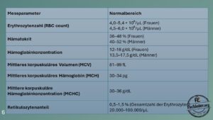
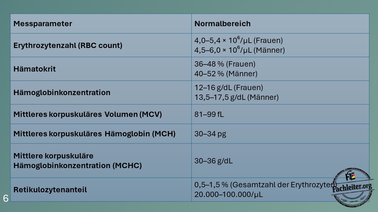
Important Laboratory Parameters
| Parameter | Normal range |
|---|---|
| Erythrocyte count (RBC) | Women: 4.0–5.4 × 10⁶/µL Men: 4.5–6.0 × 10⁶/µL |
| Hematocrit | Women: 36–48 % Men: 40–52 % |
| Hemoglobin | Women: 12–16 g/dL Men: 13.5–17.5 g/dL |
| MCV | 81–99 fL |
| MCH | 30–34 pg |
| MCHC | 30–36 g/dL |
| Reticulocytes | 0.5–1.5 % or 20,000–100,000/µL |
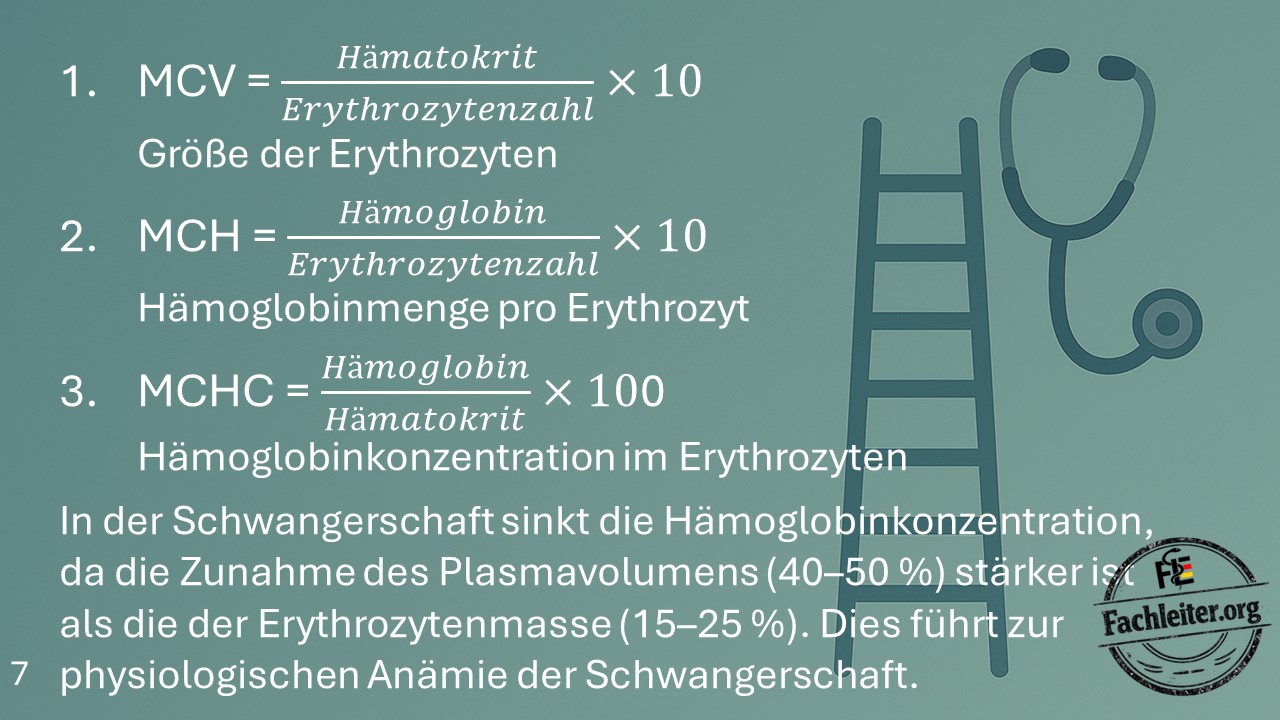
Red Cell Indices
MCV – Mean Corpuscular Volume
MCV = Hematocrit ÷ Erythrocyte count × 10
MCV describes the size of the erythrocytes.
MCH – Mean Corpuscular Hemoglobin
MCH = Hemoglobin ÷ Erythrocyte count × 10
MCH describes the hemoglobin amount per erythrocyte.
MCHC – Mean Corpuscular Hemoglobin Concentration
MCHC = Hemoglobin ÷ Hematocrit × 100
MCHC describes the hemoglobin concentration within an erythrocyte.
Physiological anemia of pregnancy
During pregnancy plasma volume increases by about 40–50 %, whereas erythrocyte mass increases only by about 15–25 %. As a result, the hemoglobin concentration physiologically decreases.
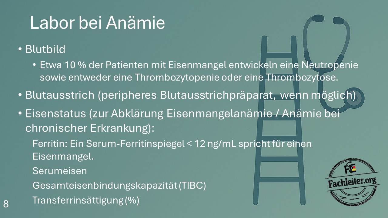
Laboratory in Anemia
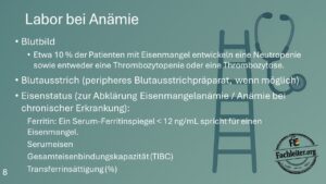
Complete blood count
In addition to anemia, the following may occur:
- Neutropenia
- Thrombocytopenia
- Thrombocytosis
About 10 % of patients with iron deficiency show such changes.
Peripheral blood smear
A blood smear provides important clues to:
- Microcytosis
- Macrocytosis
- Schistocytes
- Spherocytes
- Target cells
- Sickle cells
Iron status
To clarify an iron deficiency anemia, the following should be measured:
- Ferritin
- Serum iron
- Total iron binding capacity (TIBC)
- Transferrin saturation
A serum ferritin below 12 ng/mL strongly indicates iron deficiency.
Important: Ferritin is an acute-phase protein and can be normal or elevated in inflammation despite iron deficiency.
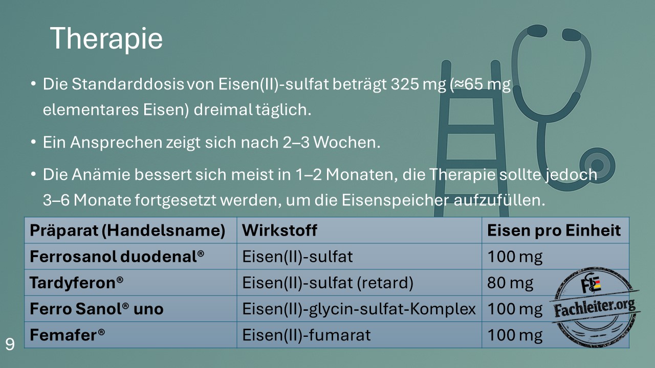
Therapy of Iron Deficiency Anemia
Treatment is guided by the cause of the anemia.
Oral iron supplementation
The classical standard dose is:
Iron(II) sulfate 325 mg ≈ 65 mg elemental iron, three times daily.
According to current recommendations, a lower dosing is often preferred, for example 40–100 mg elemental iron daily or every other day. This dosing is often better tolerated.
Therapeutic success
- Reticulocyte rise after about 7–10 days
- Increase in hemoglobin after 2–3 weeks
- Normalization usually within 1–2 months
- Continue therapy for 3–6 months to replenish iron stores
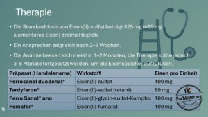
Commonly used preparations in Germany
| Brand name | Active ingredient | Elemental iron |
|---|---|---|
| Ferrosanol duodenal® | Iron(II) sulfate | 100 mg |
| Tardyferon® | Iron(II) sulfate, retard | 80 mg |
| Ferro Sanol® uno | Iron(II)-glycine-sulfate complex | 100 mg |
| Femafer® | Iron(II) fumarate | 100 mg |
Transfusion Indications
An erythrocyte transfusion is not based solely on the hemoglobin value, but primarily on the patient’s clinical situation.
- Hb < 7 g/dL: Transfusion is recommended in most hemodynamically stable adults.
- Hb 7–8 g/dL: Transfusion for symptoms or patients at increased risk (e.g. cardiovascular disease).
- Hb 8–10 g/dL: Individual decision, e.g. in active bleeding, coronary artery disease or pronounced symptoms.
- Hb > 10 g/dL: A transfusion is generally not required.
Typical symptoms of a transfusion-requiring anemia:
- Dyspnea at rest
- Chest pain
- Syncope or presyncope
- Tachycardia or hemodynamic instability
- Myocardial ischemia
When IV iron?
Intravenous iron supplementation is preferred when oral therapy is not possible, not sufficiently effective or too slow.
- Intolerance to oral iron preparations
- No sufficient effect under oral therapy
- Malabsorption syndromes (e.g. celiac disease, chronic inflammatory bowel disease, status post gastric surgery)
- Severe iron deficiency with rapid need for substitution
- Chronic renal insufficiency, especially under erythropoietin therapy
- Heart failure with iron deficiency (particularly symptomatic HFrEF)
- Active chronic inflammatory bowel disease
- Preoperatively in marked iron deficiency with planned surgery
Note: An erythrocyte transfusion replaces lost oxygen carriers but does not replenish iron stores. After a transfusion, therefore — if iron deficiency exists — additional iron supplementation should be given.
Note
- The most common cause of iron deficiency anemia in adults is chronic blood loss.
- In men and postmenopausal women, a gastrointestinal source of bleeding should always be sought.
- Ferritin is the most important laboratory parameter for detecting iron deficiency.
- MCV, MCH and MCHC help classify the anemia.
- Reticulocytes indicate bone marrow activity.
- The cause of the anemia should always be treated — not just the low hemoglobin value.