
Introduction
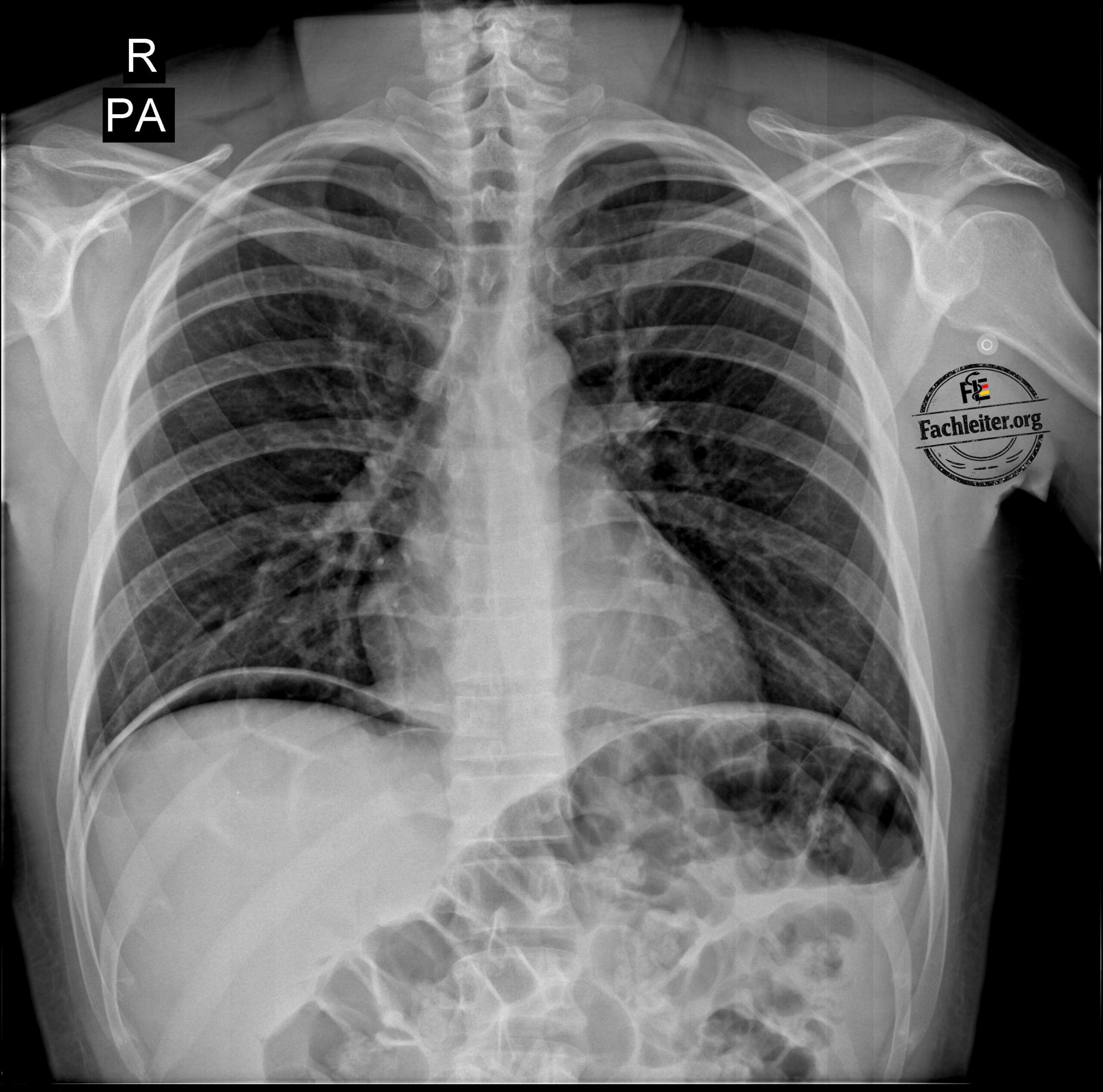
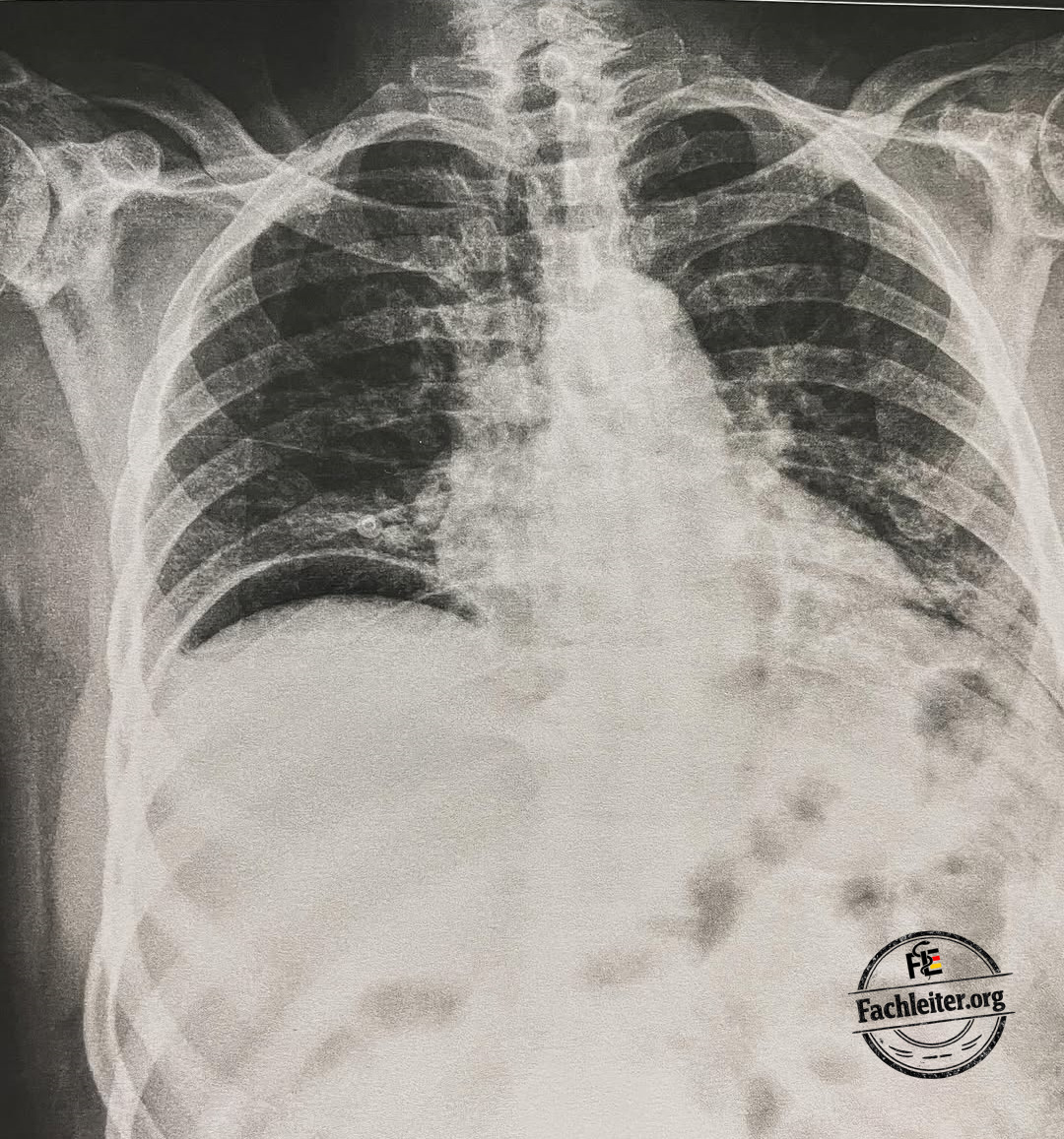
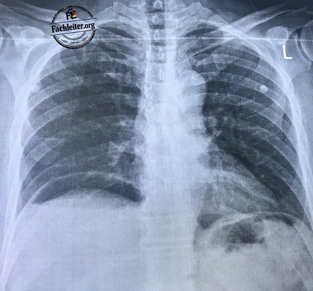
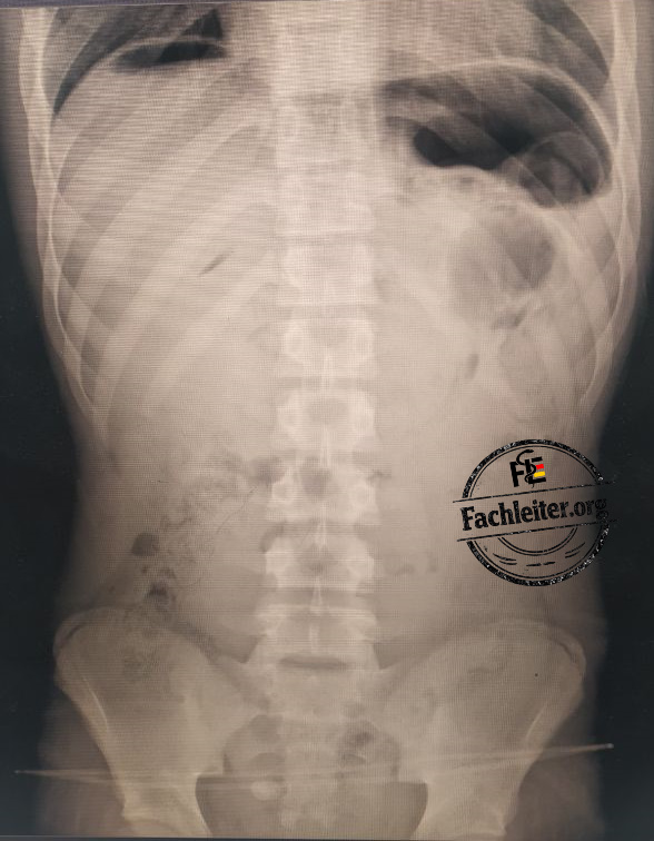
You see four X‑ray images with free air under the diaphragm.
- What is your suspected diagnosis?
- Which causes are possible?
- How would you proceed?
Free subdiaphragmatic air (pneumoperitoneum) is an important radiological finding and should — until proven otherwise — be considered an indication of a hollow-organ perforation.
What is free air under the diaphragm?
Free air under the diaphragm occurs when air from a hollow organ (e.g., stomach or intestine) enters the free peritoneal cavity.
On an upright chest or abdominal X‑ray, the air accumulates beneath the diaphragmatic domes and appears as a crescent-shaped lucency.
Common causes
1. Perforated gastric or duodenal ulcer (most common cause)
Typical symptoms:
- Sudden onset of severe upper abdominal pain
- Board‑hard abdomen (peritonism)
- Nausea and vomiting
Risk factors:
- Helicobacter pylori infection
- NSAID use
- Corticosteroids
- Smoking
- Older age
2. Bowel perforation
Typical symptoms:
- Severe, diffuse abdominal pain
- Fever
- Guarding
- Signs of sepsis
Typical symptoms (depending on cause):
- Diverticulitis: Pain in the left lower abdomen, fever, change in bowel habits
- Colon cancer: Weight loss, blood in stool, anemia, altered bowel habits
- Mesenteric ischemia: Sudden severe abdominal pain with an initially often unremarkable examination (“pain out of proportion”)
- After perforation: Suddenly increasing diffuse abdominal pain, peritonism, fever and signs of sepsis
3. Perforated appendicitis
Typical symptoms:
- Pain initially periumbilical, later in the right lower abdomen
- Fever
- Nausea and vomiting
- Peritonism
Risk factors:
- Delayed diagnosis
- Advanced age
- Diabetes mellitus
- Immunosuppression
4. Iatrogenic perforation
Typical symptoms:
- New onset abdominal pain after a procedure
- Tenderness on pressure
- Fever
- Peritoneal irritation
Risk factors:
- Colonoscopy
- Gastroscopy
- ERCP
- Laparoscopic or open surgeries
5. Traumatic hollow-organ injury
Typical symptoms:
- Abdominal pain after trauma
- Guarding
- Hemodynamic instability
- Signs of peritonitis
Risk factors:
- Blunt abdominal trauma
- Penetrating trauma
- Motor vehicle collision
- Fall
- Stab or gunshot wound
Further management
1. Clinical assessment
- Vital signs
- Hemodynamic stability
- Signs of peritonitis
- Exclude sepsis
2. History
- Onset of pain
- NSAID use
- History of ulcers
- Previous endoscopy or surgery
- Trauma
- Fever
- Vomiting
3. Laboratory tests
- Complete blood count
- CRP
- Procalcitonin
- Lactate
- Electrolytes
- Renal function
- Coagulation
- Blood type and crossmatch
4. Imaging
Gold standard:
CT abdomen with intravenous contrast
The CT can
- localize the site of perforation,
- identify the cause,
- assess the extent of free air,
- detect free fluid or abscesses.
5. Surgical consultation
If a hollow-organ perforation is suspected, general or visceral surgery should be involved early.
X‑ray images
Treatment
General initial measures
- ABCDE scheme and continuous monitoring of vital signs
- Immediate surgical consultation if a hollow-organ perforation is suspected
- Nil per os (NPO)
- Two large-bore peripheral IV lines
- Intravenous fluid therapy with crystalloid solutions
- Adequate analgesia
- Broad‑spectrum IV antibiotics as early as possible after diagnosis
- Intravenous proton pump inhibitors when upper GI perforation is suspected
- Blood tests: CBC, CRP, electrolytes, renal function, coagulation, lactate, blood type and crossmatch
- Nasogastric tube if needed (e.g., gastric decompression or ileus)
- Urinary catheter to monitor urine output in critically ill patients
- Oxygen therapy if hypoxia present
- CT abdomen with contrast, provided the patient is hemodynamically stable and no immediate emergency surgery is required