Description
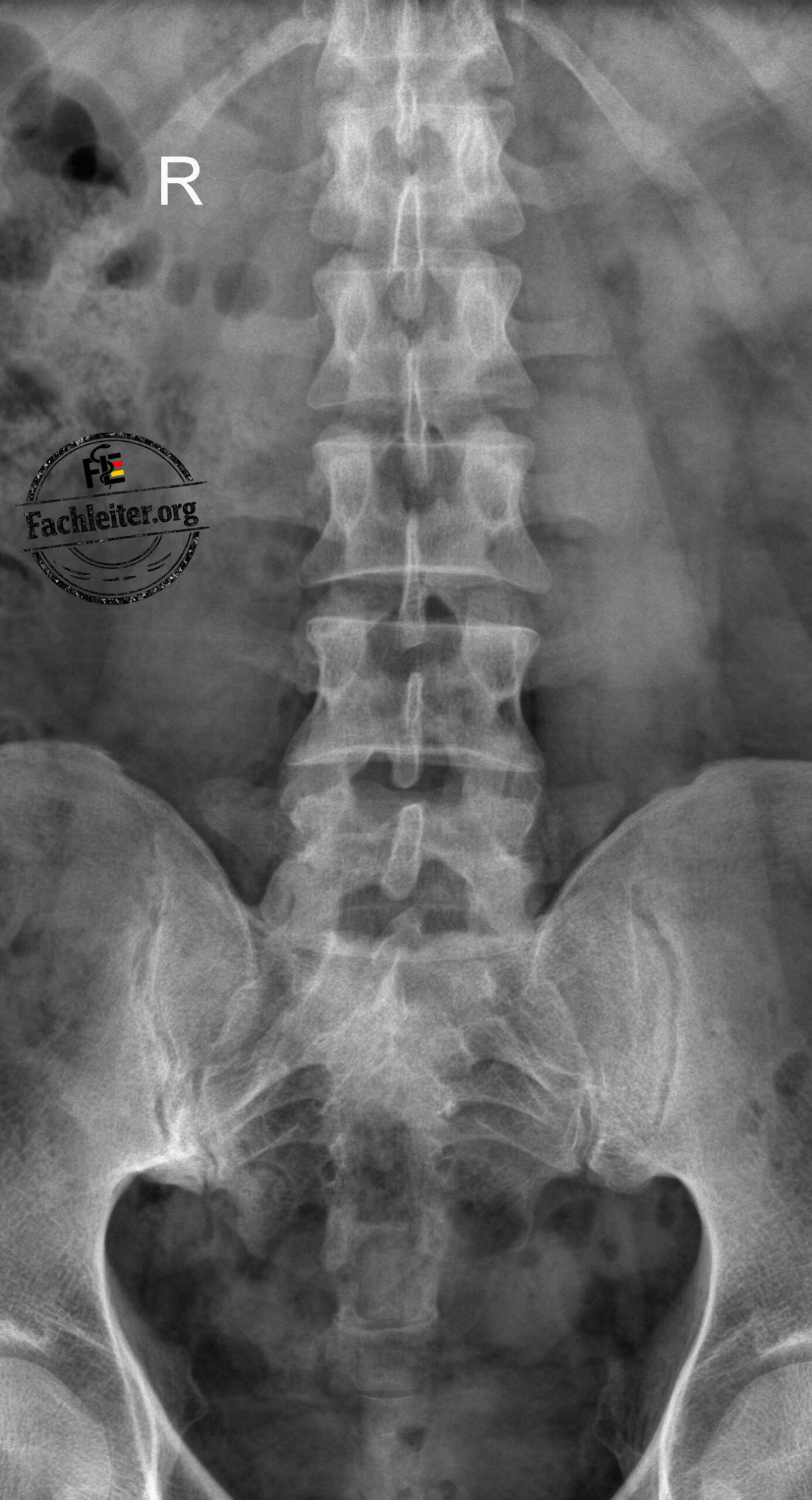
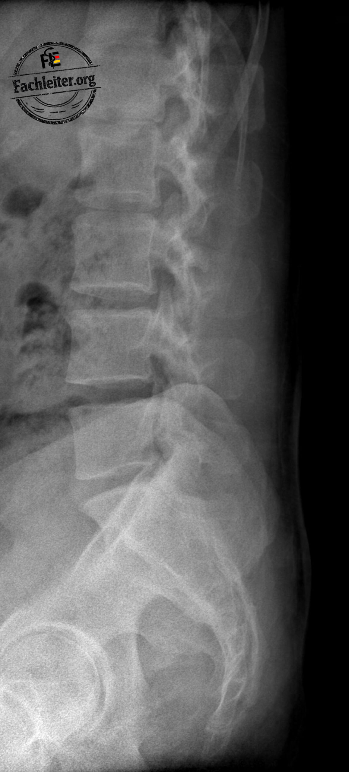
The assessment of a lumbar spine X-ray begins with a reliable anatomical orientation. In this article you will find two labeled standard views of the lumbar spine (AP and lateral), through which you can systematically learn the most important bony structures.
Marked are, among others, the vertebral bodies, pedicles, spinous and transverse processes, intervertebral disc spaces, the sacrum, the sacroiliac joints, and the hip joints. These anatomical landmarks form the basis for interpreting X-rays and facilitate the recognition of common pathologies such as fractures, spondylolisthesis, or degenerative changes.
The images are particularly suitable for medical students, physicians in training, and for preparation for the Fachsprachprüfung (FSP), knowledge examination (KP) and M3.
Anatomical structures
AP view
-
- Ribs
- Transverse processes
- Pedicles
- Spinous processes
- Intervertebral disc space L2/L3
- Vertebral body L4
- Sacrum (Sacrum)
- Sacroiliac joints (SIJ)
- Hip joint spaces
- Iliac wings (Ala ossis ilii)
Lateral view
- Lower ribs
- Vertebral body L3
- Intervertebral disc space L4/L5
- Sacrum in lateral view
- Anterior rim of the hip joint
- Posterior vertebral elements