Article text
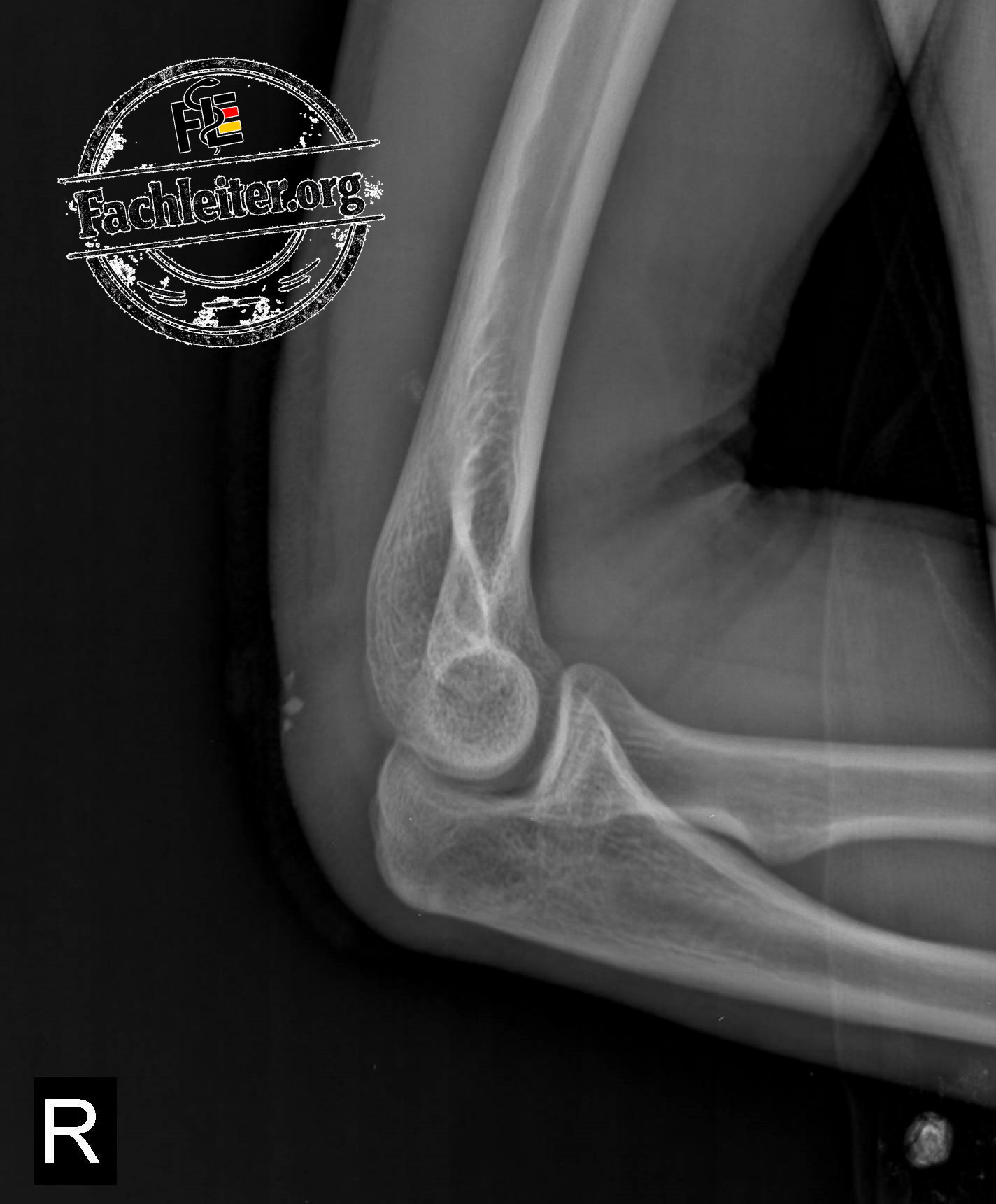
Recognizing normal anatomy is the basis of any X-ray interpretation. Those who can reliably identify the bony structures detect fractures, dislocations and malalignments much faster.
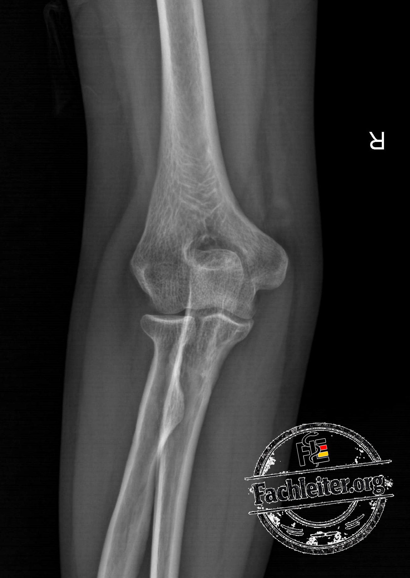
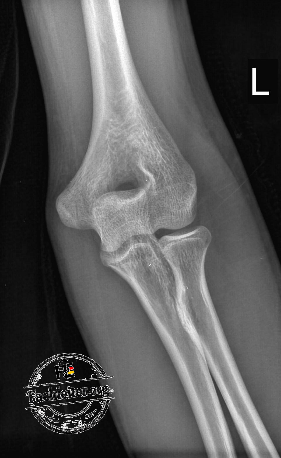
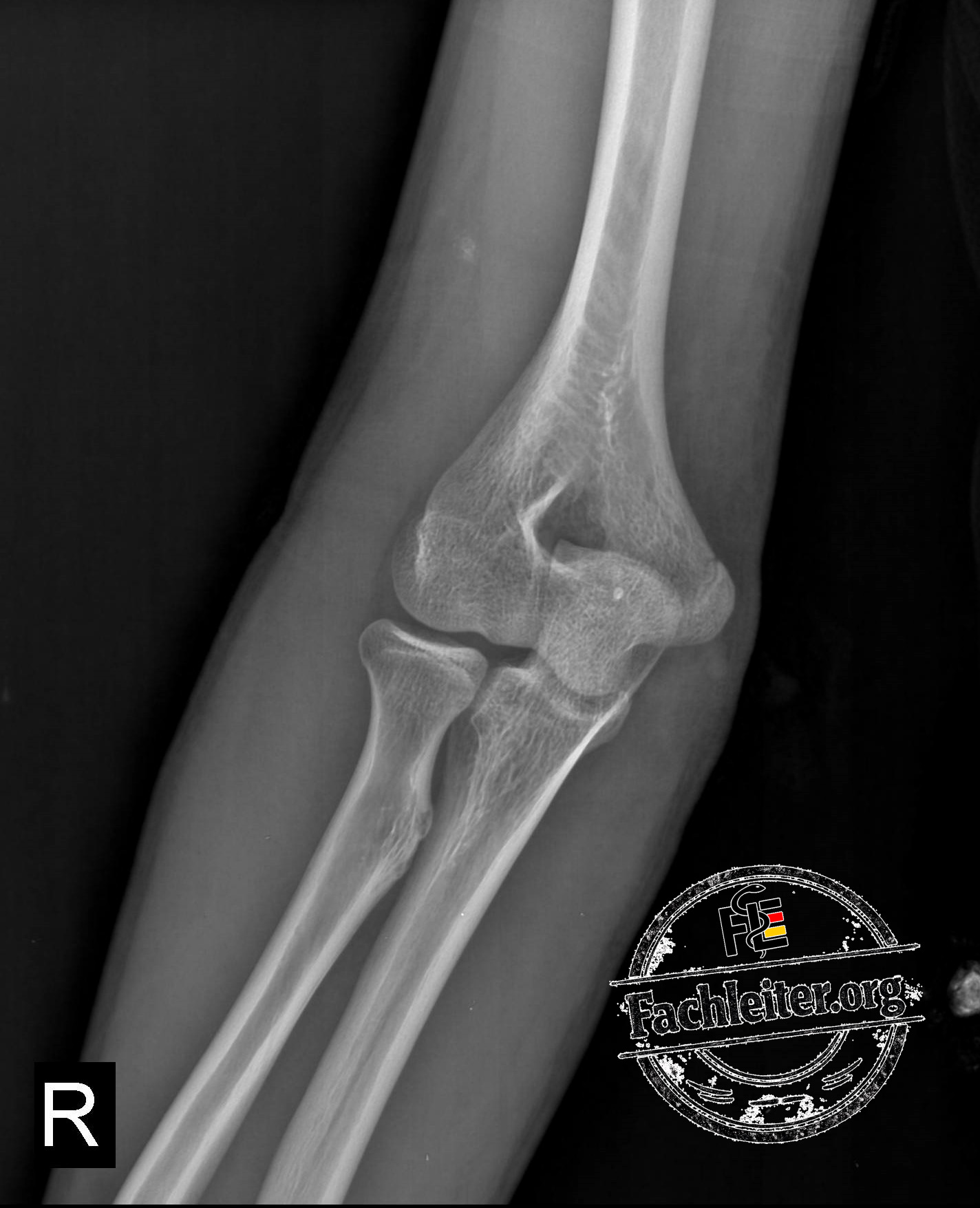
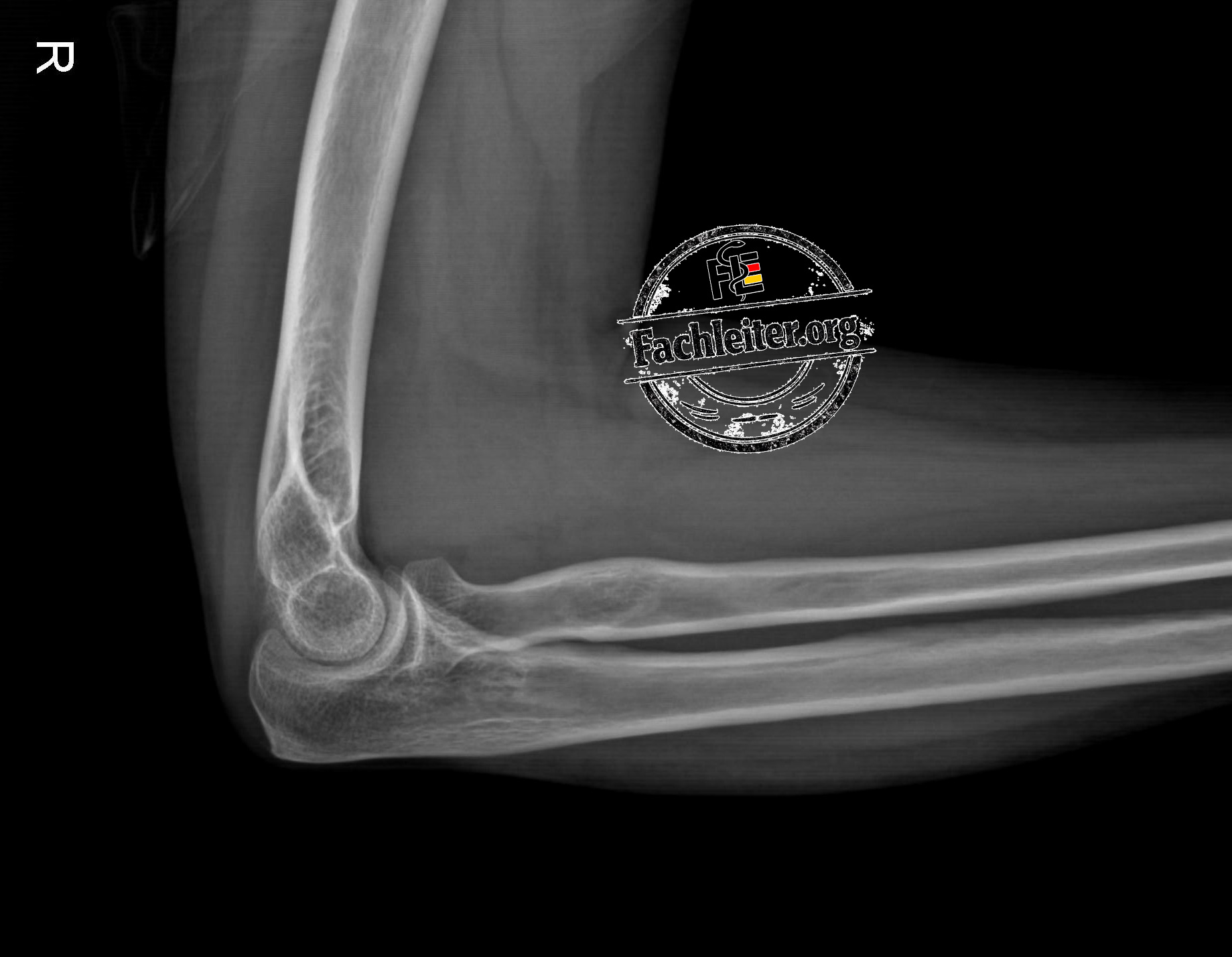
In this article, nearly all important anatomical structures of the elbow are marked on the AP and lateral views. In addition, you will find important reference lines that are regularly used in daily practice as well as in the knowledge exam, medical language exam and specialist exam.
In this article you will learn:
- Identify the distal humerus reliably
- Differentiate radius and ulna
- Identify the trochlea and capitulum
- Locate the olecranon and coronoid process
- Assess joint surfaces and joint spaces
- Recognize supracondylar ridges
- Understand the olecranon fossa and coronoid fossa
- Apply the Anterior Humeral Line (AHL)
- Assess the radiocapitellar line correctly
These anatomical landmarks form the basis for diagnosing common injuries such as:
- Radial head fractures
- Supracondylar humerus fractures
- Monteggia lesions
- Elbow dislocations
- Olecranon fractures
Tip: Learn the normal anatomy first before you interpret pathological X-rays. Those who master normal anatomy recognize abnormalities much faster.
AP
A
B
C
Lateral
A
B