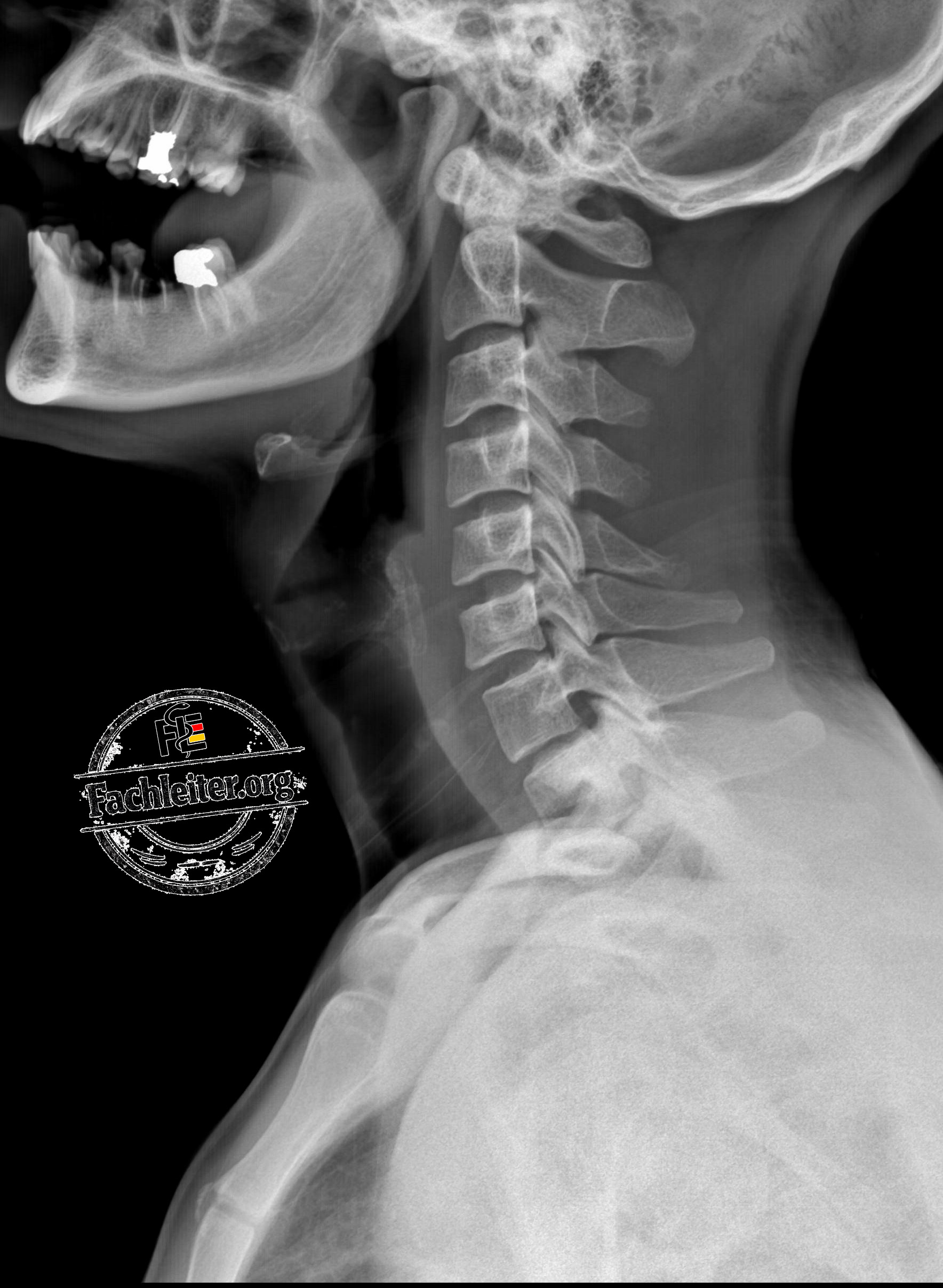
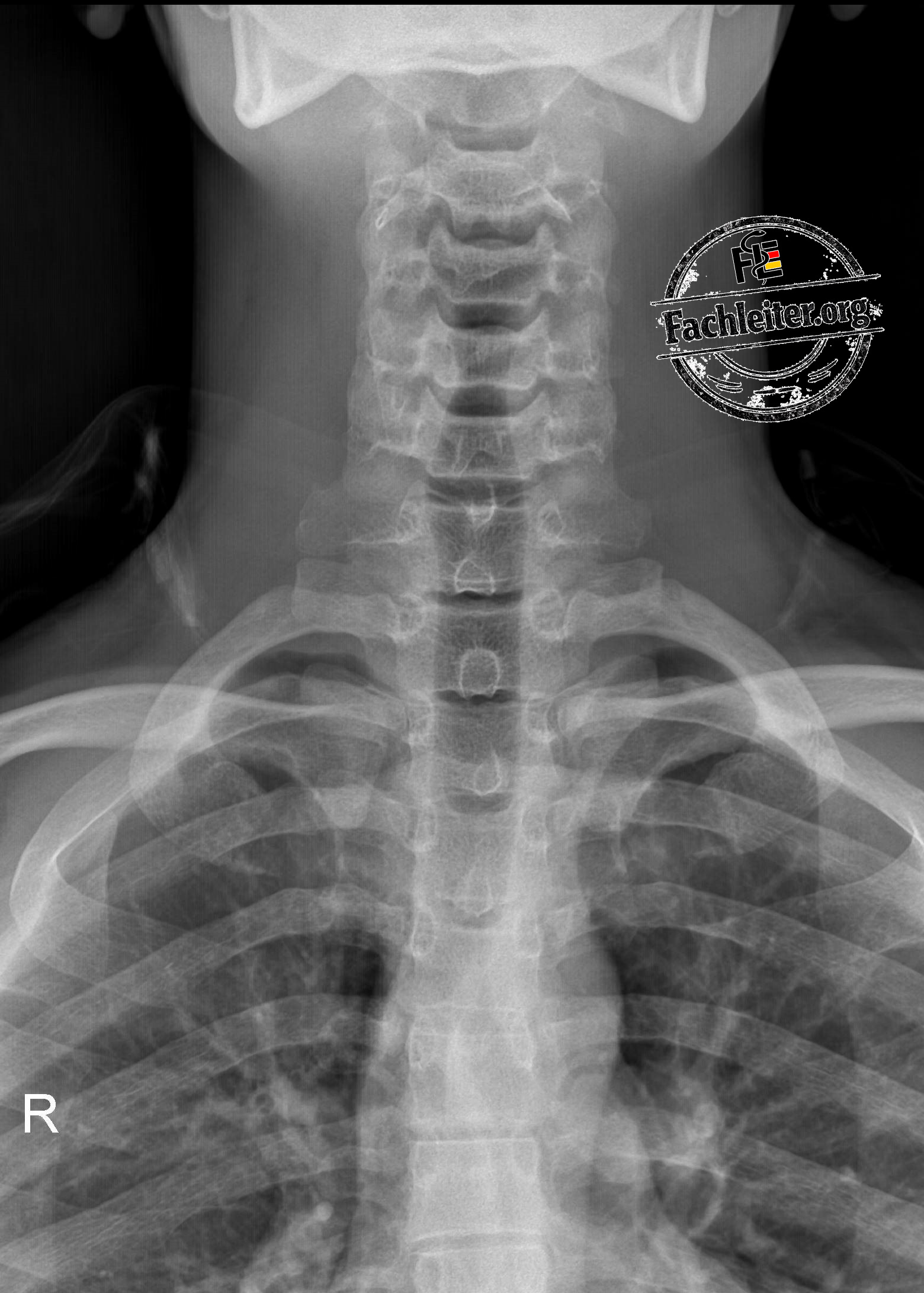
Anyone who wants to reliably assess X-rays of the neck soft tissues should first be able to recognize the normal anatomy. In this article, the most important anatomical structures of a neck soft-tissue image (AP and lateral) are labeled. The overview is particularly suitable for preparation for the knowledge exam (KP), for clinical everyday use, and for those interested in radiology.
Marked structures include:
- Mandibular angle and mandibular condyles
- Cervical spine (C1–C7)
- Atlas and Axis
- Vertebral bodies
- Spinous processes
- Transverse processes
- Pedicles
- Laminae
- Trachea
- Carina
- Thyroid gland
- Hyoid bone
- Clavicles
- Sternum (manubrium and body of the sternum)
- Lung apices
- Aortic arch
- Second rib (anterior and posterior portions)
Use the marked images to systematically learn the anatomical structures and then recognize them on unmarked X-rays.
Röntgen
a.-p.-Aufnahme (anteroposterior)
Seitliche Aufnahme (lateral)