
Normal Knee X-ray – Recognize Anatomy and Assess Systematically
Recognizing a normal radiograph is at least as important as diagnosing a fracture or other pathology. Only those who master the normal anatomy will later notice subtle abnormalities.
This interactive knee X-ray will help you learn the most important bony structures step by step.
Learning Objectives
- Safely recognize the normal anatomy of the knee joint
- Improve orientation in AP and lateral views
- Name important bony landmarks
- Learn systematic reporting of a knee X-ray
- Create a basis for recognizing fractures and degenerative changes
Röntgen
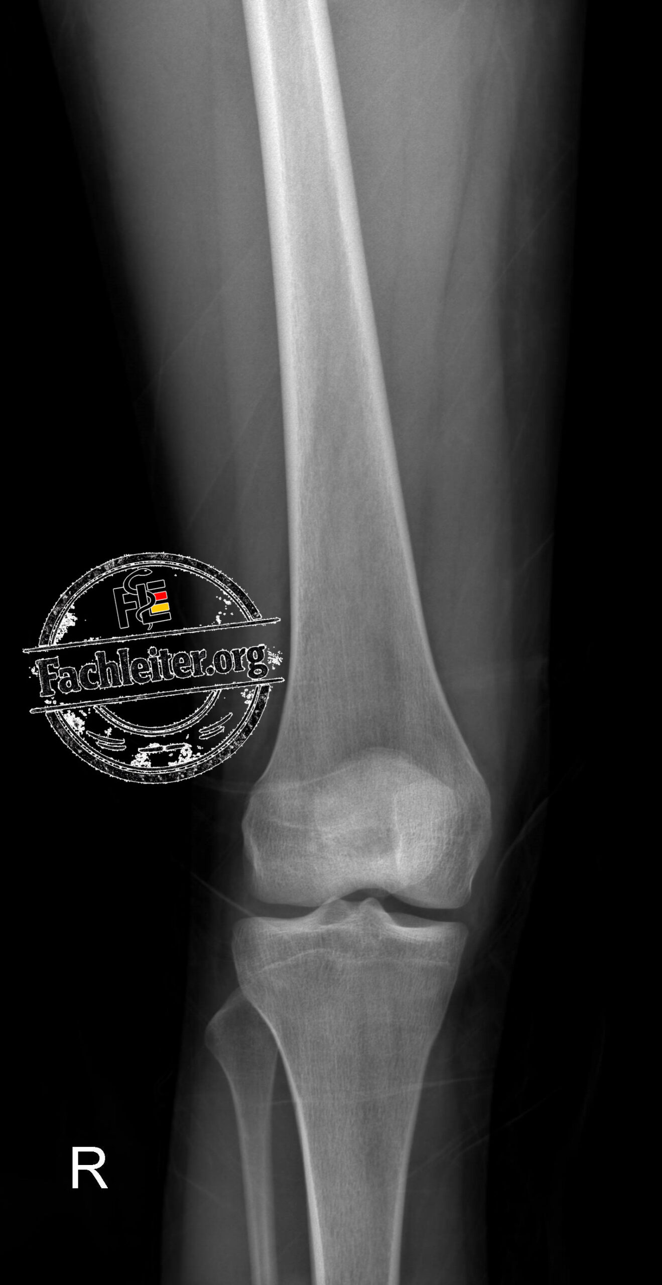
AP
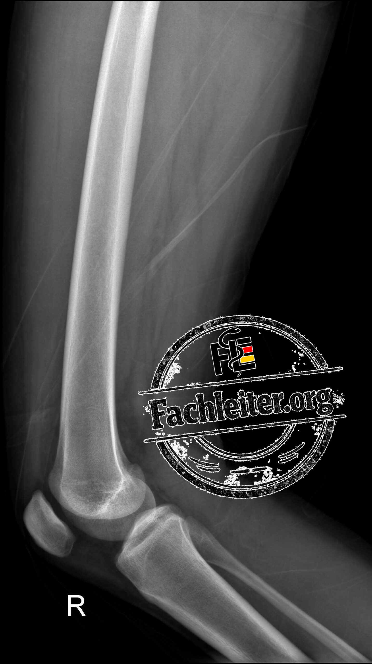
Lateral
Radiological Assessment
Examination: X-ray of the right knee in two planes (AP and lateral)
Indication:
18-year-old female patient after trauma to the right knee.
Findings
- Normal alignment of the knee joint.
- Intact cortices of the femur, tibia, fibula, and patella.
- No evidence of fracture or bony avulsion.
- No dislocation or subluxation.
- Preserved femorotibial and patellofemoral joint spaces.
- Normal joint surfaces without step-off or depression.
- Patella in normal position.
- No focal osteolytic or osteosclerotic bone lesions.
- No periosteal reaction.
- No definite evidence of a joint effusion on the lateral view.
- Unremarkable appearance of the visible soft tissues.
Assessment
- Unremarkable X-ray of the right knee in two planes.
- No evidence of an acute osseous injury.
- No fracture or dislocation.
- If symptoms persist or there is clinical suspicion of a ligament, meniscal, or cartilage injury, further MRI diagnostics are recommended.